

Is the diagnostic validity of conventional radiography for Lisfranc injury acceptable?
- PMID: 36855126
- PMCID: PMC9976526
- DOI: 10.1186/s13047-023-00608-0
Is the diagnostic validity of conventional radiography for Lisfranc injury acceptable?
Abstract
Background: Lisfranc injuries mainly involve the tarsometatarsal joint complex and are commonly misdiagnosed or missed in clinical settings. Most medical institutions prefer to use conventional radiography. However, existing studies on conventional radiographs in Lisfranc injury lack a large population-based sample, influencing the validity of the results. We aimed to determine the diagnostic validity and reliability of conventional radiography for Lisfranc injury and whether computed tomography can alter clinical decision-making.
Methods: This retrospective study included 307 patients with, and 100 patients without, Lisfranc injury from January 2017 to December 2019. Diagnosis was confirmed using computed tomography. A senior and junior surgeon independently completed two assessments of the same set of anonymised conventional radiographs at least 3 months apart. The surgeons were then asked to suggest one of two treatment options (surgery or conservative treatment) for each case based on the radiographs and subsequently on the CT images.
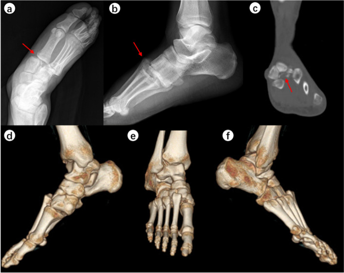
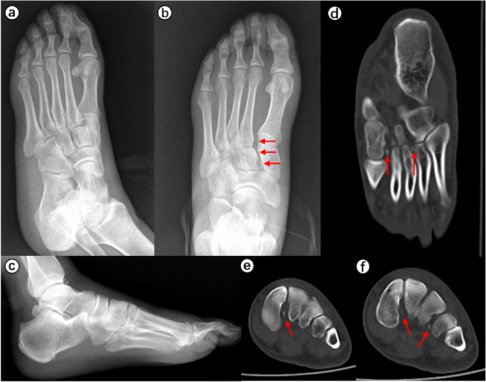
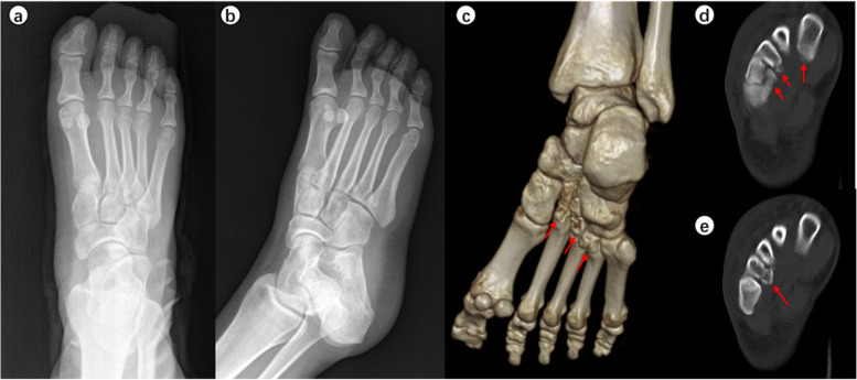
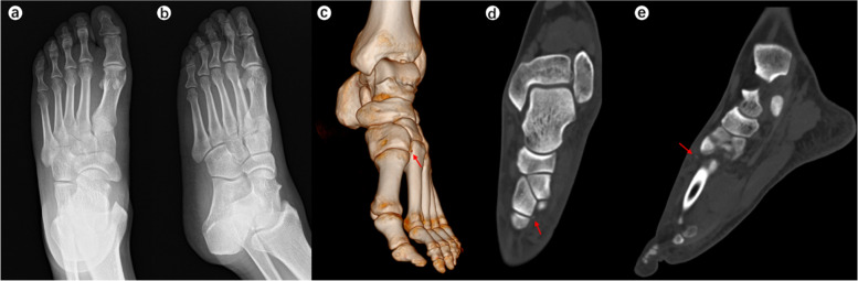
Results: All inter- and intra-observer reliabilities were moderate to very good (all κ coefficients > 0.4). The mean (range) true positive rate was 81.8% (73.9%-87.0%), true negative rate was 90.0% (85.0%-94.0%), false positive rate was 10.0% (6.0%-15.0%), false negative rate was 18.2% (13.0%-26.1%), positive predictive value was 96.1% (93.8%-97.8%), negative predictive value was 62.4% (51.5%-69.7%), classification accuracy was 83.8% (76.7%-88.2%), and balanced error rate was 14.1% (10.2%-20.5%). Three-column injuries were most likely to be recognized (mean rate, 92.1%), followed by intermediate-lateral-column injuries (mean rate, 81.5%). Medial-column injuries were relatively difficult to identify (mean rate, 60.7%). The diagnostic rate for non-displaced injuries (mean rate, 76.7%) was lower than that for displaced injuries (mean rate, 95.5%). The typical examples are given. A significant difference between the two surgeons was found in the recognition rate of non-displaced injuries (p = 0.005). The mean alteration rate was 21.9%; the senior surgeon tended to a lower rate (15.6%) than the junior one (28.3%) (p < 0.001).
Conclusions: The sensitivity, specificity, and classification accuracy of conventional radiographs for Lisfranc injury were 81.8%, 90.0%, and 83.8%, respectively. Three-column or displaced injuries were most likely to be recognized. The possibility of changing the initial treatment decision after subsequently evaluating computed tomography images was 21.9%. The diagnostic and clinical decision-making of surgeons with different experience levels demonstrated some degree of variability. Protected weight-bearing and a further CT scan should be considered if a Lisfranc injury is suspected and conventional radiography is negative.
Keywords: Clinical decision-making; Conventional radiographs; Diagnosis; Lisfranc injury; Validity.
© 2023. The Author(s).
Conflict of interest statement
The authors declare that they have no competing interests.
Figures
Similar articles
-
Inter- and intraobserver reliability of non-weight-bearing foot radiographs compared with CT in Lisfranc injuries.Arch Orthop Trauma Surg. 2020 Oct;140(10):1423-1429. doi: 10.1007/s00402-020-03391-w. Epub 2020 Mar 5. Arch Orthop Trauma Surg. 2020. PMID: 32140830 Free PMC article.
-
Imaging in Lisfranc injury: a systematic literature review.Skeletal Radiol. 2020 Jan;49(1):31-53. doi: 10.1007/s00256-019-03282-1. Epub 2019 Jul 31. Skeletal Radiol. 2020. PMID: 31368007
-
Is Pes Cavus Alignment Associated With Lisfranc Injuries of the Foot?Clin Orthop Relat Res. 2017 May;475(5):1463-1469. doi: 10.1007/s11999-016-5131-6. Epub 2016 Oct 28. Clin Orthop Relat Res. 2017. PMID: 27796800 Free PMC article.
-
Lisfranc injury: Refined diagnostic methodology using weightbearing and non-weightbearing radiographs.Injury. 2022 Jun;53(6):2318-2325. doi: 10.1016/j.injury.2022.02.040. Epub 2022 Feb 19. Injury. 2022. PMID: 35227511
-
Diagnostic challenges of Lisfranc joint injuries: A review of imaging methods.Wiad Lek. 2025;78(3):626-633. doi: 10.36740/WLek/202333. Wiad Lek. 2025. PMID: 40219893 Review.
Cited by
-
Trends in the Use of Weightbearing Computed Tomography.J Clin Med. 2024 Sep 18;13(18):5519. doi: 10.3390/jcm13185519. J Clin Med. 2024. PMID: 39337007 Free PMC article. Review.
