. 2023 Jan;16(1):82-93.
doi: 10.14202/vetworld.2023.82-93. Epub 2023 Jan 11.

Ameliorating effect of Mucuna pruriens seed extract on sodium arsenite-induced testicular toxicity and hepato-renal histopathology in rats

Affiliations

Ameliorating effect of Mucuna pruriens seed extract on sodium arsenite-induced testicular toxicity and hepato-renal histopathology in rats

Preethi Lavina Concessao et al. Vet World. 2023 Jan.

Abstract

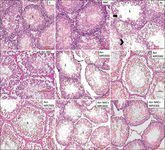
Background and aim: A significant cause of arsenic poisoning is polluted groundwater. Arsenic poisoning results in the suppression of spermatogenesis and the liver and kidneys are vulnerable to the toxic effects as well. Mucuna pruriens has been identified to have fertility-enhancing and anti-lipid peroxidation properties. Based on these properties of M. pruriens, this study aimed to investigate the efficacy of M. pruriens seed extract in reducing sodium arsenite-induced testicular impairment and hepato-renal histopathology in rats.

Materials and methods: The study was divided into two groups; short-term (45 days) and long-term (90 days) treatment groups and each group was divided into nine subgroups. Subgroups 1 and 2 served as normal and N-acetyl cysteine (NAC) controls, respectively. Subgroups 3-9 received sodium arsenite in the drinking water (50 mg/L). Subgroup-4 received NAC (210 mg/kg body weight [BW]) orally once daily. Subgroups 5-7 received aqueous seed extract of M. pruriens (350, 530, and 700 mg/kg BW, respectively) orally once daily. Subgroups 8 and 9 received a combination of NAC and aqueous seed extract (350 and 530 mg/kg BW, respectively) orally once daily. Following the treatment, animals were sacrificed and sperm parameters and DNA damage were evaluated. Testis, liver, and kidneys were analyzed for histopathology.

Results: Sodium arsenite-induced a significant reduction in sperm parameters and increase in the abnormal architecture of spermatozoa. Histology revealed tissue necrosis. The M. pruriens seed extract ameliorated the damaging effects of sodium arsenite with respect to tissue architecture and sperm parameters when coadministered.

Conclusion: Mucuna pruriens has beneficial effects against the deleterious effects of sodium arsenite on various tissues. Thus, M. pruriens (530 and 700 mg/kg BW) supplementation would reduce the adverse changes observed with sodium arsenite exposure.

Keywords: DNA damage; Mucuna pruriens; arsenic; hepato-renal; testis damage.


Conflict of interest statement

The authors declare that they have no competing interests.

Figures

Figure-1
Figure-1
Representative photographs of H and E stained sections of testis following 45 days of treatment, viewed under 100×. Scale bar=20 μm. NAC=N-acetyl cysteine, As=Arsenic, MP (350)=M. pruriens aqueous seed extract 350 mg/kg BW, MP (530)=M. pruriens aqueous seed extract 530 mg/kg body weight, MP (700)=M. pruriens aqueous seed extract 700 mg/kg body weight. Control group and NAC group showed normal organization of seminiferous tubule. Arsenic treated group showed reduced seminiferous tubule epithelial height and increased tubular lumen. The tubular basement membrane was irregular or interrupted. There was less number of spermatozoa in the lumen. As + MP (350) and As + MP (530) treated group exhibited cellular damage (arrow) and Sertoli cell damage (arrow). Higher doses of Mucuna pruriens and the combination treatment showed improved cell count, decreased structural damage. L=Lumen, E=Epithelium, S=Stroma.
Figure-2
Figure-2
Representative photographs of H and E stained sections of testis following 90 days of treatment, viewed under 100×. Scale bar=20 μm. NAC=N-acetyl cysteine, As=Arsenic, MP (350)=Mucuna pruriens aqueous seed extract 350 mg/kg body weight, MP (530)=Mucuna pruriens aqueous seed extract 530 mg/kg body weight, MP (700)=Mucuna pruriens aqueous seed extract 700 mg/kg body weight. Control group and NAC group showed normal organization of seminiferous tubule. Arsenic treated group showed reduced seminiferous tubule epithelial height and increased tubular lumen. The tubular basement membrane was irregular or interrupted (arrow). Less spermatozoa in the lumen. Provoked alterations in the seminiferous tubules were evident. Sloughing of germ cells was seen (arrow head). As+ MP (350) treated group showed intact basement membrane (arrow), but reduction in the cell count in the lumen. The higher doses of Mucuna pruriens (530 mg/kg body weight and 700 mg/kg body weight) and the combination treatments showed minimal damage and improved cell count. L=Lumen, E=Epithelium, LC=Leydig cell.
Figure-3
Figure-3
Representative photographs of H and E stained sections of liver following 45 days of treatment, viewed under 100×. Scale bar=10 μm. NAC=N-acetyl cysteine, As=Arsenic, MP (350)=Mucuna pruriens aqueous seed extract 350 mg/kg body weight, MP (530)=Mucuna pruriens aqueous seed extract 530 mg/kg body weight, MP (700)=Mucuna pruriens aqueous seed extract 700 mg/kg body weight. Normal histoarchitecture with normal hepatocytes (arrow head), sinusoids, central vein (C) and Kupffer cells were observed in control and the NAC treated groups. Arsenic treated group showed damage to the cell lining of sinusoids, increase in sinusoidal spaces and dilated central vein. Wall of the central vein was damaged. Cellular necrosis of hepatocytes was seen (double arrow). As + MP (350) treated group showed inflammatory changes in the hepatocytes along with increase in the area of sinusoids (arrow) and dilated central vein.
Figure-4
Figure-4
Representative photographs of H and E stained sections of liver following 90 days of treatment, viewed under 100×. Scale bar=10 μm. NAC=N-acetyl cysteine, As=Arsenic, MP (350)=Mucuna pruriens aqueous seed extract 350 mg/kg body weight, MP (530)=Mucuna pruriens aqueous seed extract 530 mg/kg body weight, MP (700)=Mucuna pruriens aqueous seed extract 700 mg/kg body weight. Normal histoarchitecture with normal hepatocytes (arrow head), central vein (C), sinusoids, and Kupffer cells were observed in control and the NAC treated groups. Arsenic treated group showed damage to the cell lining of sinusoids, increase in sinusoidal spaces (arrow). Cellular necrosis of hepatocytes was seen (double arrow). As + MP (350) treated group showed macrophage activity. Increased sinusoidal spaces were evident (arrow).
Figure-5
Figure-5
Representative photographs of H and E stained sections of kidney following 45 days of treatment, viewed under 100×. Scale bar=20 μm. NAC=N-acetyl cysteine, As=Arsenic, MP (350)=Mucuna pruriens aqueous seed extract 350 mg/kg body weight, MP (530)=Mucuna pruriens Mucuna pruriens aqueous seed extract 530 mg/kg body weight, MP (700)=Mucuna pruriens aqueous seed extract 700 mg/kg body weight. The kidney tissue of control and NAC treated rats showed normal renal corpuscle (G), proximal convoluted tubules (P), distal convoluted tubules (D). In the arsenic treated rats, renal cortex swelling and congestion in the renal corpuscle (arrow), showed damage to the bowman capsule, basement membrane, increased urinary space, damage to the intercellular structures (arrow head). As + NAC treated group showed inflammatory changes (arrow). As + MP (350) treated group showed infiltration of inflammatory cells (double arrow) and macrophage activity (arrow).
Figure-6
Figure-6
Representative photographs of H and E stained sections of kidney following 90 days of treatment, viewed under 100×. Scale bar=20 μm. NAC=N-acetyl cysteine, As=Arsenic, MP (350)=Mucuna pruriens aqueous seed extract 350 mg/kg body weight, MP (530)=Mucuna pruriens aqueous seed extract 530 mg/kg body weight, MP (700)=Mucuna pruriens aqueous seed extract 700 mg/kg body weight. The kidney tissue of control and NAC treated rats showed normal renal corpuscle (G), proximal convoluted tubules (P), distal convoluted tubules (D). Arsenic treated rats showed damage to the bowman capsule (arrow), basement membrane increased urinary space, damage to the intercellular structures (double arrow). As + MP (350) treated group showed damage to the cellular lining of the duct system. As + MP (530) group showed reduction in the urinary space (arrow head).

References

    1. Amitai Y, Koren G. High risk for neural tube defects;The role of arsenic in drinking water and rice in Asia. Med. Hypotheses. 2018;119(10):88–90. - PubMed
    1. Canalis A.M, Pérez R.D, Falchini G.E, Soria E.A. Experimental acute arsenic toxicity in Balb/c mice:Organic markers and splenic involvement. Biomedica. 2021;41(1):99–110. - PMC - PubMed
    1. Gordan J.J, Quastel J.H. Effects of organic arsenicals on enzyme systems. Biochem. J. 1948;42(3):337–350. - PubMed
    1. Hu Y, Li J, Lou B, Wu R, Wang G, Lu C, Wang H, Pi J, Xu Y. The role of reactive oxygen species in arsenic toxicity. Biomolecules. 2020;10(2):240. - PMC - PubMed
    1. Ratnaike R.N. Acute and chronic arsenic toxicity. Postgrad. Med. J. 2003;79(933):391–396. - PMC - PubMed

LinkOut - more resources