Review
. 2022 Dec 7;33(1):80-88.
doi: 10.1055/s-0042-1758874. eCollection 2023 Jan.

Morrison's Pouch: Anatomy and Radiological Appearance of Pathological Processes

Affiliations
Review

Morrison's Pouch: Anatomy and Radiological Appearance of Pathological Processes

Gurkawal Kaur et al. Indian J Radiol Imaging. .

Abstract

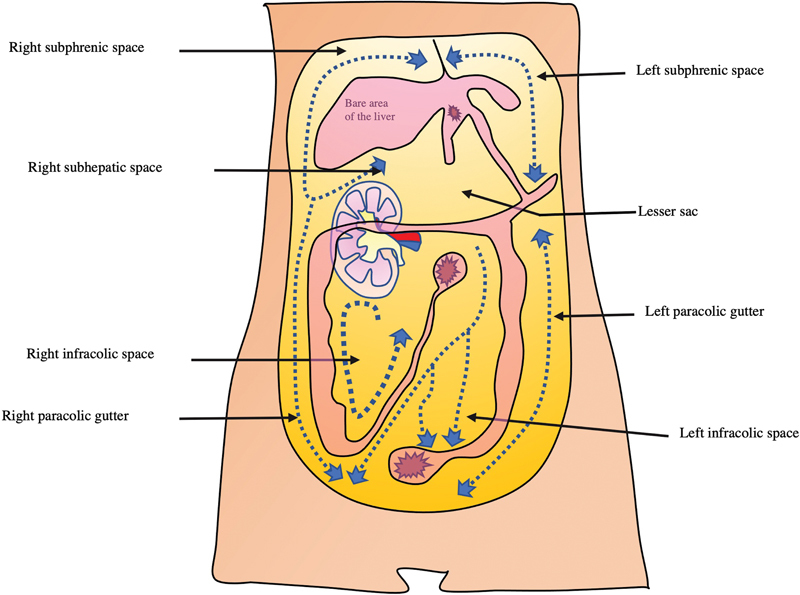
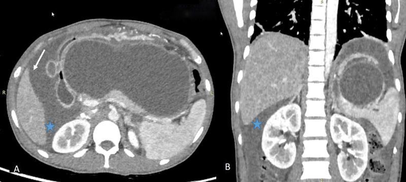
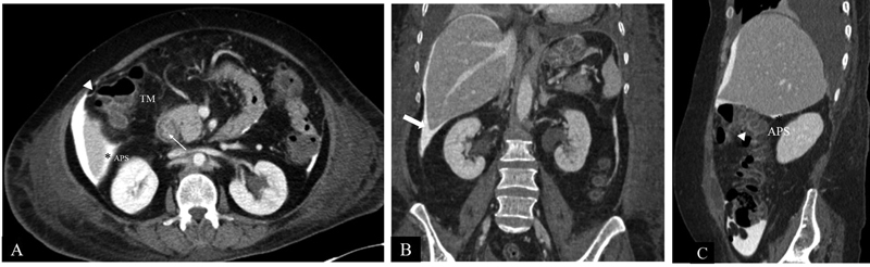
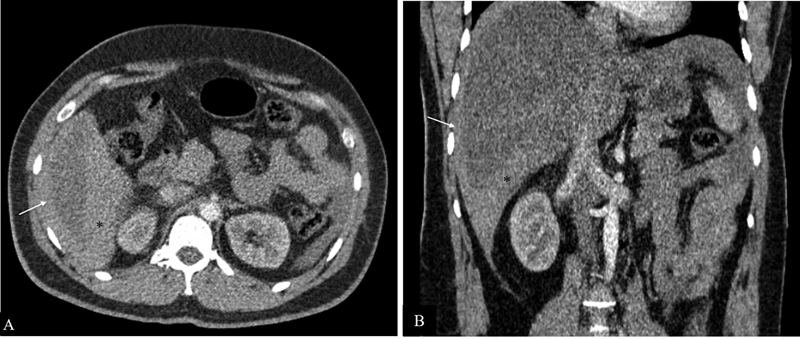
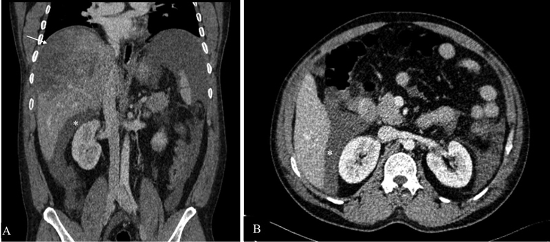
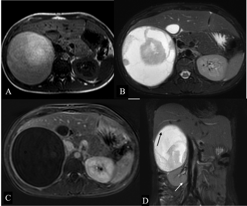
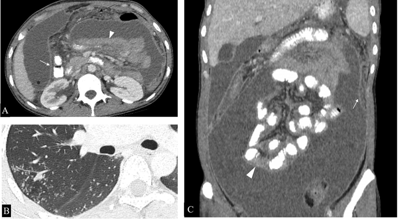
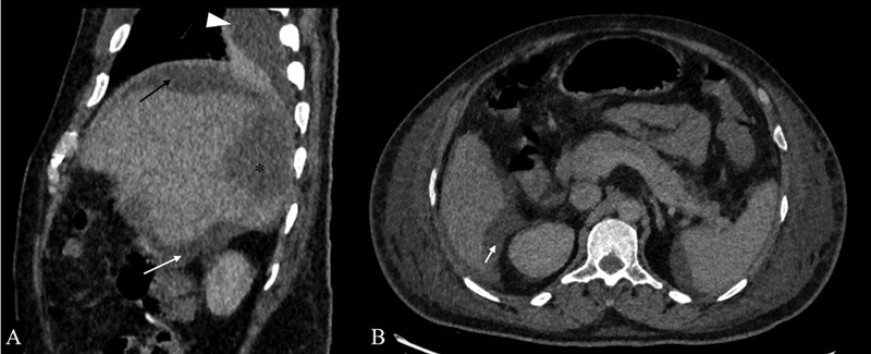
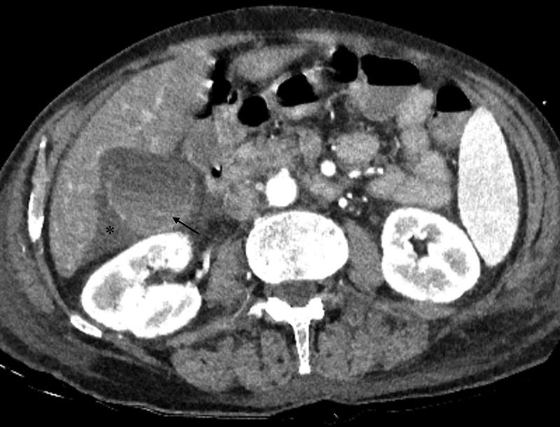
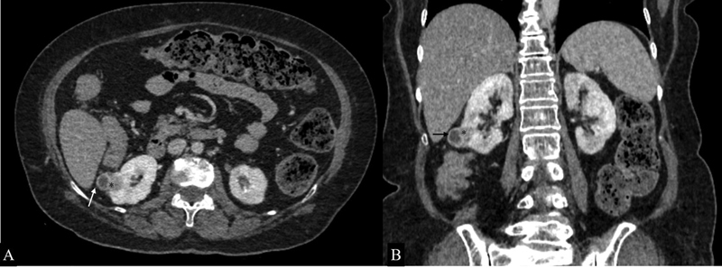
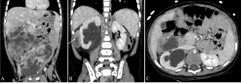
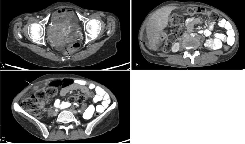
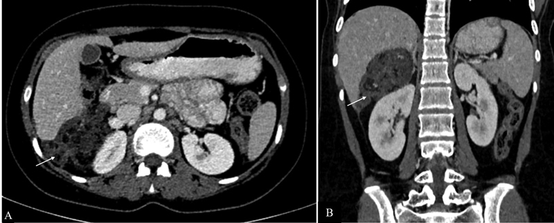
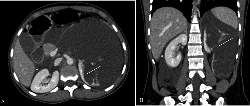
Morrison's pouch is the intraperitoneal space in the supramesocolic compartment located between the right liver lobe and right kidney. Pathological conditions that can involve this peritoneal space include fluid collections, infectious or inflammatory processes, and neoplasms. Frequent involvement by disease entities can be attributed to its dependent location, communication with the inframesocolic compartment, close proximity to the adjacent organs and peritoneal fluid dynamics. Knowledge of the appearance of pathological entities on various imaging modalities helps the radiologist in making the correct diagnosis.

Keywords: CT scan; MRI; Morrison's pouch; peritoneal space.


Conflict of interest statement

Conflict of Interest None declared.

Figures

Fig. 1
Fig. 1
Representative image of the peritoneal space. Right subhepatic space is the Morrison's pouch. Dotted line represents the direction of peritoneal flow through which the intraperitoneal spread of malignancy or infection can occur.
Fig. 2
Fig. 2
Ascites. Contrast-enhanced axial ( A ) and coronal ( B ) CT images in a 40-year-old patient in a patient with acute pancreatitis showing a large pseudocyst (central walled off cystic lesion) and ascitic fluid (arrow) accumulating in Morrison's pouch (*).
Fig. 3
Fig. 3
CT Anatomy of Morrison's pouch. Axial CECT image ( A ) shows intraperitoneal contrast outlining the peritoneum. Morrison's pouch (*) is the space between the right kidney and the right liver lobe. Hepatic flexure of the colon (arrowhead), transverse mesocolon (TM), and duodenum (line arrow) are seen anteromedially. Posteriorly, the parietal peritoneum separates the anterior pararenal space (APS) from Morrison's pouch (*). Coronal reformatted CT image ( B ) shows Morrison's pouch (*) between the inferior surface of the right lobe of the liver and the upper pole of the right kidney. Laterally, limited by parietal peritoneum (block arrow) lining the right lateral abdominal wall. Sagittal-reformatted CT image ( C ) shows Morrison's pouch (*) between the inferior surface of the right lobe of the liver and upper pole of the right kidney. Parietal peritoneum separates the anterior pararenal space (APS) from Morrison's pouch. Anteroinferiorly, hepatic flexure of the colon (arrowhead) is seen.
Fig. 4
Fig. 4
Hematoma. Contrast-enhanced arterial phase axial ( A ) and coronal ( B ) computed tomography images in a 28-year-old male patient with history of trauma showing a high-density hematoma in the perihepatic space (arrow) and in hepatorenal pouch (*).
Fig. 5
Fig. 5
Hemoperitoneum. Contrast-enhanced coronal ( A ) and axial ( B ) CT scan in a 41-year-old patient with history of road traffic accident and positive extended focused assessment with sonography in trauma (eFAST) showing grade 4 liver injury (arrow) along with high-density fluid in Morrison's pouch (*) suggestive of hemoperitoneum.
Fig. 6
Fig. 6
Adrenal hematoma. Axial T1-Weighted ( A ) and T2W FS ( B ) image showing a well-defined heterogeneously hyperintense lesion (*) in the right suprarenal location causing mass effect on the right lobe of the liver. Subtracted post contrast sequence ( C ) shows no enhancing component in this lesion. Coronal T2-weighted fat-saturated image ( D ) showing that the lesion is causing inferior displacement of the right kidney (white arrow), effacement of hepatorenal space, and mass effect on the liver (black arrow).
Fig. 7
Fig. 7
Peritoneal involvement in a microbiologically proven case of disseminated tuberculosis. ( A ). Axial CT sections show loculated ascites, smooth peritoneal thickening, and enhancement (white arrow), omental thickening (black arrow) and clumped bowel loops (arrowhead). Ascitic fluid can be seen extending into Morrison's pouch (*). ( B ) Axial section of the chest shows numerous centrilobular nodules in a tree and bud configuration (*), further supporting the imaging diagnosis of tuberculosis in this case. ( C ) Coronal reformatted image better demonstrates clumped small bowel loops (arrowhead) along with peritoneal thickening and enhancement (arrow).
Fig. 8
Fig. 8
Ruptured hepatic abscess. ( A ) Sagittal sections of contrast-enhanced CT in a 52 year-old-male patient presenting with high-grade fever and pain in the abdomen showing hepatic abscess in segments VI and VII (*) with discontinuity in hepatic surface, subdiaphragmatic (black arrow), hepatorenal pouch fluid (white arrow) and, ipsilateral pleural effusion (arrowhead). ( B ) Axial sections of the same patient better demonstrate the hepatorenal pouch fluid collection (white arrow).
Fig. 9
Fig. 9
Acute cholecystitis in a 75 year-old patient presenting with upper abdominal pain. Axial contrast-enhanced CT images show a distended gall bladder, enhancing wall, layering of the hyperdense sludge (arrow) and pericholecystic fluid extending into the hepatorenal pouch (*).
Fig. 10
Fig. 10
Renal cell carcinoma. Axial ( A ) and coronal ( B ) sections of nephrographic phase in a 55-year-old patient with incidentally detected right renal lesion on ultrasound showing a ball-type partially exophytic lesion (*) in the inferior pole of the right kidney with extension into the perinephric space (white arrow) and seen bulging into the hepatorenal pouch (black arrow).
Fig. 11
Fig. 11
Infantile fibrosarcoma in a 9-month-old male child. Coronal ( A and B ) and axial ( C ) sections of contrast-enhanced CT shows an ill-defined heterogeneously enhancing abdominopelvic soft tissue mass with large necrotic areas (*) extending into the hepatorenal pouch (arrows in B and C ).
Fig. 12
Fig. 12
Peritoneal carcinomatosis from high-grade serous adenocarcinoma of the ovary. Axial sections of intravenous and oral contrast material-enhanced computed tomographic images showing the right adnexal mass (arrowhead in A ), soft-tissue deposits in hepatorenal pouch (* in B ), and omental nodularity (white arrow in C ) along with free fluid in pelvis (black arrows).
Fig. 13
Fig. 13
Metastatic mature teratoma. Axial ( A ) and coronal ( B ) contrast-enhanced CT sections of a 16-year old girl previously treated for immature teratoma showing a soft tissue deposit in Morrison's pouch (arrow) consisting of areas of fat and calcifications. Tumor markers were normal in this patient.
Fig. 14
Fig. 14
Lymphatic malformation. Contrast-enhanced axial ( A ) and coronal ( B ) computed tomographic images showing a large insinuating cystic lesion in the retroperitoneum causing distortion of the left renal parenchyma (white arrows in A and B ), crossing the midline to reach the contralateral perirenal space bulging into hepatorenal pouch (*).

References

    1. Le O. Patterns of peritoneal spread of tumor in the abdomen and pelvis. World J Radiol. 2013;5(03):106–112. - PMC - PubMed
    1. Tirkes T, Sandrasegaran K, Patel A A. Peritoneal and retroperitoneal anatomy and its relevance for cross-sectional imaging. Radiographics. 2012;32(02):437–451. - PubMed
    1. Meyers M A. Distribution of intra-abdominal malignant seeding: dependency on dynamics of flow of ascitic fluid. Am J Roentgenol Radium Ther Nucl Med. 1973;119(01):198–206. - PubMed
    1. Hanbidge A E, Lynch D, Wilson S R.US of the peritoneum Radiographics 20032303663–684., discussion 684–685 - PubMed
    1. Anthony M P, Khong P L, Zhang J. Spectrum of (18)F-FDG PET/CT appearances in peritoneal disease. AJR Am J Roentgenol. 2009;193(06):W523-9. - PubMed