

COVID-Associated Sinonasal Mucormycosis: Radiological Pathological Correlation and Prognostic Value of MR Imaging
- PMID: 36855711
- PMCID: PMC9968527
- DOI: 10.1055/s-0042-1759639
COVID-Associated Sinonasal Mucormycosis: Radiological Pathological Correlation and Prognostic Value of MR Imaging
Abstract
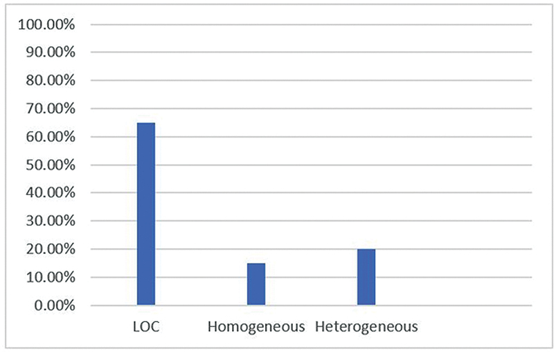
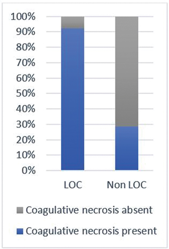
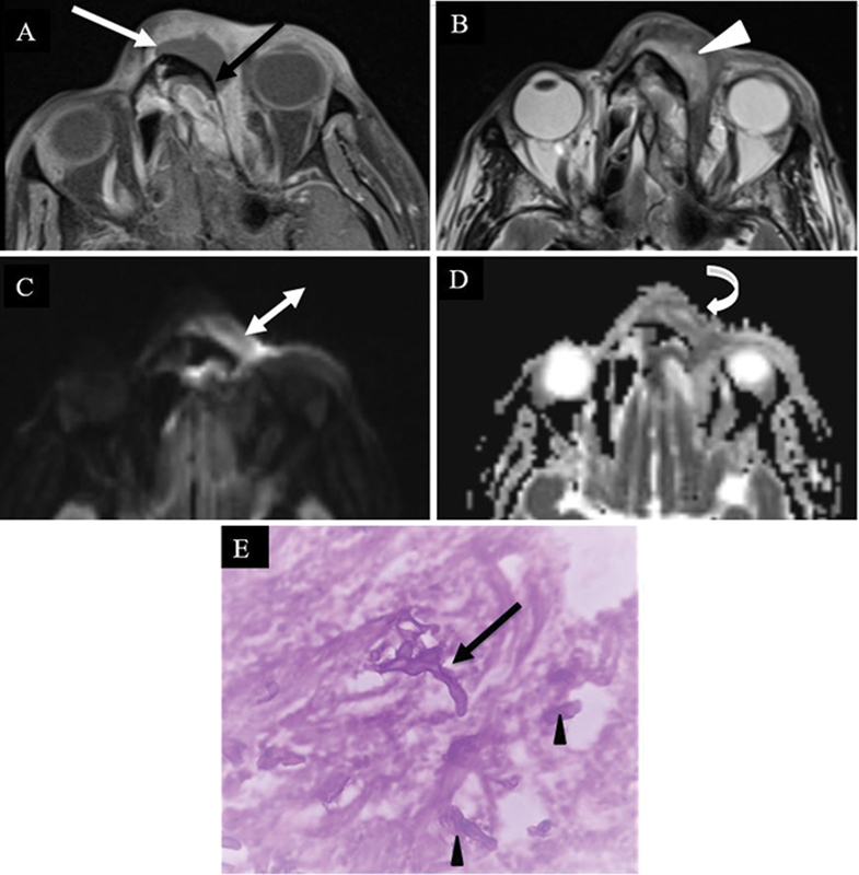
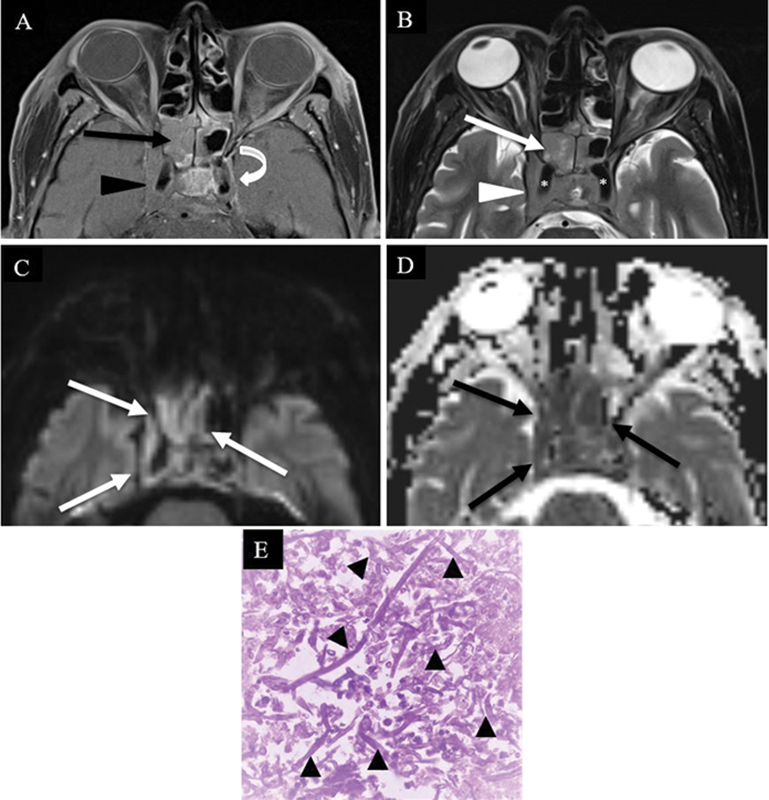
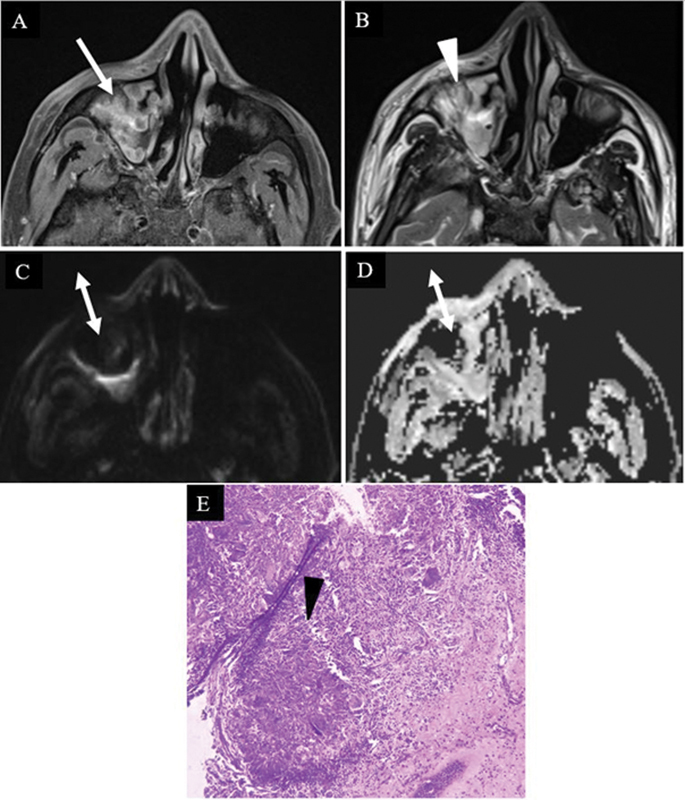
Purpose Our aim was to assess the sinonasal magnetic resonance imaging (MRI) features of acute invasive fungal rhinosinusitis (AIFRS) in coronavirus disease (COVID)-associated mucormycosis (CAM) and to correlate these with histopathology and patient outcome in terms of duration of hospital stay and survival at 10 weeks. Methods Twenty patients with histopathologically confirmed sinonasal CAM underwent MRI (including postcontrast T1-weighted and diffusion-weighted imaging). Histopathological findings (presence of coagulative necrosis, granulomatous reaction, and fungal burden) were recorded and all patients were followed up at 6 and 10 weeks. Statistical analysis was done using chi-square test and Fischer's exact test. Results Enhancement patterns seen in our subjects included homogeneous, heterogeneous, and lack of contrast enhancement (LOC), with LOC being the most common (65%). Diffusion restriction was found in 90% patients. Statistically significant correlation was found between LOC pattern and presence of coagulative necrosis ( p -value = 0.007), extent of fungal hyphae ( p -value = 0.047), and duration of hospital stay ( p -value = 0.004). Restricted diffusion was also seen to correlate with a high fungal load ( p -value = 0.007). Conclusion Our study describes the MRI findings of AIFRS in CAM and highlights the imaging features which may be surrogate markers for coagulative necrosis and fungal burden.
Keywords: MRI; coagulative necrosis; diffusion restriction; fungal load; lack of contrast enhancement.
Indian Radiological Association. This is an open access article published by Thieme under the terms of the Creative Commons Attribution-NonDerivative-NonCommercial License, permitting copying and reproduction so long as the original work is given appropriate credit. Contents may not be used for commercial purposes, or adapted, remixed, transformed or built upon. ( https://creativecommons.org/licenses/by-nc-nd/4.0/ ).
Conflict of interest statement
Conflict of Interest None declared.
Figures
Similar articles
-
Acute invasive fungal rhinosinusitis: MR imaging features and their impact on prognosis.Neuroradiology. 2018 Jul;60(7):715-723. doi: 10.1007/s00234-018-2034-0. Epub 2018 May 17. Neuroradiology. 2018. PMID: 29774383
-
Magnetic resonance imaging in COVID-19-associated acute invasive fungal rhinosinusitis - Diagnosis and beyond.J Clin Imaging Sci. 2023 Aug 9;13:23. doi: 10.25259/JCIS_46_2023. eCollection 2023. J Clin Imaging Sci. 2023. PMID: 37680251 Free PMC article.
-
Imaging of COVID-19-associated craniofacial mucormycosis: a black and white review of the "black fungus".Clin Radiol. 2021 Nov;76(11):812-819. doi: 10.1016/j.crad.2021.07.004. Epub 2021 Jul 28. Clin Radiol. 2021. PMID: 34364672 Free PMC article. Review.
-
Acute invasive fungal rhinosinusitis (AIFRS) - A histopathological analysis of expanding spectrum of fungal infections in backdrop of COVID-19 pandemic.J Family Med Prim Care. 2023 Sep;12(9):2097-2102. doi: 10.4103/jfmpc.jfmpc_629_23. Epub 2023 Sep 30. J Family Med Prim Care. 2023. PMID: 38024940 Free PMC article.
-
MR imaging spectrum in COVID associated Rhino-Orbito-Cerebral mucormycosis with special emphasis on intracranial disease and impact on patient prognosis.Eur J Radiol. 2022 Jul;152:110341. doi: 10.1016/j.ejrad.2022.110341. Epub 2022 May 6. Eur J Radiol. 2022. PMID: 35569303 Free PMC article. Review.
Cited by
-
The Radiological Spectrum of Rhino-Oculo-Cerebral Mucormycosis.Cureus. 2023 Jun 25;15(6):e40932. doi: 10.7759/cureus.40932. eCollection 2023 Jun. Cureus. 2023. PMID: 37519552 Free PMC article.
References
-
- Aribandi M, McCoy V A, Bazan C., III Imaging features of invasive and noninvasive fungal sinusitis: a review. Radiographics. 2007;27(05):1283–1296. - PubMed
-
- Therakathu J, Prabhu S, Irodi A, Sudhakar S V, Yadav V K, Rupa V. Imaging features of rhinocerebral mucormycosis: a study of 43 patients. Egypt J Radiol Nucl Med. 2018;49:447–452.
-
- deShazo R D, Chapin K, Swain R E. Fungal sinusitis. N Engl J Med. 1997;337(04):254–259. - PubMed
LinkOut - more resources
Full Text Sources