. 2023 Apr;43(4):435-454.
doi: 10.1002/cac2.12412. Epub 2023 Feb 28.

Combining radiation and the ATR inhibitor berzosertib activates STING signaling and enhances immunotherapy via inhibiting SHP1 function in colorectal cancer

Affiliations

Combining radiation and the ATR inhibitor berzosertib activates STING signaling and enhances immunotherapy via inhibiting SHP1 function in colorectal cancer

Chaofan Liu et al. Cancer Commun (Lond). 2023 Apr.

Abstract

Background: Immune checkpoint inhibitors (ICIs) targeting programmed cell death protein 1 (PD-1) and programmed death-ligand 1 (PD-L1) have shown a moderate response in colorectal cancer (CRC) with deficient mismatch repair (dMMR) functions and poor response in patients with proficient MMR (pMMR). pMMR tumors are generally immunogenically "cold", emphasizing combination strategies to turn the "cold" tumor "hot" to enhance the efficacy of ICIs. ATR inhibitors (ATRi) have been proven to cooperate with radiation to promote antitumor immunity, but it is unclear whether ATRi could facilitate the efficacy of IR and ICI combinations in CRCs. This study aimed to investigate the efficacy of combining ATRi, irradiation (IR), and anti-PD-L1 antibodies in CRC mouse models with different microsatellite statuses.

Methods: The efficacy of combining ATRi, IR, and anti-PD-L1 antibodies was evaluated in CRC tumors. The tumor microenvironment and transcriptome signatures were investigated under different treatment regimens. The mechanisms were explored via cell viability assay, flow cytometry, immunofluorescence, immunoblotting, co-immunoprecipitation, and real-time quantitative PCR in multiple murine and human CRC cell lines.

Results: Combining ATRi berzosertib and IR enhanced CD8+ T cell infiltration and enhanced the efficacy of anti-PD-L1 therapy in mouse CRC models with different microsatellite statuses. The mechanistic study demonstrated that IR + ATRi could activate both the canonical cGAS-STING-pTBK1/pIRF3 axis by increasing cytosolic double-stranded DNA levels and the non-canonical STING signaling by attenuating SHP1-mediated inhibition of the TRAF6-STING-p65 axis, via promoting SUMOylation of SHP1 at lysine 127. By boosting the STING signaling, IR + ATRi induced type I interferon-related gene expression and strong innate immune activation and reinvigorated the cold tumor microenvironment, thus facilitating immunotherapy.

Conclusions: The combination of ATRi and IR could facilitate anti-PD-L1 therapy by promoting STING signaling in CRC models with different microsatellite statuses. The new combination strategy raised by our study is worth investigating in the management of CRC.

Keywords: ATR inhibitor; DNA damage; PD-L1; SHP1; SUMOylation; cGAS-STING; colorectal cancer; immune checkpoint inhibitor; innate immunity; radiotherapy.


Conflict of interest statement

All authors have no conflicts of interests to declare.

Figures

FIGURE 1
FIGURE 1
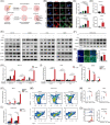
IR +ATRi increases TILs in MC38 and CT26 tumors (A) Treatment schedule for IHC and FC analysis. (B) Tumor volume curve of MC38 and CT26 tumors treated with IR or IR + ATRi. (C‐D) Representative IHC images of CD3 and CD8 staining (C) and statistical analysis (D) of MC38 tumors. (E‐F) Representative IHC images of CD3 and CD8 staining (E) and statistical analysis (F) of CT26 tumors. (G‐H) Representative flow cytometry images (G) and quantitative analysis (H) of TILs in MC38 tumors. (I‐J) Representative flow cytometry images (I) and quantitative analysis (J) of TILs in CT26 tumors. *, P < 0.05; **, P < 0.01; ***, P < 0.001; ns, not significant. Abbreviations: IR, radiation; IHC, immunohistochemistry; FC, flow cytometry; TIL, tumor‐infiltrating lymphocyte.
FIGURE 2
FIGURE 2
ATRi sensitizes CRC cells to IR, promotes mitotic reentry, and enhances innate immune signaling (A) Cell viability assay of multiple CRC cell lines treated with ATRi and IR +ATRi. (B) Immunoblot analysis of γH2AX and pCHK1 in multiple CRC cell lines treated with different regimens. (C‐D) Cell cycle distribution analysis of HCT116 and CT26 cells treated with IR and IR +ATRi. (E‐F) Representative of flow cytometry images (E) and quantitative analysis (F) of phospho‐histone H3+ cells in HCT116 and CT26 cells. (G) Number of DEGs in GO analysis of the RNA‐seq results. (H) GSEA of DEGs in the IR +ATRi group compared to the vehicle group. (I) GSEA of DEGs in the IR +ATRi group compared to the IR group. *, P < 0.05; **, P < 0.01; ***, P < 0.001; ns, not significant. Abbreviations: CRC, colorectal cancer; IR, radiation; CHK1, checkpoint kinase 1; pH3, phospho‐histone H3; RNA‐seq, RNA‐sequencing; DEG, differentially expressed gene; GO, gene ontology; GSEA, gene‐set enrichment analysis.
FIGURE 3
FIGURE 3
IR +ATRi activates the canonical cGAS‐STING signaling and promotes innate immune activation (A) Schematic diagram of micronuclei formation after IR‐induced cell cycle checkpoint and DNA repair are disrupted by ATRi. (B) IF images of dsDNA and cGAS staining in HCT116 cells. (C) Quantification of micronucleated cells in HCT116 cells and CT26 cells. (D) dsDNA quantification in HCT116 and CT26 cells after treatment with IR or IR + ATRi. (E) Immunoblot of the key proteins from the canonical cGAS‐STING axis in multiple CRC cell lines. (F) Immunoblot of pTBK1 and pSTING from CT26 tumors. (G) Representative images of pIRF3 staining in CT26 tumors and quantification of pIRF3 MFI. (H‐I) Relative expression levels of the CXCL10, CCL5 and IFNB genes at different time points in HCT116 (H) and CT26 cells (I). (J‐K) Relative expression levels of CXCL10, CCL5 and IFNB in SW480 (J) and MC38 (K) cells after different treatments. (L) Relative expression levels of Cxcl10, Ccl5 in CT26 tumors. (M‐N) Representative images (M) and statistical analysis of DCs and tumor infiltrating‐DCs (N) from flow cytometry analysis of CT26 tumors. (O) Representative images of CD86 staining and statistical analysis of CD86 MFI in CT26 tumors. *, P < 0.05; **, P < 0.01; ***, P < 0.001; ns, not significant. Abbreviations: IR, radiation; cGAS, cyclic GMP‐AMP synthase; STING, stimulator of interferon genes; TBK1, TANK binding kinase 1; IRF3, interferon regulatory factor 3; dsDNA, double stranded DNA; IF, immunofluorescence; MFI, mean fluorescence intensity; CXCL10, C‐X‐C motif chemokine ligand 10; CCL5, C‐C motif chemokine ligand 5; IFNB, interferon beta 1; DC, dendritic cell; TIDC, tumor‐infiltrating dendritic cell.
FIGURE 4
FIGURE 4
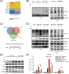
IR +ATRi activates the non‐canonical STING‐p65 signaling by inhibiting SHP1 (A) Immunoblot analysis of pSTAT1 and pP65 in multiple CRC cell lines after treatments. () Representative IF images of P65 staining in HCT116 cells and quantitative analysis of nuclear P65 MFI in HCT116 cells after treatments. (C) Heatmap of genes related to type I interferon signaling and innate immune signaling from RNA‐seq results. (D) Co‐IP analysis of SHP1‐interacted STING and TRAF6 under different treatment regimens. (E) Co‐IP analysis of STING‐interacted SHP1 and TRAF6 under different treatment regimens. (F) Immunoblot analysis of pP65, pIRF3 and pSTING after SHP1 agonist SC43 treatment in HCT116 and STING‐knockdown HCT116 cells. (G) Relative expression levels of CXCL10, CCL5 and IFNB after treatment with SHP1 agonist SC43 in HCT116 and STING‐knockdown HCT116 cells. *, P < 0.05; **, P < 0.01; ***, P < 0.001; ns, not significant. Abbreviations: IR, radiation; STAT1, signal transducer and activator of transcription 1; MFI, mean fluorescence intensity; SHP1, src homology 2 (SH2) domain containing protein tyrosine phosphatase‐1; IF, immunofluorescence; RNA‐seq, RNA sequencing; IP, immunoprecipitation; co‐IP, co‐immunoprecipitation; TRAF6, TNF receptor associated factor 6; STING, stimulator of interferon genes; IRF3, interferon regulatory factor 3; CXCL10, C‐X‐C motif chemokine ligand 10; CCL5, C‐C motif chemokine ligand 5; IFNB, interferon beta 1; NC, negative control.
FIGURE 5
FIGURE 5
IR + ATRi inhibits SHP1 function via promoting the SUMOylation of SHP1 at serine 127 (A) Heatmap of SHP1‐conjugated proteins in HCT116 cells from mass spectrometry data. *, differently altered proteins. (B) SUMOylation analysis of SHP1 in HCT116 cells after treatment with IR + ATRi. (C) Venn diagram of common SUMOylation sites predicted by SUMOplot analysis program, JASA, and GPS‐SUMO. (D) Co‐IP analysis of SUMO1 conjugation and the SHP1 phosphatase substrates STAT3, TRAF6 and STING in SHP1‐WT and SHP1‐K127R‐Mut HCT116 cells. (E) Immunoblot analysis of key STING signaling proteins in SHP1‐WT and SHP1‐K127R‐Mut HCT116 cells under different treatments. (F) Relative mRNA levels of CXCL10, CCL5 and IFNB in SHP1‐WT and SHP1‐K127R‐Mut HCT116 cells under different treatment different regimens. *, P < 0.05; **, P < 0.01; ***, P < 0.001; ns, not significant. Abbreviations: IR, radiation; SHP1, src homology 2 (SH2) domain containing protein tyrosine phosphatase‐1; IP, immunoprecipitation; SUMO1, small ubiquitin like modifier 1; STAT3, signal transducer and activator of transcription 3; TRAF6, TNF receptor associated factor 6; STING, stimulator of interferon genes; IRF3, interferon regulatory factor 3; CXCL10, C‐X‐C motif chemokine ligand 10; CCL5, C‐C motif chemokine ligand 5; IFNB, Interferon Beta 1, WT, wild type.
FIGURE 6
FIGURE 6
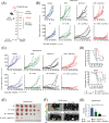
IR +ATRi +anti‐PD‐L1 exhibits optimal antitumor efficacy and prolonged survival in CT26 and MC38 tumors (A) Treatment schedule for the subcutaneous tumor models. (B) Tumor volume curve of CT26 tumors treated with IR (n = 6), ATRi (n = 6), anti‐PD‐L1 (n = 6), or combination therapies (n = 6 in each group). (C) Tumor volume curve of MC38 tumors treated with IR (n = 6), ATRi (n = 6), anti‐PD‐L1 (n = 6), or combination therapies (n = 6 in each group). (D) Survival analysis of mice bearing CT26 (n = 6) or MC38 tumors (n = 6). (E) Views of tumors derived from the flanks of MC38‐tumor bearing mice (n = 6). (F‐G) Bioluminescence imaging of MC38 tumors (F; n = 6) and statistical analysis (G; n = 6) of total flux of bioluminescence imaging. *, P < 0.05; **, P < 0.01; ***, P < 0.001. Abbreviations: IR, radiation; PD‐L1, programmed death‐ligand 1.
FIGURE 7
FIGURE 7
Abstract figure showing the antitumor efficacy of IR +ATRi +anti‐PD‐L1 and the mechanism IR + ATRi activates the canonical cGAS‐STING‐pTBK1‐pIRF3 axis by inducing of cytosolic dsDNA, and the non‐canonical axis by abrogating SHP1‐mediated TRAF6/STING‐p65 complex inhibition, through SUMOylation of SHP1. By boosting the STING‐type I interferon signaling, IR + ATRi promotes immune activation, turning the cold tumor hot, and in turn facilitate the efficacy of anti‐PD‐L1 therapy. The red lines indicate the effects of ATRi. Abbreviations: IR, radiation; PD‐L1, programmed death‐ligand 1; SHP1, src homology 2 (SH2) domain containing protein tyrosine phosphatase‐1; IP, immunoprecipitation; SUMO1, small ubiquitin like modifier 1; TRAF6, TNF Receptor Associated Factor 6; STING, stimulator of interferon genes; TBK1, TANK Binding Kinase 1; IRF3, interferon regulatory factor 3; DDR, DNA damage response; dsDNA, double stranded DNA; DDR, DNA damage response; cGAS, cyclic GMP‐AMP synthase; IFN, interferon.

Similar articles

Cited by

References

    1. Siegel RL, Miller KD, Jemal A. Cancer statistics, 2020. CA: A Cancer Journal for Clinicians. 2020;70(1):7‐30. - PubMed
    1. Qiu H, Cao S, Xu R. Cancer incidence, mortality, and burden in China: a time‐trend analysis and comparison with the United States and United Kingdom based on the global epidemiological data released in 2020. Cancer Commun (Lond). 2021;41(10):1037‐48. - PMC - PubMed
    1. van der Geest LGM, Jt Lam‐Boer, Koopman M, Verhoef C, Elferink MAG, de Wilt JHW. Nationwide trends in incidence, treatment and survival of colorectal cancer patients with synchronous metastases. Clinical & experimental metastasis. 2015;32(5):457‐65. - PubMed
    1. Wang Y, Wang M, Wu HX, Xu RH. Advancing to the era of cancer immunotherapy. Cancer Commun (Lond). 2021;41(9):803‐29. - PMC - PubMed
    1. Killock D. Immunotherapy: PD‐1 blockade exploiting MMR deficiency. Nature Reviews Clinical Oncology. 2017;14(8):459. - PubMed

Publication types