. 2023 Mar 1;11(2):e0485422.
doi: 10.1128/spectrum.04854-22. Online ahead of print.

Development of a Novel Chikungunya Virus-Like Replicon Particle for Rapid Quantification and Screening of Neutralizing Antibodies and Antivirals

Affiliations

Development of a Novel Chikungunya Virus-Like Replicon Particle for Rapid Quantification and Screening of Neutralizing Antibodies and Antivirals

Hui-Chung Lin et al. Microbiol Spectr. .

Abstract

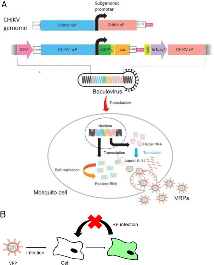
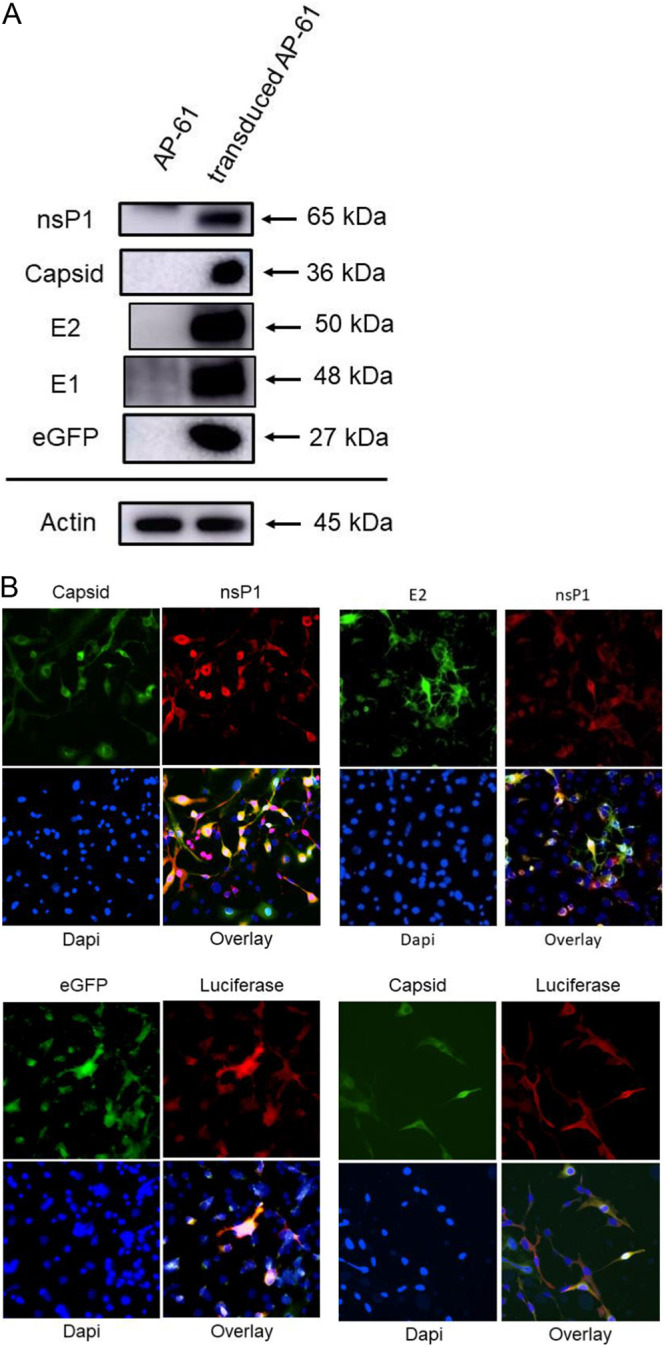
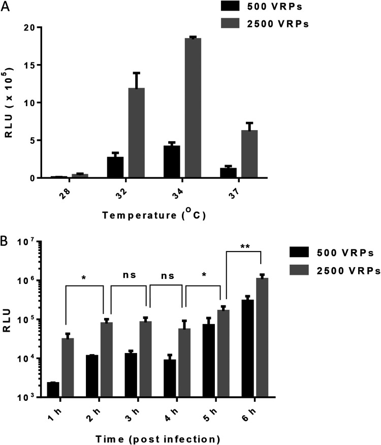
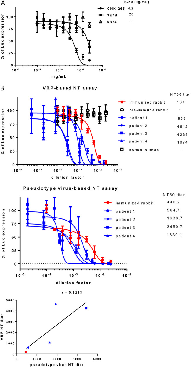
Chikungunya fever is a mosquito-transmitted infectious disease that induces rash, myalgia, and persistent incapacitating arthralgia. At present, no vaccines or antiviral therapies specific to Chikungunya virus (CHIKV) infection have been approved, and research is currently restricted to biosafety level 3 containment. CHIKV-like replicon particles (VRPs) are single-cycle infectious particles containing viral structure proteins, as well as a defective genome to provide a safe surrogate for living CHIKV to facilitate the testing of vaccines and antivirals. However, inefficient RNA transfection and the potential emergence of the competent virus through recombination in mammalian cells limit VRP usability. This study describes a transfection-free system for the safe packaging of CHIK VRP with all necessary components via transduction of mosquito cell lines using a single baculovirus vector. We observed the release of substantial quantities of mosquito cell-derived CHIK VRP (mos-CHIK VRP) from baculovirus-transduced mosquito cell lines. The VRPs were shown to recapitulate viral replication and subgenomic dual reporter expression (enhanced green fluorescent protein [eGFP] and luciferase) in infected host cells. Interestingly, the rapid expression kinetics of the VRP-expressing luciferase reporter (6 h) makes it possible to use mos-CHIK VRPs for the rapid quantification of VRP infection. Treatment with antivirals (suramin or 6-azauridine) or neutralizing antibodies (monoclonal antibodies [MAbs] or patient sera) was shown to inhibit mos-CHIK VRP infection in a dose-dependent manner. Ease of manufacture, safety, scalability, and high throughput make mos-CHIK VRPs a highly valuable vehicle for the study of CHIKV biology, the detection of neutralizing (NT) antibody activity, and the screening of antivirals against CHIKV. IMPORTANCE This study proposes a transfection-free system that enables the safe packaging of CHIK VRPs with all necessary components via baculovirus transduction. Those mosquito cell-derived CHIK VRP (mos-CHIK VRPs) were shown to recapitulate viral replication and subgenomic dual reporter (enhanced green fluorescent protein [eGFP] and luciferase) expression in infected host cells. Rapid expression kinetics of the VRP-expressing luciferase reporter (within hours) opens the door to using mos-CHIK VRPs for the rapid quantification of neutralizing antibody and antiviral activity against CHIKV. To the best of our knowledge, this is the first study to report a mosquito cell-derived alphavirus VRP system. Note that this system could also be applied to other arboviruses to model the earliest event in arboviral infection in vertebrates.

Keywords: Chikungunya virus; baculovirus; mosquito cell; virus replicon particle.


Conflict of interest statement

The authors declare no conflict of interest.

Figures

FIG 1
FIG 1
Schematic illustration showing the Chikungunya virus (CHIKV)-like replicon particle (VRP) production cycle in mosquito cells using a single baculovirus vector. (A) A recombinant baculovirus bearing both DNA cassettes of the CHIKV replicon dual reporter (enhanced green fluorescent protein [eGFP] and luciferase [Luc]) under the control of a cytomegalovirus (CMV) promoter (a shuttle promoter), as well as a helper gene (CHIKV structural protein [sP]) under a hr1pag1 promoter (an insect-specific promoter). Transduction using a single baculovirus vector delivers these genes (the components required for the packaging of VRP) into the nucleus of a mosquito cell for the transcription of self-amplifying replicon RNA and 26S RNA. The VRPs were packaged in a mosquito cell and released. HDR, hepatitis delta virus ribozyme; nsP, nonstructural proteins; T2A, Thosea asigna virus 2A self-cleaving peptides; UTR, untranslated region of CHIKV genome. (B) Single-round infectious VRP infects susceptible cells and expresses reporter genes but does not produce a new VRP for reinfection.
FIG 2
FIG 2
Characterization of the recombinant baculovirus. (A) Western blot analysis. AP-61 cells were transduced with the baculovirus at a multiplicity of infection (MOI) of 20. Total cell lysates were subjected to Western blot analysis using anti-CHIKV nsP1, capsid, E2, E1, or eGFP (actin served as a loading control) antibodies (shown on the left). The protein sizes (kDa) of markers are indicated by arrows on the right. (B) Colocalization of capsid, E2, nsP1, eGFP, and Luc in transduced mosquito cells. Transduced AP-61 cells were costained using anti-CHIKV capsid/anti-CHIKV nsP1 antibodies (upper left), anti-CHIKV E2/anti-CHIKV nsP1 antibodies (upper right), anti-eGFP/anti-Luc antibodies (lower left), or anti-CHIKV capsid/anti-Luc antibodies (lower right) with 4′,6-diamidino-2-phenylindole (DAPI); overlay figures are presented.
FIG 3
FIG 3
Package of CHIKV VRPs with dual reporter from mosquito cells. (A) Treating with patient serum and the effects on VRP infection. Vero cells were infected with CHIKV VRPs in the presence of sera of a CHIKV patient or normal individual and incubated for 20 h. The upper panels show the fluorescein isothiocyanate (FITC) channel, and the lower panels show the bright field. (B) Western blot analysis. Vero cells infected with the VRPs at an MOI of 0.05. Total cell lysate was subjected to Western blot analysis using anti-CHIKV nsP1, capsid (included transduced AP-61 cell lysate from Fig. 2A as a positive control), or eGFP (actin served as a loading control) antibodies (shown on the left). Protein sizes (kDa) of markers are indicated by arrows on the right. (C) Colocalization of nsP1, eGFP, and Luc in VRP-infected cells. Panel C shows immunofluorescence imagining of VRP-infected Vero cells: anti-CHIKV capsid/anti-CHIKV nsP1 antibodies (left panels), anti-Luc/anti-eGFP antibodies (middle panels), or anti-CHIKV nsP1/anti-eGFP antibodies (right panels) with DAPI. Merged figures are presented in the lower right panels. White arrows indicate double-positive cells for immunofluorescence assays (IFAs). (D) Viral RNA amplification in VRP-infected cells. Vero cells were infected with VRPs at an MOI of 0.1. Following incubation for 1 or 24 h, the total cellular RNA was harvested and subjected to quantitative reverse transcription (qRT)-PCR. Error bars indicate the standard deviation (SD). Statistical significance was analyzed using Student’s t test. ****, P < 0.0001.
FIG 4
FIG 4
eGFP expression of CHIK VRP as a function of temperature and production kinetics. (A) VRP titrations obtained at various temperature. The panel shows morphology of eGFP-positive cells incubated at various temperatures (left; 28, 34, and 37°C). eGFP-positive cells at various temperature were counted, and the VRP titer was calculated (right). (B) AP-61 cells or C6/36 cells were transduced using the recombinant baculovirus at MOIs of 5 and 20 in triplicate. Culture supernatant was collected at intervals of 3 days between days 1 and 14 and subjected to VRP titration. Error bars indicate SD.
FIG 5
FIG 5
Luc expression as a function of temperature or expression kinetics. (A) Temperature dependency assay. Vero cells were infected (in triplicate) with VRPs at a density of 500 or 2,500 infectious units (IU)/well and incubated at various temperature for 1 h, whereupon the culture medium was refreshed, and incubation was continued for another 5 h at the same corresponding temperature. The cells were harvested to quantify Luc activity (relative light units [RLU]). (B) Time-course evaluation at 34°C. Vero cells were infected (in triplicate) at a density of 500 or 2,500 IU/well and incubated for 1 h, whereupon the culture medium was refreshed and incubation was continued for another 5 h at 34°C. The cells were harvested at interval of 1 h to quantify Luc activity (RLU). Error bars indicate SD. Statistical significance was analyzed using Student’s t test. *, P < 0.05; **, P < 0.01.
FIG 6
FIG 6
Rapid measurement of antibody-mediated neutralization using mosquito cell-derived CHIK VRP (mos-CHIK VRP). (A) Monoclonal antibodies (MAbs). mos-CHIK VRP were preincubated with either CHK-265 (a CHIKV-neutralizing Ab), 3E7B (another CHIKV-neutralizing Ab), or 6B6C (negative control) at 37°C for 1 h. The resulting mixtures was then used to infect Vero cell via incubation at 34°C for 1 h. Luc activity was measured after incubation at 34°C for another 5 h and normalized to VRP-infected cells. Nonlinear regression analysis was used to calculate the 50% infective concentration (IC50) values. Error bars indicate the SD of values averaged from experiments performed in triplicate. (B) Antisera. Samples included the sera from four CHIKV patients and serum from an immunized rabbit. Negative control included serum from a preimmunized rabbit and normal human serum. Nonlinear regression analysis was used to calculate the NT50 values. Error bars indicate the SD of values averaged from experiments performed in triplicate. Neutralization titers of sera following with mos-CHIK VRP or CHIKV-pseudotyped virus. The correlation calculated using the Pearson correlation coefficient was 0.7 < r < 0.9 (strong correlation).
FIG 7
FIG 7
Rapid quantification and screening of antivirals using mos-CHIK VRP. (A) Rapid antiviral quantification using a Luc reporter. (Left) VRP-infected Vero cells treated with 6-azauridine (6-AU) at indicated concentrations. VRPs were mixed with suramin at different concentration and then infected Vero cells. (Right) The infected Vero cells were treated with suramin at indicated concentration. Luc activity was measured after incubation at 34°C for 5 h and normalized to VRP-infected cell (black lines). Cytotoxicity of 6-AU or suramin on Vero cells (dotted lines) was determined using 3-(4,5-dimethyl-2-thiazolyl)-2,5-diphenyl-2H-tetrazolium bromide (MTT) assay. The data points show the means ± SD of experiments conducted in triplicate, while the error bars indicate the SD. (B) Screening of antivirals using the eGFP reporter. In accordance with panel A, VRP-infected Vero cells were treated with either 6-AU (upper panel) or suramin (lower panel) at the indicated concentrations. The cells were photographed after incubation at 28°C for additional 20 h.

Similar articles

Cited by

References

    1. Silva LA, Dermody TS. 2017. Chikungunya virus: epidemiology, replication, disease mechanisms, and prospective intervention strategies. J Clin Invest 127:737–749. doi:10.1172/JCI84417. - DOI - PMC - PubMed
    1. Pathak H, Mohan MC, Ravindran V. 2019. Chikungunya arthritis. Clin Med 19:381–385. doi:10.7861/clinmed.2019-0035. - DOI - PMC - PubMed
    1. Tsetsarkin KA, Vanlandingham DL, McGee CE, Higgs S. 2007. A single mutation in Chikungunya virus affects vector specificity and epidemic potential. PLoS Pathog 3:e201. doi:10.1371/journal.ppat.0030201. - DOI - PMC - PubMed
    1. Chen R, Plante JA, Plante KS, Yun R, Shinde D, Liu J, Haller S, Mukhopadhyay S, Weaver SC. 2021. Lineage divergence and vector-specific adaptation have driven Chikungunya virus onto multiple adaptive landscapes. mBio 12:e0273821. doi:10.1128/mBio.02738-21. - DOI - PMC - PubMed
    1. Burt FJ, Chen W, Miner JJ, Lenschow DJ, Merits A, Schnettler E, Kohl A, Rudd PA, Taylor A, Herrero LJ, Zaid A, Ng LFP, Mahalingam S. 2017. Chikungunya virus: an update on the biology and pathogenesis of this emerging pathogen. Lancet Infect Dis 17:e107–e117. doi:10.1016/S1473-3099(16)30385-1. - DOI - PubMed