. 2023 Apr;37(4):e22829.
doi: 10.1096/fj.202201564R.

Mitigating the detrimental developmental impact of early fetal alcohol exposure using a maternal methyl donor-enriched diet

Affiliations

Mitigating the detrimental developmental impact of early fetal alcohol exposure using a maternal methyl donor-enriched diet

Mélanie Breton-Larrivée et al. FASEB J. 2023 Apr.

Abstract

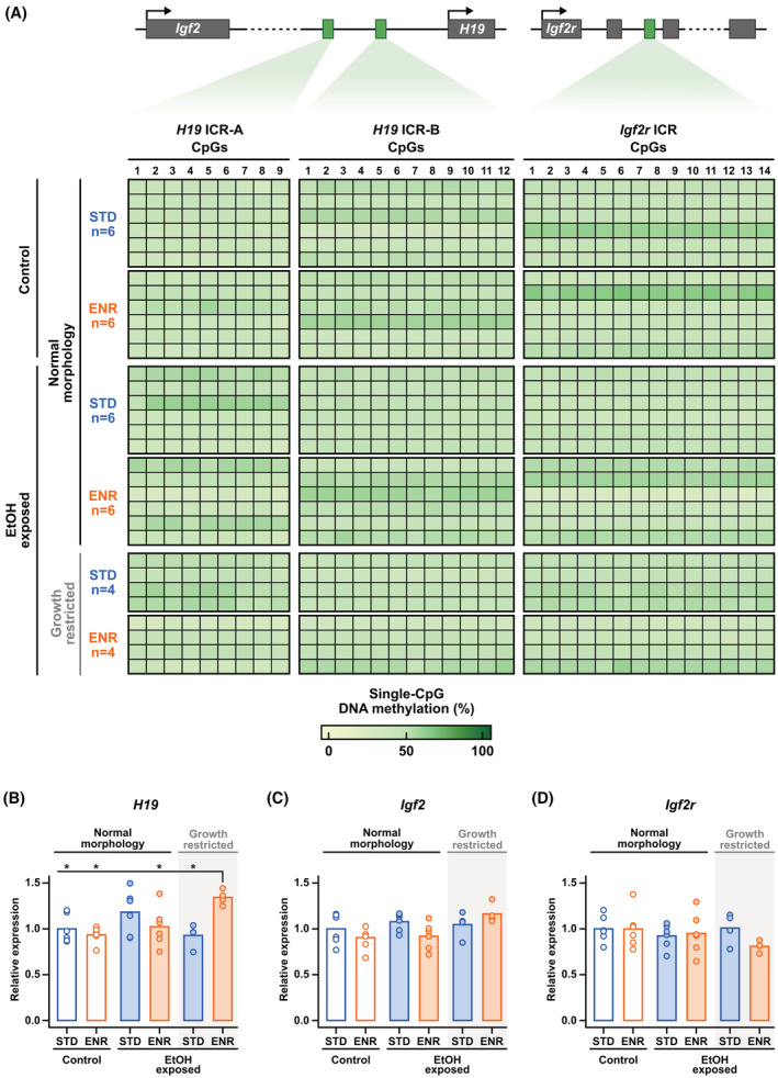
Fetal alcohol exposure at any stage of pregnancy can lead to fetal alcohol spectrum disorder (FASD), a group of life-long conditions characterized by congenital malformations, as well as cognitive, behavioral, and emotional impairments. The teratogenic effects of alcohol have long been publicized; yet fetal alcohol exposure is one of the most common preventable causes of birth defects. Currently, alcohol abstinence during pregnancy is the best and only way to prevent FASD. However, alcohol consumption remains astoundingly prevalent among pregnant women; therefore, additional measures need to be made available to help protect the developing embryo before irreparable damage is done. Maternal nutritional interventions using methyl donors have been investigated as potential preventative measures to mitigate the adverse effects of fetal alcohol exposure. Here, we show that a single acute preimplantation (E2.5; 8-cell stage) fetal alcohol exposure (2 × 2.5 g/kg ethanol with a 2h interval) in mice leads to long-term FASD-like morphological phenotypes (e.g. growth restriction, brain malformations, skeletal delays) in late-gestation embryos (E18.5) and demonstrate that supplementing the maternal diet with a combination of four methyl donor nutrients, folic acid, choline, betaine, and vitamin B12, prior to conception and throughout gestation effectively reduces the incidence and severity of alcohol-induced morphological defects without altering DNA methylation status of imprinting control regions and regulation of associated imprinted genes. This study clearly supports that preimplantation embryos are vulnerable to the teratogenic effects of alcohol, emphasizes the dangers of maternal alcohol consumption during early gestation, and provides a potential proactive maternal nutritional intervention to minimize FASD progression, reinforcing the importance of adequate preconception and prenatal nutrition.

Keywords: DNA methylation; DOHaD; FASD; embryonic development; environmental exposure; fetal alcohol exposure; maternal nutrition; methyl donors.


Figures

FIGURE 1
FIGURE 1
Mouse model combining a maternal methyl donor‐enriched diet and a single acute preimplantation alcohol exposure. CB57BL/6 females were fed a standard or a methyl donor‐enriched (folic acid, choline, betaine, and vitamin B12) diet for 4 weeks to stabilize serum nutrient levels prior to mating with CB57BL/6 males kept on a standard diet; Respective maternal diets were continued throughout gestation. Pregnant females were subjected to a single acute alcohol exposure (2.5 g/kg EtOH) or 0.15 M saline solution (control) by two subcutaneous injections of equivalent volumes with a 2‐h interval to target E2.5 stage embryos (~8‐cell stage). E18.5 embryos were harvested for morphological analysis, skeletal staining, and analysis of DNA methylation and gene expression of imprinted genes in forebrain tissue. Created with BioRender.com.
FIGURE 2
FIGURE 2
Maternal methyl donor supplementation alleviates late‐gestation abnormal morphometrics induced by early fetal alcohol exposure. Morphometric measurements of E18.5 embryos subjected to a maternal standard diet (control; n = 68, ethanol‐exposed; n = 70) or a maternal methyl donor‐enriched diet (control; n = 69, ethanol‐exposed n = 75). Statistical analysis: Shapiro–Wilk test followed by either non‐parametric tests (Kruskal–Wallis test and Pairwise Wilcoxon Rank Sum tests) or parametric tests (Levene's test and one‐way anova with pairwise comparisons and, if required, Welch's correction) with Benjamini–Hochberg p‐value adjustment. *p ≤ .05, **p ≤ .01, ***p ≤ .001. See associated Tables S2 and S3.
FIGURE 3
FIGURE 3
Maternal methyl donor‐enriched diet protects embryos from late‐gestation morphological defects associated with early fetal alcohol exposure. Total frequency of morphological defects (A) and of the specific types of morphological defects (growth restriction, brain anomalies, eye defects, and other defects) (B) in E18.5 embryos subjected to a maternal standard diet (control; n = 68, ethanol‐exposed; n = 70) or a maternal methyl donor‐enriched diet (control; n = 69, ethanol‐exposed n = 75). See associated Table S4. Statistical analysis (A): pairwise chi‐squared tests with Yates continuity correction *p ≤ .05, **p ≤ .01, ***p ≤ .001. (C) Images of E18.5 morphologically normal embryos and embryos with various morphological defects associated with ethanol exposure for both maternal diet conditions. (D) Comparison of the number of affected litters, number of E18.5 embryos per litter, and frequency of E18.5 embryos with morphological defects per litter between the four conditions. See associated Table S5.
FIGURE 4
FIGURE 4
Growth restriction linked to early fetal alcohol exposure is accompanied by ossification delays and mild craniofacial hypoplasia in both standard and enriched maternal diet conditions. Total frequency of ossification delays (A) and of occipital bone and 5th sternebrae ossification delays specifically (B) in E18.5 embryos with normal morphology (control and ethanol‐exposed) and with ethanol exposure‐associated growth restriction in both standard and enriched maternal diet conditions. See associated Table S6. Statistical analysis (A): pairwise chi‐squared tests with Yates continuity correction. (C) E18.5 embryo skeletal staining images showing examples of normal ossification, ossification delays (occipital bone (III, XII), 5th sternebrae (VI, XV)), and mild craniofacial hypoplasia (shorter head and snout length (IX, XVIII)). (D–F) Body weight, head length, and snout length of E18.5 embryos stained for skeletal analysis. See associated Tables S6 and S7. Statistical analysis (D–F): Shapiro–Wilk test followed by either non‐parametric tests (Kruskal–Wallis test and pairwise Wilcoxon rank sum tests) or parametric tests (Levene's test and one‐way anova with pairwise comparisons and, if required, Welch's correction) with Benjamini–Hochberg p‐value adjustment. Number of embryos: standard maternal diet; control (normal morphology: n = 6) and ethanol‐exposed embryos (normal morphology: n = 11, growth‐restricted: n = 7), enriched maternal diet; control (normal morphology: n = 5) and ethanol‐exposed embryos (normal morphology: n = 10, growth‐restricted: n = 6). *p ≤ .05, **p ≤ .01, ***p ≤ .001.
FIGURE 5
FIGURE 5
Early fetal alcohol exposure does not affect DNA methylation at the H19 and Igf2r ICRs in forebrain of late‐gestation embryos. (A) DNA methylation levels of single‐CpGs in two regions of the H19 ICR (H19 ICR‐A/B) and at the Igf2r ICR in E18.5 morphologically normal embryos (control or EtOH‐exposed) and growth‐restricted EtOH‐exposed embryos subjected to a standard (STD) or enriched (ENR) maternal diet. See associated Figure S3. (B–D) Relative expression levels (normalized to Hprt1 and STD‐Control) of H19, Igf2, and Igf2r, respectively, in same embryos used for DNA methylation analysis. Gene expression statistical analysis (B–D): Shapiro–Wilk test followed by Levene's Test and one‐way anova with pairwise comparisons, Welch's correction (if required), and Benjamini–Hochberg p‐value adjustment. *p ≤ .05. Number of embryos: Standard maternal diet: control (normal morphology: n = 6) and ethanol‐exposed embryos (normal morphology: n = 6, growth‐restricted: n = 4); Enriched maternal diet: control (normal morphology: n = 6) and ethanol‐exposed embryos (normal morphology: n = 6, growth‐restricted: n = 4).

Similar articles

Cited by

References

    1. Jones KL, Smith DW. Recognition of the fetal alcohol syndrome in early infancy. Lancet. 1973;302(7836):999‐1001. - PubMed
    1. Hoyme HE, Kalberg WO, Elliott AJ, et al. Updated clinical guidelines for diagnosing fetal alcohol spectrum disorders. Pediatrics. 2016;138(2):e20154256. - PMC - PubMed
    1. May PA, Blankenship J, Marais AS, et al. Maternal alcohol consumption producing fetal alcohol spectrum disorders (FASD): quantity, frequency, and timing of drinking. Drug Alcohol Depend. 2013;133(2):502‐512. - PMC - PubMed
    1. Wozniak JR, Riley EP, Charness ME. Clinical presentation, diagnosis, and management of fetal alcohol spectrum disorder. Lancet Neurol. 2019;18(8):760‐770. - PMC - PubMed
    1. Popova S, Dozet D, Shield K, Rehm J, Burd L. Alcohol's impact on the fetus. Nutrients. 2021;13(10):3452. - PMC - PubMed

Publication types

Grants and funding