. 2023 Jun 16;108(7):1585-1601.
doi: 10.1210/clinem/dgad098.

Aggressive Pituitary Tumors and Pituitary Carcinomas: From Pathology to Treatment

Affiliations

Aggressive Pituitary Tumors and Pituitary Carcinomas: From Pathology to Treatment

Pia Burman et al. J Clin Endocrinol Metab. .

Erratum in

Abstract

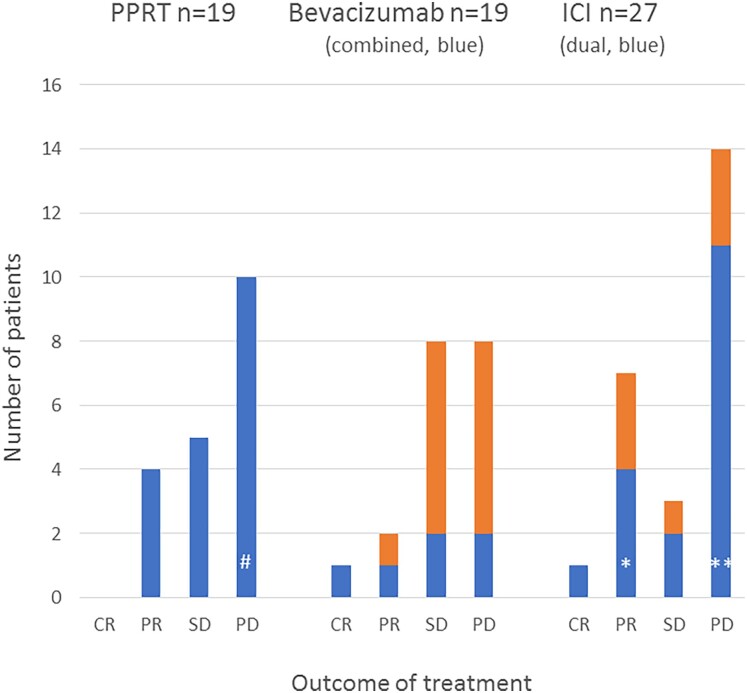
Aggressive pituitary tumors (APTs) and pituitary carcinomas (PCs) are heterogeneous with regard to clinical presentation, proliferative markers, clinical course, and response to therapy. Half of them show an aggressive course only many years after the first apparently benign presentation. APTs and PCs share several properties, but a Ki67 index greater than or equal to 10% and extensive p53 expression are more prevalent in PCs. Mutations in TP53 and ATRX are the most common genetic alterations; their detection might be of value for early identification of aggressiveness. Treatment requires a multimodal approach including surgery, radiotherapy, and drugs. Temozolomide is the recommended first-line chemotherapy, with response rates of about 40%. Immune checkpoint inhibitors have emerged as second-line treatment in PCs, with currently no evidence for a superior effect of dual therapy compared to monotherapy with PD-1 blockers. Bevacizumab has resulted in partial response (PR) in few patients; tyrosine kinase inhibitors and everolimus have generally not been useful. The effect of peptide receptor radionuclide therapy is limited as well. Management of APT/PC is challenging and should be discussed within an expert team with consideration of clinical and pathological findings, age, and general condition of the patient. Considering that APT/PCs are rare, new therapies should preferably be evaluated in shared standardized protocols. Prognostic and predictive markers to guide treatment decisions are needed and are the scope of ongoing research.

Trial registration: ClinicalTrials.gov NCT04244708.

Keywords: ATRX; Ki67- index; PRRT; TP53; bevacizumab; immunotherapy; temozolomide.


Figures

Figure 1.
Figure 1.
Proportion of tumor subtypes according to hormone secretion in 247 patients with aggressive pituitary tumors (47 PC) in comparison with a reference population. Combined data on APT/PC from the 2 European Society of Endocrinology surveys (6, 8). The reference population from a national study of the prevalence of pituitary tumors (13) and of nonfunctioning pituitary tumors in a surgical series (15).
Figure 2.
Figure 2.
A 74-year-old man with an invasive lactotroph macroadenoma and high prolactin (PRL) levels responding to cabergoline. Subsequently, the tumor changed its behavior and progressed despite increasing doses of cabergoline. After developing headache and ophthalmoplegia, the patient was referred to our clinic for pituitary surgery. Upper, Response to cabergoline (mg/wk) over time, imaging and PRL levels (mU/L). Lower, Pathology showed a highly proliferative tumor. (Left) mitotic count 41/10 high-power magnification fields (reference ≤ 2); mitoses, arrows, (right) Ki67 30% (reference < 3%).
Figure 3.
Figure 3.
Time from diagnosis to clinically aggressive behavior in 97 patients with APT/PC. Data from (8).
Figure 4.
Figure 4.
A, A 63-year-old man diagnosed 2007 with Cushing syndrome, UFC 5.4 × ULN, adrenocorticotropin (ACTH) 16 (reference < 10 pmol/L), and an invasive macroadenoma (Ki67 1%, focally 5%-7%, no “atypical” features). B, The tumor 4 years later (Ki67 5%-6%) after 3 pituitary surgeries, radiotherapy, and adrenalectomy. During the last year, ACTH was reported as above the upper level of assay measurement (> 400 pmol/L). The patient developed anemia and was referred to the university hospital. ACTH was 15 060 pmol/L after dilution of the serum; a bone marrow biopsy showed widespread infiltration of tumor cells (C, hematoxylin-eosin, magnification 200×) staining for ACTH (D, ACTH, 400×).
Figure 5.
Figure 5.
A 61-year-man was referred in 2010 because of panhypopituitarism (not diabetes insipidus). Upper, Pituitary imaging showed a macroadenoma, largest diameter 32 mm. Histopathology was compatible with hormone-negative pituitary adenoma. A, Hematoxylin-eosin staining, and B, chromogranin A was expressed; staining for pituitary transcription factors could not be performed at that time. The tumor was not controlled by 2 surgeries and radiotherapy. Temozolomide had no effect. Octreotide scintigraphy showed strong uptake in the pituitary tumor, not elsewhere. Treatment with octreotide led to relief of headache but the tumor continued to grow; the largest diameter increased to 72 mm 2.5 years after diagnosis. 177Lu DOTATATE was given but the patient died shortly afterwards. At reexamination of the tumor, pituitary transcription factors (Pit-1, TPIT, SF-1) were negative (T-PIT staining presented in C). D, A subset of the tumor cells spread throughout the tumor stained for CDX2, suggesting that the tumor represents a metastasis from another neuroendocrine tumor, in the first place from the gastrointestinal tract, although other primary locations cannot be excluded (34, 35). At reevaluation of the octreotide scintigraphy, there was a faint uptake in the right pulmonary hilus, possibly a lymph node metastasis. All microphotographs taken at 200× magnification.
Figure 6.
Figure 6.
Ki67 indices at first surgery in 150 patients with aggressive pituitary tumors. Combined data from the 2 European Society of Endocrinology surveys (6, 8).
Figure 7.
Figure 7.
Evolution of corticotroph tumors in 2 women with initially similar features but different ATRX status: a 33-year-old woman, 3 op + radiotherapy (RT). At the first op KI67 was 2.5%, mitotic count less than 2/10 high-power field (HPF), p53 neg. A, Magnetic resonance imaging (MRI) and B, immunohistochemistry with positive ATRX staining (retained expression) in the tumor cells. At follow-up 10 years later she seems cured. A 38-year-old woman, 3 op + RT + bilateral adrenalectomy. C, MRI of the pituitary tumor at diagnosis. At first op Ki67 was less than 1%, in a hot spot 7%, mitotic count less than 2/10 HPF, p53 neg. D, immunohistochemistry was negative for ATRX (loss of expression) in the tumor cells. Metastases were confirmed 7 years after the first surgery. Both microphotographs taken at 400× magnification. Illustrations for the ATRX-negative case, lower row, are provided by courtesy of Dr Britt Edén Engström, Uppsala University Hospital, Uppsala, Sweden.
Figure 8.
Figure 8.
Outcome of temozolomide (TMZ) monotherapy and of TMZ concurrent with radiotherapy (Stupp). CR, complete regression; PD, progressive disease; PR, partial regression; SD, stable disease. Data on TMZ monotherapy (8) and on the Stupp protocol (6, 8).
Figure 9.
Figure 9.
Response to treatment with peptide receptor radionuclide therapy (PRRT), bevacizumab, and immune checkpoint blockade, the overall experience. #Imaging data not reported, died 1 year later, *hormonal CR in 3 patients, **dissociated responses, see Table 4.

References

    1. Dekkers OM, Karavitaki N, Pereira AM. The epidemiology of aggressive pituitary tumors (and its challenges). Rev Endocr Metab Disord. 2020;21(2):209‐212. - PMC - PubMed
    1. Raverot G, Burman P, McCormack A, et al. . Clinical practice guidelines for the management of aggressive pituitary tumours and carcinomas. Eur J Endocrinol. 2018;178(1):G1‐G24. - PubMed
    1. WHO Classification of Tumours Editorial Board . Endocrine and Neuroendocrine Tumours [Internet]. International Agency for Research on Cancer; 2022 (WHO Classification of Tumours Series, 5th ed; vol. 8). https://tumourclassification.iarc.who.int/chapters/36
    1. Chanson P, Dormoy A, Dekkers OM. Use of radiotherapy after pituitary surgery for non-functioning pituitary adenomas. Eur J Endocrinol. 2019;181(1):D1‐D13. - PubMed
    1. Tampourlou M, Ntali G, Ahmed S, et al. . Outcome of nonfunctioning pituitary adenomas that regrow after primary treatment: a study from two large UK centers. J Clin Endocrinol Metab. 2017;102(6):1889‐1897. - PubMed

Publication types

MeSH terms

Associated data