. 2023 Sep;33(3):709-719.
doi: 10.1007/s00062-023-01265-3. Epub 2023 Mar 1.

Diagnostic Accuracy of Epilepsy-dedicated MRI with Post-processing

Affiliations

Diagnostic Accuracy of Epilepsy-dedicated MRI with Post-processing

Horst Urbach et al. Clin Neuroradiol. 2023 Sep.

Abstract

Purpose: To evaluate the diagnostic accuracy of epilepsy-dedicated 3 Tesla MRI including post-processing by correlating MRI, histopathology, and postsurgical seizure outcomes.

Methods: 3 Tesla-MRI including a magnetization-prepared two rapid acquisition gradient echo (MP2RAGE) sequence for post-processing using the morphometric analysis program MAP was acquired in 116 consecutive patients with drug-resistant focal epilepsy undergoing resection surgery. The MRI, histopathology reports and postsurgical seizure outcomes were recorded from the patient's charts.

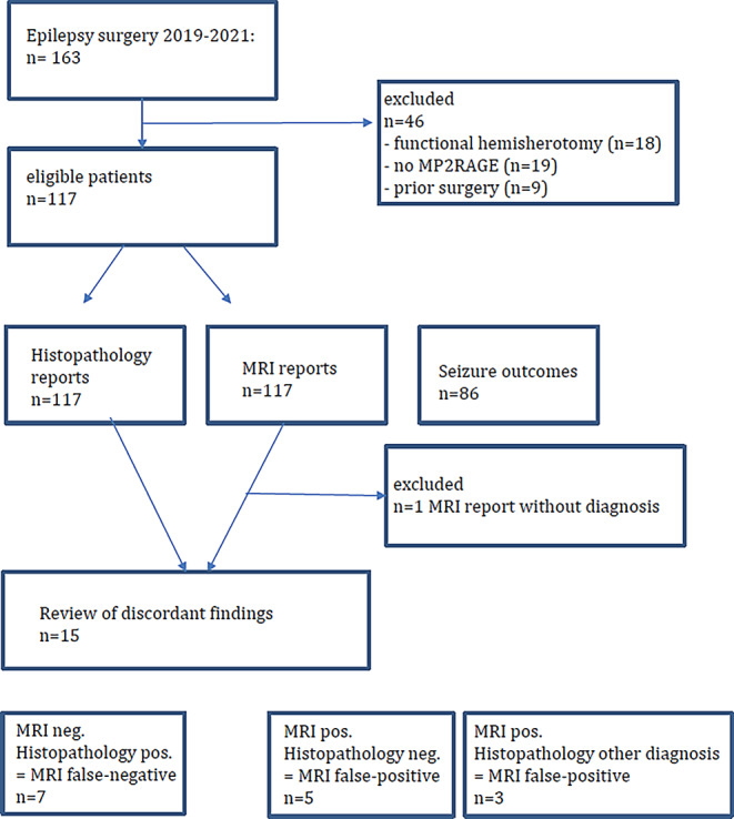
Results: The MRI and histopathology were concordant in 101 and discordant in 15 patients, 3 no hippocampal sclerosis/gliosis only lesions were missed on MRI and 1 of 28 focal cortical dysplasia (FCD) type II associated with a glial scar was considered a glial scar only on MRI. In another five patients, MRI was suggestive of FCD, the histopathology was uneventful but patients were seizure-free following surgery. The MRI and histopathology were concordant in 20 of 21 glioneuronal tumors, 6 cavernomas, and 7 glial scars. Histopathology was negative in 10 patients with temporal lobe epilepsy, 4 of them had anteroinferior meningoencephaloceles. Engel class IA outcome was reached in 71% of patients.

Conclusion: The proposed MRI protocol is highly accurate. No hippocampal sclerosis/gliosis only lesions are typically MRI negative. Small MRI positive FCD can be histopathologically missed, most likely due to sampling errors resulting from insufficient harvesting of tissue.

Keywords: Epilepsy; Lesion; MP2RAGE; MRI; Post-processing.


Conflict of interest statement

H. Urbach, C. Scheiwe, M.J. Shah, J.M. Nakagawa, M. Heers, M.V. San Antonio-Arce, D.-M. Altenmueller, A. Schulze-Bonhage, H.-J. Huppertz, T. Demerath and S. Doostkam declare that they have no competing interests.

Figures

Fig. 1
Fig. 1
Study flowchart
Fig. 2
Fig. 2
A 12-year-old girl presented with daily sensory and bilateral tonic seizures since the age of 6 years. An MRI at the age of 10 years was considered normal (a: arrow). MRI at the age of 11 years revealed a FCD in the depth of the left superior frontal sulcus (be) with a thickened cortex and a transmantle sign (d: arrow). Post-processing of the MP2RAGE images using the MAP tool highlights the lesion histopathologically characterized as FCD type IIB (c, e). MRI after 3 months shows the resection of the FCD (f). Outcome after 6 months was Engel 1B
Fig. 3
Fig. 3
A 14-year-old girl presented with sleep-related, hyperkinetic, tonic and clonic seizures associated with fear since the age of 5. MRI at the age of 5 did not disclose a lesion (a). MRI at the age of 13 showed a slight FLAIR hyperintensity at the gray-white matter border (b,d,f: arrow) highlighted by post-processing of MP2RAGE images using the MAP tool (e,g)
Fig. 4
Fig. 4
A 14-year-old girl presented with hypermotor seizures since the age of 11. MRI showed a hyperintense left-sided cingulate cortex (a,c,e: arrow). The lesion was found by scrolling through the co-registered MP2RAGE images (b: arrow). The junction parameter map displays the gray-white matter blurring (d: arrow). Histopathology revealed gliotic changes, a gray-white matter blurring, some ectopic white matter neurons, no dysplastic neurons, no balloon cells. MRI after 3 months confirmed that the lesion was resected (f). The patient was seizure-free 12 months following surgery
Fig. 5
Fig. 5
FCD type IIA in the depth of the right inferior frontal sulcus in a 5-year-old girl with focal motor, tonic and atonic seizures. The FCD shows a funnel-shaped hyperintensity tapering towards the right frontal horn (visible on FLAIR (a) and on T2-weighted (b) images: arrow). The initial histopathological diagnosis of a multinodular vacuolating tumor was revised due to the characteristic MRI pattern also highlighted by post-processing (c). Furthermore, the intracortical black sign is described as a pathognomonic feature of a FCD IIB (d: arrows)
Fig. 6
Fig. 6
Left-sided hippocampal sclerosis ILAE type 1 (c: blue arrow) with associated gray-white matter blurring (a,b: hollow arrows) in a 38-year-old patient who had fever-related seizures at the age of 8 months. Right-sided hippocampal sclerosis ILAE type 1 (f: blue arrow) in a 50-year-old patient with temporal lobe seizures since the age of 13 years. Coronal FLAIR (d) and post-processed MP2RAGE images (e) showed no associated gray-white matter blurring

References

    1. Von Oertzen J, Urbach H, Jungbluth S, Kurthen M, Reuber M, Fernández G, Elger CE. Standard magnetic resonance imaging is inadequate for patients with refractory focal epilepsy. J Neurol Neurosurg Psychiatry. 2002;73:643–647. doi: 10.1136/jnnp.73.6.643. - DOI - PMC - PubMed
    1. Bien CG, Szinay M, Wagner J, Clusmann H, Becker AJ, Urbach H. Characteristics and surgical outcomes of patients with refractory magnetic resonance imaging-negative epilepsies. Arch Neurol. 2009;66:1491–1499. doi: 10.1001/archneurol.2009.283. - DOI - PubMed
    1. Nguyen DK, Mbacfou MT, Nguyen DB, Lassonde M. Prevalence of non lesional focal epilepsy in an adult epilepsy clinic. Can J Neurol Sci. 2013;40:198–202. doi: 10.1017/S0317167100013731. - DOI - PubMed
    1. Duncan JS, Winston GP, Koepp MJ, Ourselin S. Brain imaging in the assessment for epilepsy surgery. Lancet Neurol. 2016;15:420–433. doi: 10.1016/S1474-4422(15)00383-X. - DOI - PMC - PubMed
    1. Muhlhofer W, Tan YL, Mueller SG, Knowlton R. MRI-negative temporal lobe epilepsy—what do we know? Epilepsia. 2017;58:727–742. doi: 10.1111/epi.13699. - DOI - PubMed

Supplementary concepts