Review
. 2023 May;41(3):388-403.
doi: 10.1007/s00774-023-01403-4. Epub 2023 Mar 1.

Myeloma bone disease: pathogenesis and management in the era of new anti-myeloma agents

Affiliations
Review

Myeloma bone disease: pathogenesis and management in the era of new anti-myeloma agents

Jumpei Teramachi et al. J Bone Miner Metab. 2023 May.

Abstract

Introduction: Multiple myeloma (MM) is a malignancy of plasma cells with characteristic bone disease. Despite recent great strides achieved in MM treatment owing to the implementation of new anti-MM agents, MM is still incurable and bone destruction remains a serious unmet issue in patients with MM.

Approach: In this review, we will summarize and discuss the mechanisms of the formation of bone disease in MM and the available preclinical and clinical evidence on the treatment for MM bone disease.

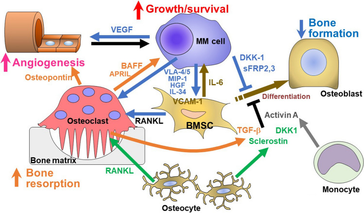
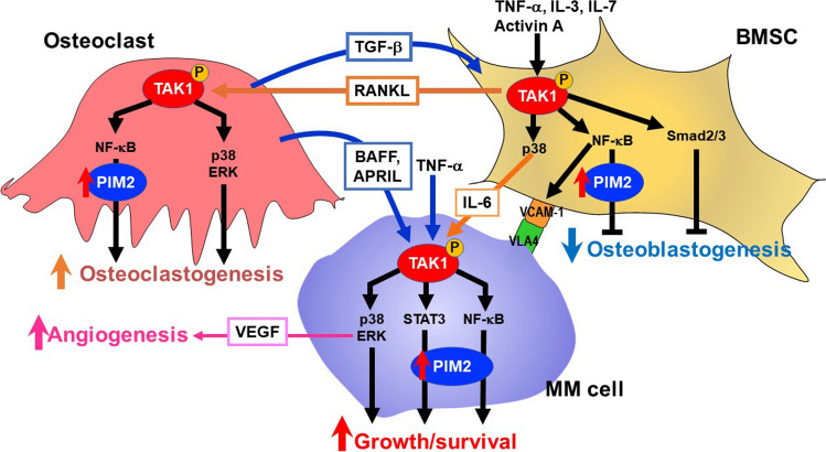
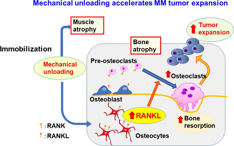
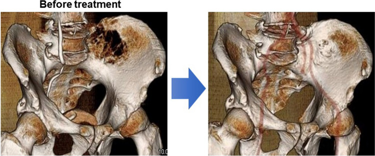
Conclusions: MM cells produce a variety of cytokines to stimulate receptor activator of nuclear factor-κB ligand-mediated osteoclastogenesis and suppress osteoblastic differentiation from bone marrow stromal cells, leading to extensive bone destruction with rapid loss of bone. MM cells alter the microenvironment through bone destruction where they colonize, which in turn favors tumor growth and survival, thereby forming a vicious cycle between tumor progression and bone destruction. Denosumab or zoledronic acid is currently recommended to be administered at the start of treatment in newly diagnosed patients with MM with bone disease. Proteasome inhibitors and the anti-CD38 monoclonal antibody daratumumab have been demonstrated to exert bone-modifying activity in responders. Besides their anti-tumor activity, the effects of new anti-MM agents on bone metabolism should be more precisely analyzed in patients with MM. Because prognosis in patients with MM has been significantly improved owing to the implementation of new agents, the therapeutic impact of bone-modifying agents should be re-estimated in the era of these new agents.

Keywords: Bone-modifying agents; Daratumumab; Myeloma bone disease; Proteasome inhibitors; Receptor activator of nuclear factor-κB ligand.


Conflict of interest statement

M.A. received research funding from Chugai Pharmaceutical, GlaxoSmithKline, Sanofi K.K., Kyowa Kirin, Nippon Shinyaku, Teijin Pharma, and Ono Pharmaceutical, and honoraria from Daiichi Sankyo, Janssen Pharmaceutical K.K., Takeda Pharmaceutical, Sanofi K.K., Bristol-Myers Squibb, and Ono Pharmaceutical. Other authors have no competing financial interests to declare.

Figures

Fig. 1
Fig. 1
Images of bone disease in patients with MM. A Myeloma tumor cells accumulate in the bone marrow. B Multiple compression fractures in lumbar vertebrae. C Multiple radiolucent lesions without ossification, known as “punched-out lesions” in a skull X-p, implying enhanced bone resorption along with impaired calcification. D Bone fractures in long bone occur in an advanced case
Fig. 2
Fig. 2
Bone destruction by factors over-produced by MM cells and/or their surrounding microenvironment in bone in MM. MM cells enhance osteoclastogenesis and suppress osteoblastic differentiation from bone marrow stromal cells (BMSCs), leading to skewing of the cellular microenvironment in the bone marrow. Cytokines aberrantly over-produced by MM cells, including MIP-1, HGF and IL-34 as well as MM cell adhesion (VLA4/5-VCAM-1) up-regulate RANKL and IL-6 production in BMSCs to enhance osteoclastogenesis and MM cell growth/survival. Osteoclasts enhance MM cell growth/survival. MM cells enhance angiogenesis in concert with osteoclasts. In addition, factors over-produced by MM cells and/or their surrounding microenvironment in bone such as soluble Wnt inhibitors, IL-3, IL-7, TNF-α, activin A and TGF-β suppress osteoblastic differentiation. RANKL and sclerostin are over-produced by osteocytes. Therefore, multiple factors act together to eventually develop extensive bone destruction along with MM tumor expansion
Fig. 3
Fig. 3
The TAK1-PIM2 pathway in the mutual interaction between MM cells and the bone microenvironment. PIM2 is a novel pro-survival mediator for MM cells. Interaction with the MM bone marrow microenvironment potentiates PIM2 expression in MM cells through activation of the JAK2/STAT3 pathway by IL-6 and the NF-κB pathway by TNF family cytokines, TNF-α, BAFF, and APRIL, to promote MM cell growth and survival. At the same time, PIM2 is induced in osteoclasts and bone marrow stromal cells (BMSCs) though the interaction with MM cells to cause bone destruction. TAK1 is overexpressed and activated upstream of PIM2 in MM cells, BMSCs and osteoclasts through mutual interaction between these cells in the bone marrow. Besides PIM2 upregulation, TAK1 mediates a wide range of intracellular signaling pathways, including VEGF production via ERK in MM cells and the expression of VCAM-1 and RANKL in BMSCs. Therefore, TAK1 activation is vital for MM cell growth and survival and bone destruction
Fig. 4
Fig. 4
Mechanical unloading accelerates MM tumor expansion. The mechanical unloading not only induce muscle atrophy but also bone loss with up-regulation of RANKL expression in osteocytes and thereby osteoclastogenesis in the bone marrow. The increased and activated osteoclasts then enhance MM growth and dissemination in and outside of the bone. These results suggest the importance of mechanical loading in maintaining bone mass and suppression of tumor expansion in MM, and also the importance of inhibition of RANKL activity for immobilized patients in a bed-ridden state or paralysis
Fig. 5
Fig. 5
Proportion of MM patients without SRE. Time to first SRE on denosumab was retrospectively analyzed in 84 MM patients treated with proteasome inhibitor-based regimens between June 2012 and August 2022 in Tokushima University Hospital. The present study was approved by the Institutional Review Board of Tokushima University (permission number 3086-2)
Fig. 6
Fig. 6
Bone recovery in responders to bortezomib. A newly diagnosed patient was treated with bortezomib and dexamethasone and achieved a very good partial response after 2 cycles of the treatment. Bone formation appeared in bone defective lesions in the iliac bone (right)

References

    1. Silbermann R, Roodman GD. Myeloma bone disease: pathophysiology and management (in eng) J Bone Oncol. 2013;2:59–69. doi: 10.1016/j.jbo.2013.04.001. - DOI - PMC - PubMed
    1. Pecherstorfer M, Seibel MJ, Woitge HW, Horn E, Schuster J, Neuda J, Sagaster P, Köhn H, Bayer P, Thiébaud D, Ludwig H. Bone resorption in multiple myeloma and in monoclonal gammopathy of undetermined significance: quantification by urinary pyridinium cross-links of collagen (in eng) Blood. 1997;90:3743–3750. doi: 10.1182/blood.V90.9.3743. - DOI - PubMed
    1. Hashimoto T, Abe M, Oshima T, Shibata H, Ozaki S, Inoue D, Matsumoto T. Ability of myeloma cells to secrete macrophage inflammatory protein (MIP)-1alpha and MIP-1beta correlates with lytic bone lesions in patients with multiple myeloma (in eng) Br J Haematol. 2004;125:38–41. doi: 10.1111/j.1365-2141.2004.04864.x. - DOI - PubMed
    1. Pearse RN, Sordillo EM, Yaccoby S, Wong BR, Liau DF, Colman N, Michaeli J, Epstein J, Choi Y. Multiple myeloma disrupts the TRANCE/ osteoprotegerin cytokine axis to trigger bone destruction and promote tumor progression (in eng) Proc Natl Acad Sci USA. 2001;98:11581–11586. doi: 10.1073/pnas.201394498. - DOI - PMC - PubMed
    1. Choi SJ, Cruz JC, Craig F, Chung H, Devlin RD, Roodman GD, Alsina M. Macrophage inflammatory protein 1-alpha is a potential osteoclast stimulatory factor in multiple myeloma (in eng) Blood. 2000;96:671–675. doi: 10.1182/blood.V96.2.671. - DOI - PubMed