. 2023 Mar 1;34(3):374-384.
doi: 10.1681/ASN.0000000000000061. Epub 2023 Jan 13.

Membranous Nephropathy in Syphilis is Associated with Neuron-Derived Neurotrophic Factor

Affiliations

Membranous Nephropathy in Syphilis is Associated with Neuron-Derived Neurotrophic Factor

Sanjeev Sethi et al. J Am Soc Nephrol. .

Abstract

Significance statement: Syphilis is a common worldwide sexually transmitted infection. Proteinuria may occur in patients with syphilis. Membranous nephropathy (MN) is the most common cause of proteinuria in syphilis. The target antigen of MN in syphilis is unknown. This study shows that MN in syphilis is associated with a novel target antigen called neuron-derived neurotrophic factor (NDNF). NDNF-associated MN has distinctive clinical and pathologic manifestations and NDNF appears to be the target antigen in syphilis-associated MN.

Background: Syphilis is a common sexually transmitted infection. Membranous nephropathy (MN) is a common cause of proteinuria in syphilis. The target antigen is not known in most cases of syphilis-associated MN.

Methods: We performed laser microdissection of glomeruli and mass spectrometry (MS/MS) in 250 cases (discovery cohort) of phospholipase A2 receptor-negative MN to identify novel target antigens. This was followed by immunohistochemistry/confocal microscopy to localize the target antigen along the glomerular basement membrane (GBM). Western blot analyses using IgG eluted from frozen biopsy tissue were performed to detect binding to target antigen.

Results: MS/MS studies of the discovery cohort revealed high total spectral counts of a novel protein, neuron-derived neurotrophic factor (NDNF), in three patients: one each with syphilis and hepatitis B, HIV (syphilis status not known), and lung tumor. Next, MS/MS studies of five cases of syphilis-MN (validation cohort) confirmed high total spectral counts of NDNF (average 45±20.4) in all (100%) cases. MS/MS of 14 cases of hepatitis B were negative for NDNF. All eight cases of NDNF-associated MN were negative for known MN antigens. Electron microscopy showed stage I MN in all cases, with superficial and hump-like deposits without GBM reaction. IgG1 was the dominant IgG subtype on MS/MS and immunofluorescence microscopy. Immunohistochemistry/confocal microscopy showed granular staining and colocalization of NDNF and IgG along GBM. Western blot analyses using eluate IgG of NDNF-MN showed binding to both nonreduced and reduced NDNF, while IgG eluate from phospholipase A2 receptor-MN showed no binding.

Conclusion: NDNF is a novel antigenic target in syphilis-associated MN.


Figures

Figure 1
Figure 1
Discovery and validation MS/MS cohorts. Initial discovery cohort of 250 cases of phospholipase A2 receptor–negative MN revealed three cases of NDNF-associated MN. One patient had syphilis and hepatitis B. The syphilis status was not known in the remaining two cases. A validation cohort of 14 cases of hepatitis B MN was negative for NDNF in all cases, while a validation cohort of five cases of syphilis MN was positive for NDNF in all cases. MN, membranous nephropathy; MS/MS, microdissection and mass spectrometry; NDNF, neuron-derived neurotrophic factor.
Figure 2
Figure 2
Proteomic Identification of NDNF in PLA2R-negative MN. Glomeruli were microdissected and analyzed using mass spectrometry as described in methods; (A) shows detection of NDNF in eight cases of PLA2R-negative MN (top row). Cases 8 and 9 are the same patient who was biopsied twice. Numbers in green boxes represent TSC of MS/MS matches to a respective protein. All nine biopsies show high TSC for NDNF and IgG1, IgG2, IgG3 subtypes, and baseline spectral counts of PLA2R were also detected in eight of nine biopsies. For comparison, the pooled TSC from six control cases (time-zero protocol transplant biopsies) are also shown; NDNF is not present in the control cases. Case 10 is a patient with diffuse proliferative glomerulonephritis in a patient with active syphilis. (B) Representative sequence coverage map of NDNF from one case. Amino acids highlighted in bold letters over yellow background are the amino acids detected. (C) An example of MS/MS spectra match to a sequence from NNDF. Example MS/MS spectra of ion 833.90 [M+2H]2++ matched to the NDNF peptide sequence TcSSATVAWLGTQER. MN, membranous nephropathy; MS/MS, microdissection and mass spectrometry; NDNF, neuron-derived neurotrophic factor; PLA2R, phospholipase A2 receptor; TSC, total spectral counts.
Figure 3
Figure 3
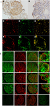
Immunohistochemical (IHC) staining and confocal IF staining for NDNF. (I) Discovery cohort. (A–B) IHC staining showing granular staining for NDNF along GBM (A—patient 3, B—patient 2). (C–H) Confocal IF (patient 1) showing NDNF in red (C and F) and IgG in green (D and G) and merged image in yellow (E and H). (C–E) Show the entire glomerulus and (F–H) show enlarged images of selected capillary loops to focus on the colocalization of NDNF and IgG. (II) Validation cohort. Confocal microscopy of validation cohort (case 4–7). Each row is one case, top row—case 5, second row—case 5, third row—case 6, and fourth row—case 7. First column is NDNF, second column is IgG, third column is overlay of NDNF and IgG, and fourth column is selected high power of NDNF-IgG overlay of few capillary loops. Note the IgG in serum staining as lakes within come capillary loops. Bottom row are control cases—a case of PLA2R-associated MN showing no staining for NDNF (column 1), granular IgG staining (column 2), and overlay showing only IgG staining (column 3). An additional case of EXT1/2-associated MN showing no staining for NDNF is also shown (column 4). EXT1/2, exostosin 1/exostosin 2; IF, immunofluorescence; MN, membranous nephropathy; NDNF, neuron-derived neurotrophic factor.
Figure 4
Figure 4
Western blot analysis showing IgG from eluate of NDNF-associated MN bind to nonreduced and reduced NDNF (2000 ng loaded in each lane). Control: Nonreduced and reduced NDNF are detected by rabbit anti-human NDNF (1:1000) at approximately 44 kDa (arrow) (1A and 1B). Eluate: Nonreduced and reduced NDNF are detected using eluate from NDNF-associated MN using a secondary anti-human IgG (1:5000) (2A and 2B). The binding is not detected using eluate from PLA2R-associated MN (3A and 3B). Near-infrared fluorescence was detected at the 700 and 800 nm channel in the Odyssey Infrared Imaging System (LI-COR© Biosciences, Lincoln, NE). The reading was performed for 10 minutes in both channels (total 20 minutes). NDNF, neuron-derived neurotrophic factor; PLA2R, phospholipase A2 receptor.
Figure 5
Figure 5
Representative kidney biopsy findings in NDNF-associated MN. (A) Light microscopy showing thickened glomerular capillary walls, with no proliferative features (periodic acid–Schiff stain 40×). (B–H) Immunofluorescence microscopy showing bright (B) granular capillary wall staining for IgG, (C) C3, (E) IgG1, (F) IgG2, (G) IgG3, but no staining for (D) PLA2R and (H) IgG4. Electron microscopy (I–L) showing superficial hump-like deposits with minimal or no glomerular basement membrane reaction in all cases (I–J case 3, K—case 2, L—case 1). (A–J) are from case 3. NDNF, neuron-derived neurotrophic factor; PLA2R, phospholipase A2 receptor.
Figure 6
Figure 6
Representative electron microscopy from validation cases showing subepithelial hump-like deposits.

References

    1. Hook EW. Syphilis. Lancet. 2017;389(10078):1550-1557. doi:10.1016/s0140-6736(16)32411-4 - DOI - PubMed
    1. Dyer O. Syphilis and gonorrhoea cases soar in US. BMJ. 2022;378:o2355. doi:10.1136/bmj.o2355 - DOI - PubMed
    1. Centers for Disease Control and Prevention. Preliminary 2021 STD Surveillance Data. 2022. https://www.cdc.gov/std/statistics/2021/default.htm.
    1. Shettigar R, Schollum J, Putt T, Chan L, Lau M, Walker R. Renal manifestations of syphilis. Intern Med J. 2021;51(7):1160-1167. doi:10.1111/imj.15407 - DOI - PubMed
    1. de Almeida Araújo S, Giordano LF, de Britto Rosa AA, do Carmo PAS, Honorato-Sampaio K. Membranous glomerulonephritis secondary to syphilis. Braz J Infect Dis. 2015;19(4):442-443. doi:10.1016/j.bjid.2015.03.003 - DOI - PMC - PubMed

Publication types