

Implication of 99mTc-sum IL-2 SPECT/CT in immunotherapy by imaging of tumor-infiltrating T cells
- PMID: 36858461
- PMCID: PMC9980373
- DOI: 10.1136/jitc-2022-005925
Implication of 99mTc-sum IL-2 SPECT/CT in immunotherapy by imaging of tumor-infiltrating T cells
Abstract
Background: Although immune checkpoint blockade (ICB) and adoptive T cell transfer (ACT) therapy have achieved impressive clinical outcomes, majority of patients do not respond to immunotherapy. Tumor-infiltrating T cells, a critical factor to immunotherapy, is dynamically changing. Therefore, a reliable real-time in vivo imaging system for tumor-infiltrating T cells, but not immunohistochemical analyses, will be more valuable to predict response and guide immunotherapy. In this study, we developed a new SPECT/CT imaging probe 99mTc-sum IL-2 targeting the IL-2Rβ/IL-2Rγ (CD122/CD132) receptor on tumor-infiltrating T cells, and evaluated its application in predicting the immune response to anti-PD-L1 (αPD-L1) therapy as well as tracking infused T cells in ACT therapy.
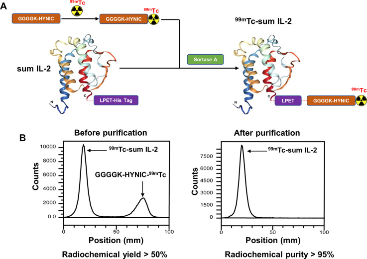
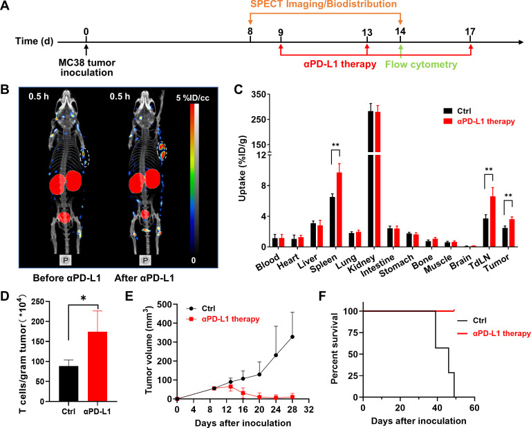
Methods: The binding affinity of the super mutated IL-2 (sum IL-2) in various T cell subtypes was measured. Sum IL-2 was subsequently labeled with 99mTc through Sortase-A mediated site-specific transpeptidation. SPECT/CT imaging and biodistribution studies of 99mTc-sum IL-2 were performed in a MC38 mouse model. Wild type IL-2 (IL-2) was used as control in the above studies. Finally, we evaluated 99mTc-sum IL-2 SPECT/CT for the detection of tumor-infiltrating T cells in the context of αPD-L1 immunotherapy and ACT therapy.
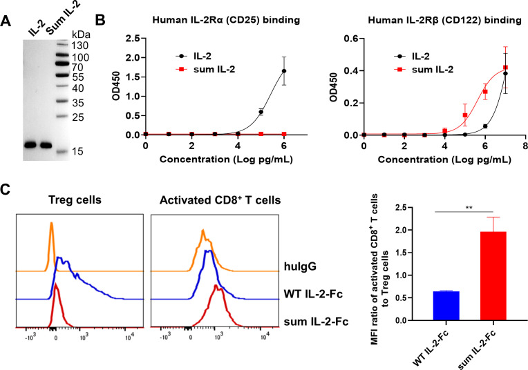
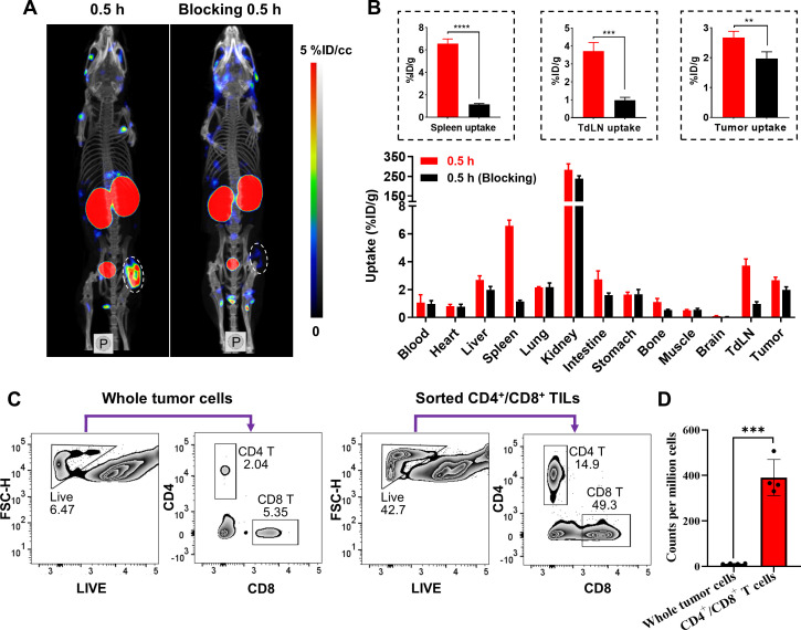
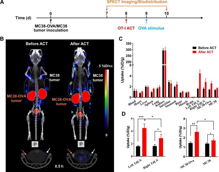
Results: Sum IL-2 preferentially bound to CD8+ T cells, especially activated CD8+ T cells, while IL-2 showed biased binding to Treg cells. As a result, 99mTc-sum IL-2 could detect tumor-infiltrating T cells. In the MC38 tumor model, SPECT/CT imaging showed the increased tumor uptake of 99mTc-sum IL-2 after αPD-L1 treatment, suggesting that the treatment significantly increased tumor-infiltrating T cells, resulting in a correspondingly significant curative effect. In addition, 99mTc-sum IL-2 SPECT/CT could also track the infiltration of antigen-specific cytotoxic CD8+ T cells during ACT therapy.
Conclusion: 99mTc-sum IL-2 has great clinical potential for non-invasive and specific SPECT/CT imaging of tumor-infiltrating T cells as well as for timely prediction and evaluation of the therapeutic efficacy of ICB and ACT therapy.
Keywords: SPECT; immunotherapy; lymphocytes, tumor-infiltrating.
© Author(s) (or their employer(s)) 2023. Re-use permitted under CC BY-NC. No commercial re-use. See rights and permissions. Published by BMJ.
Conflict of interest statement
Competing interests: None declared.
Figures

Similar articles
-
Nuclear imaging-guided PD-L1 blockade therapy increases effectiveness of cancer immunotherapy.J Immunother Cancer. 2020 Nov;8(2):e001156. doi: 10.1136/jitc-2020-001156. J Immunother Cancer. 2020. PMID: 33203663 Free PMC article.
-
Nanobody-mediated SPECT/CT imaging reveals the spatiotemporal expression of programmed death-ligand 1 in response to a CD8+ T cell and iNKT cell activating mRNA vaccine.Theranostics. 2023 Oct 9;13(15):5483-5500. doi: 10.7150/thno.85106. eCollection 2023. Theranostics. 2023. PMID: 37908728 Free PMC article.
-
A Pretargeted Imaging Strategy for Immune Checkpoint Ligand PD-L1 Expression in Tumor Based on Bioorthogonal Diels-Alder Click Chemistry.Mol Imaging Biol. 2020 Aug;22(4):842-853. doi: 10.1007/s11307-019-01441-3. Mol Imaging Biol. 2020. PMID: 31741201
-
99mTc-Interleukin-2.2006 Sep 15 [updated 2006 Oct 12]. In: Molecular Imaging and Contrast Agent Database (MICAD) [Internet]. Bethesda (MD): National Center for Biotechnology Information (US); 2004–2013. 2006 Sep 15 [updated 2006 Oct 12]. In: Molecular Imaging and Contrast Agent Database (MICAD) [Internet]. Bethesda (MD): National Center for Biotechnology Information (US); 2004–2013. PMID: 20641844 Free Books & Documents. Review.
-
99mTc-succinimidyl-6-hydrazinopyridine-3-carboxylate-interleukin 12.2007 Oct 5 [updated 2007 Nov 13]. In: Molecular Imaging and Contrast Agent Database (MICAD) [Internet]. Bethesda (MD): National Center for Biotechnology Information (US); 2004–2013. 2007 Oct 5 [updated 2007 Nov 13]. In: Molecular Imaging and Contrast Agent Database (MICAD) [Internet]. Bethesda (MD): National Center for Biotechnology Information (US); 2004–2013. PMID: 20641943 Free Books & Documents. Review.
Cited by
-
Molecular probes for in vivo optical imaging of immune cells.Nat Biomed Eng. 2025 May;9(5):618-637. doi: 10.1038/s41551-024-01275-7. Epub 2025 Feb 21. Nat Biomed Eng. 2025. PMID: 39984703 Review.
-
Effect of autologous dendritic cell cytokine-induced killer on refractory metastatic colorectal cancer: a matched case-control comparative study.Front Immunol. 2024 Feb 27;15:1329615. doi: 10.3389/fimmu.2024.1329615. eCollection 2024. Front Immunol. 2024. PMID: 38476223 Free PMC article.
-
Current status and future prospects of molecular imaging in targeting the tumor immune microenvironment.Front Immunol. 2025 Jan 22;16:1518555. doi: 10.3389/fimmu.2025.1518555. eCollection 2025. Front Immunol. 2025. PMID: 39911388 Free PMC article. Review.
References
-
- Couzin-Frankel J. Cancer immunotherapy. American Association for the Advancement of Science, 2013.
Publication types
MeSH terms
Substances
LinkOut - more resources
Full Text Sources
Medical
Research Materials