. 2023 Apr 15;63(4):141-151.
doi: 10.2176/jns-nmc.2022-0331. Epub 2023 Mar 1.

Cerebrospinal Fluid Production and Absorption and Ventricular Enlargement Mechanisms in Hydrocephalus

Affiliations

Cerebrospinal Fluid Production and Absorption and Ventricular Enlargement Mechanisms in Hydrocephalus

Shigeki Yamada et al. Neurol Med Chir (Tokyo). .

Abstract

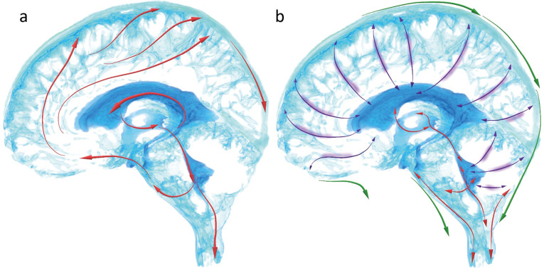
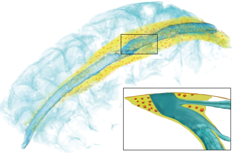
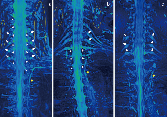
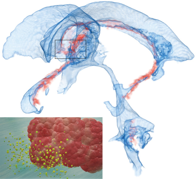
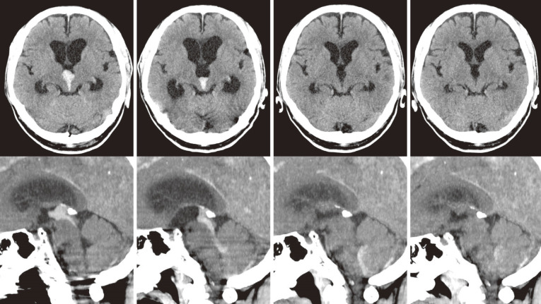
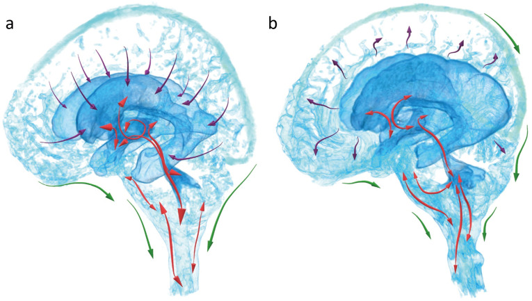
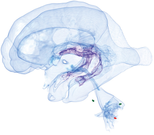
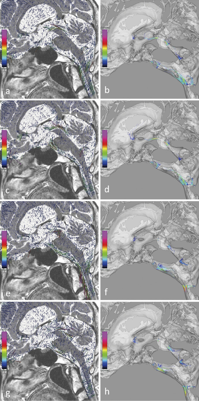
Cerebrospinal fluid (CSF) production and absorption concept significantly changed in the early 2010s from "third circulation theory" and "classical bulk flow theory" to a whole new one as follows: First, CSF is mainly produced from interstitial fluid excreted from the brain parenchyma, and CSF produced from the choroid plexus plays an important role in maintaining brain homeostasis. Second, CSF is not absorbed in the venous sinus via the arachnoid granules, but mainly in the dural lymphatic vessels. Finally, the ventricles and subarachnoid spaces have several compensatory direct CSF pathways at the borders attached to the choroid plexus, e.g., the inferior choroidal point of the choroidal fissure, other than the foramina of Luschka and Magendie. In idiopathic normal pressure hydrocephalus (iNPH), the lateral ventricles and basal cistern are enlarged simultaneously due to the compensatory direct CSF pathways. The average total intracranial CSF volume increased from about 150 mL at 20 years to about 350 mL at 70 years due to the decrease in brain volume with aging and further increased above 400 mL in patients with iNPH. CSF movements are composed of a steady microflow produced by the rhythmic wavy movement of motile cilia on the ventricular surface and dynamic pulsatile flow produced by the brain and cerebral artery pulsation, respiration, and head movement. Pulsatile CSF movements might totally decrease with aging, but it in the ventricles might increase at the foramina of Magendie and Luschka dilation. Aging CSF dynamics are strongly associated with ventricular dilatation in iNPH.

Keywords: cerebrospinal fluid dynamics; chronic hydrocephalus; ventricle dilatation.


Conflict of interest statement

All authors declare that they have no commercial or financial relationships of any kind that could be construed as potential conflicts of interest.

Figures

Fig. 1
Fig. 1
Change in the common concept of cerebrospinal fluid (CSF) dynamics (production and absorption). a: Classical third circulation theory (bulk flow theory). CSF produced from the choroid plexus in the lateral ventricles flows from the lateral ventricles to the third ventricle through the foramen of Monro and from the third ventricle to the fourth ventricle through the cerebral aqueduct and flows out through the foramina of Magendie and Luschka into the posterior cranial fossa subarachnoid space (red arrow). Subsequently, CSF ascends to the basal cistern, flows from the basal cistern to the Sylvian fissure and interhemispheric fissure, and is finally absorbed into the blood through the arachnoid granules and drained extracranially from the venous sinus (red arrows). b: Current CSF dynamics concept. Both ventricle and subarachnoid-space CSF is mainly produced from the interstitial fluid accompanied with waste products accumulated in the brain via the glymphatic system (purple arrow). CSF in the subarachnoid space is mainly absorbed from the dural lymphatic vessels running parallel to the venous sinus and nerve holes in the skull base and drains extracranially (green arrows). CSF moves in a pulsatile fashion driven by cerebral circulation and respiration and has a complex movement driven by exercise. In addition, only CSF in the ventricles has a steady microflow produced by the motile cilia along the ventricular wall surfaces (red arrows).
Fig. 2
Fig. 2
Relationship between parasagittal dura and arachnoid granules around venous sinus. The parasagittal dura (yellow) defined as the dura mater surrounding the superior sagittal sinus in the parietal region has many recessed arachnoid granules (orange). CSF migrates relatively freely from the subarachnoid space to the parasagittal dura (yellow) through the arachnoid granules (orange). However, there was no direct CSF migration to the superior sagittal sinus through the arachnoid granules, and CSF was absorbed by the intradural lymphatic vessels running parallel to the superior sagittal sinus and drained extracranially.
Fig. 3
Fig. 3
CSF absorption from subarachnoid space in cervicothoracic region. Fluid surrounding the spinal subarachnoid space and brachial plexus in the cervicothoracic region is observed on 3D SPACE T2-weighted short-tau inversion recovery (STIR) MRI. In healthy volunteers in the 40s age (a), CSF in the spinal subarachnoid space and the dural sleeve around the nerve roots (white arrowhead) and fluid in the spinal epidural space at the origin of the nerve root sheath of the brachial plexus (white arrow), thoracic duct (yellow arrow), and cervical lymph nodes (yellow circle) are observed. The non-CSF fluid in the epidural spaces is reduced in healthy volunteers in the 70s age (b) and even more reduced in iNPH patients in the 70s age (c).
Fig. 4
Fig. 4
Choroid plexus role. CSF produced by the choroid plexus releases proteins such as hormones and cytokines that act on the paraventricular organs and nuclei on the third ventricle surface, including the pineal gland, hypothalamus, and suprachiasmatic nucleus to control autonomic nervous system, circadian rhythm, emotion, and stress response homeostasis.
Fig. 5
Fig. 5
Acute hydrocephalus due to cerebral aqueduct obstruction by small hematoma. A head CT scan was performed again 6 h after admission since the patient’s consciousness worsened, and ventricles were found severely enlarged due to cerebral aqueduct occlusion by a small hematoma in the third ventricle. Emergency ventricular drainage was prepared, but the patient’s consciousness suddenly improved, so ventricular drainage was not performed. A head CT scan performed 12 h after admission showed ventricular size reduction and cerebral aqueduct hematoma moved down into the fourth ventricle and foramen of Magendie. A head CT scan performed 36 h after admission showed that the ventricular size was almost normalized and the patient fully recovered and did not develop chronic hydrocephalus.
Fig. 6
Fig. 6
Two types of normal pressure hydrocephalus. a: Secondary normal pressure hydrocephalus (sNPH). In most cases with hydrocephalus in concurrent with subarachnoid hemorrhage, acute hydrocephalus begins at the onset of subarachnoid hemorrhage and becomes chronic. Chronic sNPH is caused by extensive adhesion and obstruction of the subarachnoid spaces, which prevents the interstitial fluid draining from the brain into the subarachnoid space and draining all of them into the ventricles (purple arrows). As circumferential enlargement of the ventricles progresses, the surrounding brain and subarachnoid spaces are further compressed, and the pulsations driven by cerebral circulation are concentrated inside the ventricles, resulting in a marked increase in the CSF pulsation in the ventricles (red arrows). CSF in the subarachnoid spaces around the parietal convexity region is markedly reduced and absorption from the intradural lymphatics around the venous sinus is reduced, draining from the skull base or posterior cranial fossa and spinal dura mater (green arrow). b: Idiopathic normal pressure hydrocephalus (iNPH). The primary pathophysiology of iNPH is age-related CSF drainage impairment with decreased absorption from the dural lymphatics around the venous sinus or skull base (green arrows), the foramina of Magendie and Luschka are enlarged, and the CSF pulsation in the foramen magnum is easily transmitted into the ventricles (red arrow). Ventricular enlargement causes degeneration and desquamation of the ventricular walls, resulting in CSF permeation into the periventricular white matter becoming interstitial fluid (purple arrow).
Fig. 7
Fig. 7
Choroidal fissure acting as an overflow device during ventricular enlargement. The ventricles communicate with the subarachnoid space via the foramen of Magendie (red arrow) on the caudal side of the fourth ventricle and via the foramina of Luschka (green arrow) on both sleeves in normal conditions. In iNPH, as the total intracranial CSF volume increases, the tenia attached at the base of the choroid plexus are stretched, resulting in an open compensatory direct CSF pathway between the subarachnoid space and medial surface of the lateral ventricular inferior horns or superior surface of the third ventricle (purple), similar to an overflow device when water is about to overflow from a sink.
Fig. 8
Fig. 8
CSF movement on 4D flow MRI in iNPH. Fast upward flows from the fourth to the third ventricle through the cerebral aqueduct and from the extracranial subarachnoid space to the ventral subarachnoid of the brainstem through the foramen magnum (a-d) are observed, followed by fast downward flows from the ventral subarachnoid space of the brainstem to the extracranial subarachnoid space through the foramen magnum (e, f) and then fast downward flows from the third to the fourth ventricle through the cerebral aqueduct (g, h) in the 2D view of flow velocity vectors (left) and the 3D view of streamlines (right).

Similar articles

Cited by

References

    1. Cushing H: The third circulation and its channels. Lancet 2: 547, 1925
    1. Milhorat TH: The third circulation revisited. J Neurosurg 42: 628-645, 1975 - PubMed
    1. McComb JG: Recent research into the nature of cerebrospinal fluid formation and absorption. J Neurosurg 59: 369-383, 1983 - PubMed
    1. Iliff JJ, Wang M, Liao Y, et al. : A paravascular pathway facilitates CSF flow through the brain parenchyma and the clearance of interstitial solutes, including amyloid beta. Sci Transl Med 4: 147ra111, 2012 - PMC - PubMed
    1. Louveau A, Smirnov I, Keyes TJ, et al. : Structural and functional features of central nervous system lymphatic vessels. Nature 523: 337-341, 2015 - PMC - PubMed