Review
. 2023 Mar 1;14(3):176.
doi: 10.1038/s41419-023-05672-9.

Alpha-synuclein in Parkinson's disease and other synucleinopathies: from overt neurodegeneration back to early synaptic dysfunction

Affiliations
Review

Alpha-synuclein in Parkinson's disease and other synucleinopathies: from overt neurodegeneration back to early synaptic dysfunction

Paolo Calabresi et al. Cell Death Dis. .

Abstract

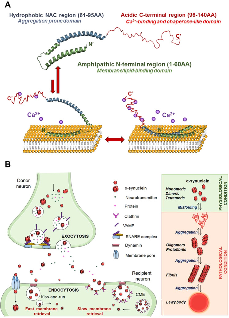
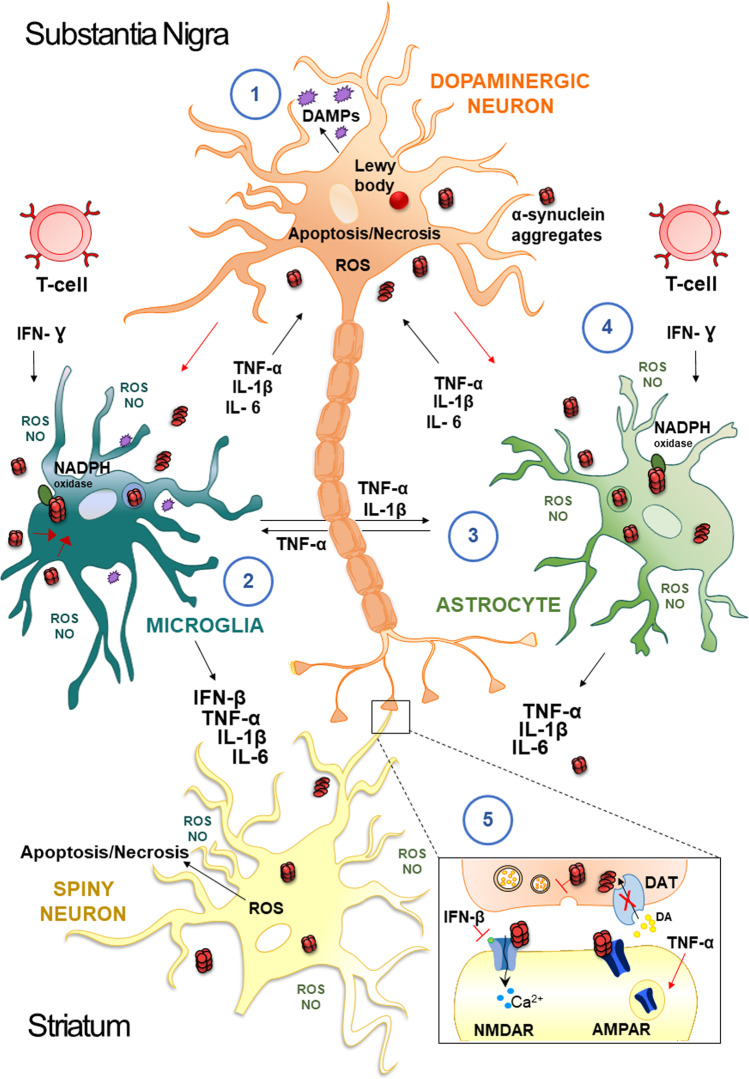
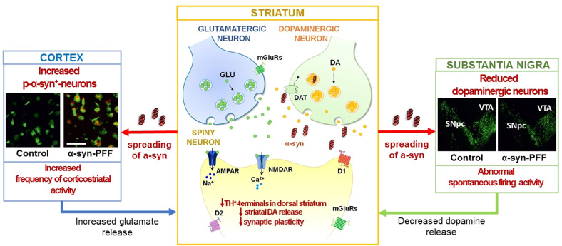
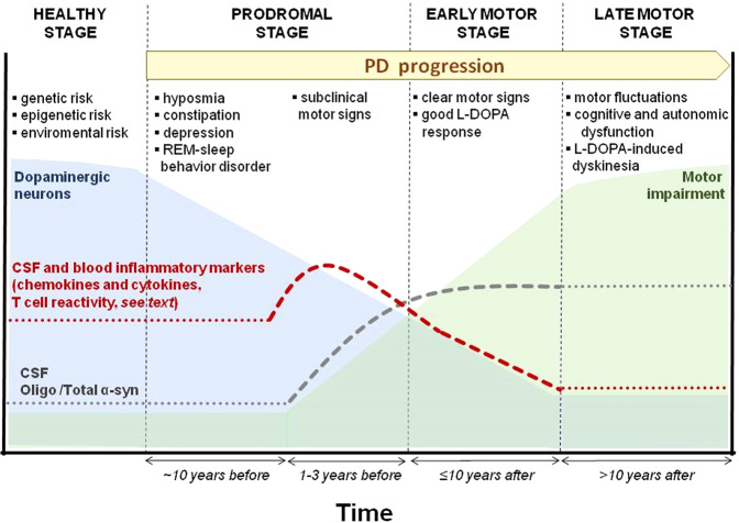
Although the discovery of the critical role of α-synuclein (α-syn) in the pathogenesis of Parkinson's disease (PD) is now twenty-five years old, it still represents a milestone in PD research. Abnormal forms of α-syn trigger selective and progressive neuronal death through mitochondrial impairment, lysosomal dysfunction, and alteration of calcium homeostasis not only in PD but also in other α-syn-related neurodegenerative disorders such as dementia with Lewy bodies, multiple system atrophy, pure autonomic failure, and REM sleep behavior disorder. Furthermore, α-syn-dependent early synaptic and plastic alterations and the underlying mechanisms preceding overt neurodegeneration have attracted great interest. In particular, the presence of early inflammation in experimental models and PD patients, occurring before deposition and spreading of α-syn, suggests a mechanistic link between inflammation and synaptic dysfunction. The knowledge of these early mechanisms is of seminal importance to support the research on reliable biomarkers to precociously identify the disease and possible disease-modifying therapies targeting α-syn. In this review, we will discuss these critical issues, providing a state of the art of the role of this protein in early PD and other synucleinopathies.


Conflict of interest statement

PC received/receives research support, speaker honoraria, and support to attend national and international conferences (not related to the present study) from: Abbvie, Bial, Bayer Schering, Biogen-Dompè, Biogen-Idec, Eisai, Lilly, Lundbeck, Lusofarmaco, Merck-Serono, Novartis, Sanofi-Genzyme, Teva, UCB Pharma, Zambon. The other authors reported no funding from any institution, including personal relationships, interests, grants, employment, affiliations, patents, inventions, honoraria, consultancies, royalties, stock options/ownership, or expert testimony for the last 12 months biomedical financial interests or potential conflicts of interest.

Figures

Fig. 1
Fig. 1. Modular structure of α-syn and its dynamic interactions with membrane lipids in physiological and pathological conditions.
A Alpha-syn is structurally characterized by three modular regions: the N-terminal (green), characterized by amphipathic repetitions, is responsible for interactions with membranes; the hydrophobic NAC (blue), is relevant for aggregation, and the acidic C-terminal (red), is involved in Ca2+ binding and chaperon-like activity. In the presence of low concentrations of Ca2+, α-syn is stably anchored to the lipid surface via its N-terminal region only. With higher Ca2+ concentrations, also the NAC region, but not the C-terminus, shows lipid-binding properties, suggesting that it plays a different role in modulating the affinity of α-syn for cellular membranes. B Left panel: in control conditions, α-syn is important for regulating protein and neurotransmitter release by promoting SNARE complex formation and vesicle docking during the exocytosis process. Extracellular α-syn can affect recipient neurons by fast (i.e., kiss-and-run mode) and slow (i.e., clathrin-mediated endocytosis, CME) modes of membrane retrieval, as several endocytotic pathways can coexist in a single synapse. Interestingly, α-syn is associated with the CME of synaptic vesicles due to its role in sensing and stabilizing the curved membranes. Right panel: under physiological conditions (green), α-syn exists in different conformations balance between unstructured soluble monomeric and tetrameric forms. Under pathological conditions (light red), when the balance between α-syn generation and clearance is disrupted, α‐syn aggregates into oligomers, protofibrils, and fibrils, which further bring to the formation of protein inclusions called LB.
Fig. 2
Fig. 2. Mechanisms underlying inflammation in PD.
Neuropathological hallmarks of PD are the presence of intracellular inclusions containing α-syn aggregates and the death of dopaminergic neurons in the SNpc of the midbrain. Damage-Associated Molecular Patterns (DAMPs), are endogenous danger molecules released from damaged or dying cells, resulting in microglia activation and persistent neuroinflammation (1). Extracellular aggregates of α-syn released by neurons activate glial cells; the internalization of α-syn is followed by activation of NADPH oxidase with the production of ROS/NO, and by the release of proinflammatory cytokines (TNF-α, IL-1β, IL-6, IFN-β) (2). In addition, activated-microglial cells produce mediators such as TNF-α and IL-1β that activate astrocytes, which in turn secrete other cytokines (TNF-α, IL-1β, IL-6) (3). These factors act on dopaminergic neurons of the SNpc and spiny neurons of the striatum and further activate microglia, amplifying the inflammatory response in a positive feedback loop. In particular, IFN-γ is secreted by T cells and is a cytokine involved in the death of DA neurons in the development of PD, which is responsible for enhancing the activation of the surrounding glial cells (4). In this scenario, soluble immune molecules can influence the modulation of synaptic transmission and plasticity (5). For example, INF-β influences NMDAR-mediated synaptic currents in spiny neurons. Conversely, TNF-α drives the internalization of AMPARs and reduces corticostriatal synaptic strength and results in a preferential removal of Ca2+-permeable AMPARs.
Fig. 3
Fig. 3. Presynaptic and postsynaptic dysfunctions induced by intrastriatal injection of α-syn-PFFs in the rat model.
Left panel: in the cortical areas, a consistent proportion of p-α-syn+ neurons was detected in α-syn-PFFs-injected rats. Analysis of spontaneous synaptic currents indicates an increased frequency of the spontaneous excitatory postsynaptic current in target neurons of the dorsal striatum that brings to a state of hyperglutamatergic activity. Right panel: the SNpc in α-syn-PFF rats presents a reduced number of dopaminergic neurons as displayed by a decrease of TH+-immunofluorence, associated with an abnormal increase in spontaneous firing activity. Center panel: in the dorsolateral striatum, α-syn-PFFs injection leads to profound alterations of the corticostriatal long-term plasticity, in the SPNs. A significant decrease of TH+ fibers and a reduced release of endogenous dopamine from SNpc terminals are also observed.
Fig. 4
Fig. 4. Hypothetical sequences of signs and biomarkers in PD stages.
In the healthy presymptomatic stage, the number of dopaminergic cells of SNpc slowly decreases (light blue curve), and clinical symptoms are absent. In prodromal PD, neurodegeneration starts with a rapid decrease in nigral neurons survival rate (light blue), non-motor symptoms, and signs of neurodegeneration are evident; peripheral inflammatory markers begin to increase (red dashed line). Subsequently, there is an increase in inflammatory markers, which decreases gradually in the early motor stage (red dashed line). The oligo α-syn/total α-syn ratio (gray dotted line) is elevated in both prodromal and early motor stages. In addition, the evolution of the motor impairment is represented by a green curve, especially during the late motor stage, when long-term complications of dopaminergic therapy emerge (motor fluctuations and dyskinesia). The dotted lines represent a hypothetical estimation. The duration of the healthy stage is unknown, while the prodromal stage can range between 10 and 15 years.

References

    1. Polymeropoulos MH, Lavedan C, Leroy E, Ide SE, Dehejia A, Dutra A, et al. Mutation in the alpha-synuclein gene identified in families with Parkinson’s disease. Science. 1997;276:2045–7. doi: 10.1126/science.276.5321.2045. - DOI - PubMed
    1. Spillantini MG, Schmidt ML, Lee VM, Trojanowski JQ, Jakes R, Goedert M. Alpha-synuclein in Lewy bodies. Nature. 1997;388:839–40. doi: 10.1038/42166. - DOI - PubMed
    1. Goldstein DS, Isonaka R, Lamotte G, Kaufmann H. Different phenoconversion pathways in pure autonomic failure with versus without Lewy bodies. Clin Auton Res. 10.1007/s10286-021-00829-y 2021. - PMC - PubMed
    1. McKeith IG, Boeve BF, Dickson DW, Halliday G, Taylor JP, Weintraub D, et al. Diagnosis and management of dementia with Lewy bodies: fourth consensus report of the DLB Consortium. Neurology. 2017;89:88–100. doi: 10.1212/WNL.0000000000004058. - DOI - PMC - PubMed
    1. Krismer F, Wenning GK. Multiple system atrophy: insights into a rare and debilitating movement disorder. Nat Rev Neurol. 2017;13:232–43. doi: 10.1038/nrneurol.2017.26. - DOI - PubMed

Publication types

Substances