. 2023 Oct;30(5):1922-1934.
doi: 10.1007/s12350-023-03214-6. Epub 2023 Mar 1.

Attacking the Achilles heel of cardiac amyloid nuclear scintigraphy: How to reduce equivocal and false positive studies

Affiliations

Attacking the Achilles heel of cardiac amyloid nuclear scintigraphy: How to reduce equivocal and false positive studies

Zainab Al Taha et al. J Nucl Cardiol. 2023 Oct.

Abstract

Background: Planar and single-photon emission computed tomography (SPECT) nuclear imaging techniques with bone seeking radiotracers have been increasingly adopted for diagnosis of ATTR cardiac amyloidosis. However, inherent limitations of these techniques due to lack of anatomical landmarks have been recognized, with consequent high numbers of equivocal or false positive cases. SPECT/computed tomography (CT) fusion imaging offers a significant advantage to overcome these limitations by substantially reducing inaccurate interpretations. The authors present the results of a 3-year imaging quality improvement project that focused on reducing the high number of equivocal studies that were noted in the first two years of the amyloidosis program, comparing SPECT only to SPECT/CT fusion technique.

Methods: A retrospective, systematic analysis of 176 patient records was performed to test the premise that SPECT/CT fusion imaging has the potential to reduce equivocal and false positive results.

Results: Of a total of 176 patients, 35 equivocal (19.8%), 32 (18.18%) strongly suggestive, and 109 (61.93%) not suggestive cases were identified. Recognizing that this was not consistent with the international data, the authors set out on a comprehensive quality assessment project to reduce the number of equivocal and false positive cases. In patients who initially underwent SPECT only (Group A; n = 78), the addition of SPECT/CT fusion resulted in the net reclassification of 73% of cases: 100% of equivocal cases (n = 35) were reclassified to not suggestive (n = 34) or strongly suggestive (n = 1). 73% of strongly suggestive cases (n = 30) were reclassified to not suggestive (n = 22) while 8 strongly suggestive cases were confirmed as true positives. 13 not suggestive cases remained negative after SPECT/CT fusion. In cases where SPECT/CT fusion was utilized from the beginning (Group B; n = 98), there were no reclassification of any of the cases when these cases were reprocessed as a control group.

Conclusion: Addition of SPECT/CT imaging reduces the false positive or equivocal studies and increases the diagnostic accuracy of the test. All false positive and equivocal studies were eliminated using the fusion technique. Utilizing the fusion imaging technique increases the spatial resolution, with the ability to localize myocardial uptake and accurately differentiate from blood pool, which is a major source of error.

Keywords: Amyloid heart disease; CT; Image Reconstruction; SPECT; Technical.


Conflict of interest statement

IR is a full-time employee at Pfizer Gulf FZ-LLC. ZAT, DA, HS, HA, and SB do not disclose any conflict of interest or relationship with industry in the past two years. HS and HA contributed equally to the development of this manuscript.

Figures

Figure 1
Figure 1
Timeline of cardiac amyloidosis program at CCAD
Figure 2
Figure 2
Total cohort: 176. Group A: 78 cases that had SPECT only in the beginning. After reprocessing with SPECT/CT fusion, a total of 57 cases were reclassified; 35 equivocal cases were reclassified as either strongly suggestive (n = 1) or not suggestive (n = 34) and 22 strongly suggestive cases were reclassified as not suggestive (false positives). Group B: 98 cases that had SPECT/CT fusion done properly from the beginning, being the more advanced technique; resulted in zero cases being reclassified during the “re-analysis”; served as the control arm
Figure 3
Figure 3
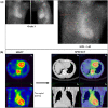
(A) Three-hour planar imaging shows Grade 1 visual tracer uptake and H/CL ratio of 1.24; interpreted as “equivocal for ATTR amyloidosis”. Visual assessment of planar images further demonstrates relatively less myocardial uptake compared to the ribs. (B) SPECT alone demonstrates an empty looking LV cavity, which is shown to be the thickened septal wall on the fused CT images. Also note that the inferior lateral border of the LV wall is not demonstrating any PYP uptake with SPECT/CT fusion. SPECT/CT reclassifies the case from equivocal to “not suggestive”
Figure 4
Figure 4
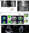
(A) Three-hour planar imaging showing Grade 1 tracer uptake and H/CL of 1.28; interpreted as “equivocal for ATTR amyloidosis”. Note the physiologic kidney uptake of PYP. (B) SPECT demonstrates empty looking LV cavity and a focal uptake in the rib on the right. There is also an appearance of apical sparing (pseudo-apical sparing). Note the rib fracture and enlarged RA and left atrium (LA). SPECT/CT demonstrates no PYP uptake in the LV myocardium, specifically in the thickened posterior wall. SPECT/CT reclassifies case from equivocal to “not suggestive.” (C) ECHO shows posterior wall thickness of 1.5 cm. Note dilated RA and LA
Figure 5
Figure 5
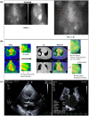
Pseudo-apical sparing (A) Three-hour planar imaging shows Grade 2 tracer uptake and H/CL ratio of 1.36; interpreted as “strongly suggestive of ATTR amyloidosis”. (B) SPECT appears to demonstrate LV wall tracer uptake, sparing the apex. SPECT/CT shows no PYP uptake in the myocardium, but rather confirms LV and RV blood pool. Note dilated RV and thickened posterior wall (1.2 cm). SPECT/CT reclassifies case from strongly suggestive to not suggestive. (C) Four chamber ECHO clearly indicates enlarged RA and RV. Posterior wall is 1.2 cm, septum is 1.2 cm
Figure 6
Figure 6
True Apical sparing (A) Three-hour planar imaging shows Grade 3 tracer uptake and H/CL ratio of 1.60; interpreted as “strongly suggestive of ATTR amyloidosis”. B Apical sparing is evident in the SPECT as well as SPECT/CT. Note thickened LV myocardium in SPECT and SPECT/CT images. SPECT/CT fusion confirms classification as strongly suggestive of ATTR amyloidosis (see Appendix A, Case 7)
Figure 7
Figure 7
A Three-hour planar imaging demonstrates difference in H/CL ratio calculation when enlarged RA is included in ROI vs when RA and diaphragm is excluded. Note lower H/CL ratio when RA is included. B SPECT fails to clearly distinguish between the septum and the enlarged RA and RV. SPECT/CT images clearly show enlarged RA and distinguish septum, RA and RV. SPECT/CT images demonstrates no PYP uptake in the myocardium, changing the classification from strongly suggestive to not suggestive. C Sternum and vertebral body are reference landmarks to accurately fuse the SPECT and CT images. D On the left, SPECT and CT images are inaccurately fused. On the right, SPECT and CT images are precisely fused. With precise registration (on the right), non-tracer avid infero-posterial wall (axial slice, right upper corner) and anterior wall (coronal slice, right lower corner) increase in thickness and are clearly distinguishable as non-tracer avid regions. In the misregistered SPECT/CT images (on the left), the inferior wall appears approximately 3–4 mm; with accurate registration it appears approximately 1.5 mm. Note: the sternum and vertebral body are reference landmarks to be used for accurate registration of SPECT and CT images, which are less effected by the significant inherent respiratory motion. Ribs should not be used as reference landmark given much greater respiratory movement
Figure 8
Figure 8
(A) Three-hours planar imaging vs six-hour planar imaging: H/CL ratio of 1.23 and 1.17 respectively, Grade 1 uptake in both planar images, demonstrating that 6 h delayed imaging does not add value. B SPECT/CT at three hours demonstrates absence of myocardial PYP uptake. SPECT/CT reclassifies case from equivocal to not suggestive

References

    1. Li W, Uppal D, Wang YC, Xu X, Kokkinidis DG, Travin MI, et al. Nuclear imaging for the diagnosis of cardiac amyloidosis in 2021. Diagnostics. 2021;11(6):996. doi: 10.3390/diagnostics11060996. - DOI - PMC - PubMed
    1. Bokhari S, Castaño A, Pozniakoff T, Deslisle S, Latif F, Maurer MS. 99m Tc-pyrophosphate scintigraphy for differentiating light-chain cardiac amyloidosis from the transthyretin-related familial and senile cardiac amyloidoses. Circulation. 2013;6(2):195–201. - PMC - PubMed
    1. Dorbala S, Ando Y, Bokhari S, Dispenzieri A, Falk RH, Ferrari VA, et al. ASNC/AHA/ASE/EANM/HFSA/ISA/SCMR/SNMMI expert consensus recommendations for multimodality imaging in cardiac amyloidosis: Part 1 of 2-evidence base and standardized methods of imaging. J Card Fail. 2019;25(11):e1–e39. doi: 10.1016/j.cardfail.2019.08.001. - DOI - PubMed
    1. Flotats A, Knuuti J, Gutberlet M, Marcassa C, Bengel FM, Kaufmann PA, et al. Hybrid cardiac imaging: SPECT/CT and PET/CT. A joint position statement by the European Association of Nuclear Medicine (EANM), the European Society of Cardiac Radiology (ESCR) and the European Council of Nuclear Cardiology (ECNC) Eur J Nucl Med Mol Imaging. 2011;38(1):201–12. doi: 10.1007/s00259-010-1586-y. - DOI - PubMed
    1. Dorbala S, Ando Y, Bokhari S, Dispenzieri A, Falk RH, Ferrari VA, et al. Addendum to ASNC/AHA/ASE/EANM/HFSA/ISA/SCMR/SNMMI Expert Consensus Recommendations for Multimodality Imaging in Cardiac Amyloidosis: Part 1 of 2-Evidence Base and Standardized Methods of Imaging. J Card Fail. 2021. - PubMed

Publication types

MeSH terms