. 2023 Jul 3;146(7):2730-2738.
doi: 10.1093/brain/awad068.

Variants in ATP5F1B are associated with dominantly inherited dystonia

Affiliations

Variants in ATP5F1B are associated with dominantly inherited dystonia

Alessia Nasca et al. Brain. .

Abstract

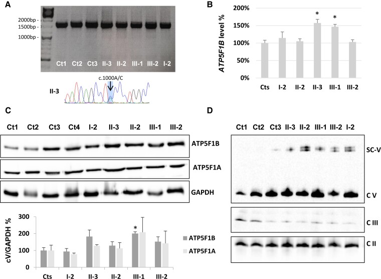
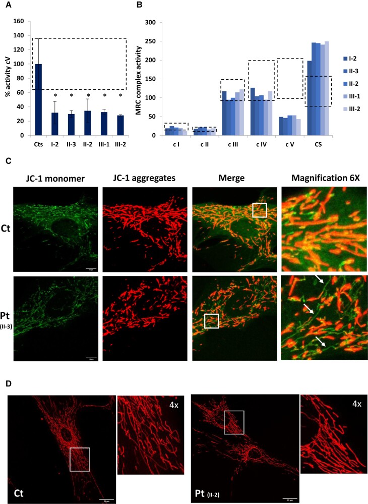
ATP5F1B is a subunit of the mitochondrial ATP synthase or complex V of the mitochondrial respiratory chain. Pathogenic variants in nuclear genes encoding assembly factors or structural subunits are associated with complex V deficiency, typically characterized by autosomal recessive inheritance and multisystem phenotypes. Movement disorders have been described in a subset of cases carrying autosomal dominant variants in structural subunits genes ATP5F1A and ATP5MC3. Here, we report the identification of two different ATP5F1B missense variants (c.1000A>C; p.Thr334Pro and c.1445T>C; p.Val482Ala) segregating with early-onset isolated dystonia in two families, both with autosomal dominant mode of inheritance and incomplete penetrance. Functional studies in mutant fibroblasts revealed no decrease of ATP5F1B protein amount but severe reduction of complex V activity and impaired mitochondrial membrane potential, suggesting a dominant-negative effect. In conclusion, our study describes a new candidate gene associated with isolated dystonia and confirms that heterozygous variants in genes encoding subunits of the mitochondrial ATP synthase may cause autosomal dominant isolated dystonia with incomplete penetrance, likely through a dominant-negative mechanism.

Keywords: ATP5F1B; case report; dystonia; incomplete penetrance; mitochondrial ATP synthase.


Conflict of interest statement

The authors report no competing interests.

Figures

Figure 1
Figure 1
Family pedigrees, genetic and structural analysis. (A) Pedigrees of the families, with segregation data on the c.1000A>C and c.1445T>C variants. Black symbols indicate the affected individuals. (B) Electropherograms of the ATP5F1B regions containing the c.1000A>C and c.1445T>C variants in different members from Families A and B. (C) Alignment of ATP5F1B protein homologues shows the conservation of the mutated amino acids (p.Thr334 and p.Val482) in different species, including human, mouse, chicken, zebrafish, fly, yeast (UniProt identifiers are given for the studied species). (D) Three-dimensional structural modelling (PDB: AF-P06576-F1-model_v3) shows that the mutant Pro334 (left) causes the loss of polar contacts (dotted black lines indicated by arrows) with surrounding amino acids of the subunit β as well as changes in steric hindrance while the mutant Ala482 (right) causes the loss of hydrophobic bonds (dotted blue lines indicated by arrows) with surrounding amino acids and increased flexibility of the C-terminal α helix (in dark red). (E) Structure (PDB: 1NBM) of the F1 module of bovine complex V with the hexamer formed by 3 α (azure) and 3 β (blue) subunits and a central stalk constituted by the subunit γ (yellow): red residues (indicated by arrows) correspond to mutant amino acids Pro334 and Ala482. Pro334 is located close to α and γ subunits, possibly impairing the contact between β and these subunits while Ala482 is located in the outer part of the hexamer.
Figure 2
Figure 2
ATP5F1B transcript and protein analysis on mutant fibroblasts from Family A. (A) Amplification of the ATP5F1B transcript in cDNA, retro-transcribed from available Family A members and control fibroblasts RNA. Bottom: An electropherogram of the region containing the ATP5F1B c.1000A>C variant, with overlapping peaks confirming biallelic expression. (B) Graph reporting ATP5F1B transcript level normalized to a housekeeping gene (ACTN or GAPDH), being 100% the mean value of controls (Cts). Three replicates (with different primer pairs) were assessed and reported as means ± standard deviation. Asterisks indicate statistical significance by t-test (P < 0.001). (C) SDS gel electrophoresis of fibroblasts from available family members and four controls (Ct1–4). For immunoblotting, we used antibodies against ATP5F1B (complex V subunit β), ATP5F1A (complex V subunit α) and GAPDH (as loading control). The graph reports the densitometric analysis (means ± standard deviation) of three independent experiments. All comparisons ‘patient versus controls’ were not significant, except III-1 versus controls for ATP5F1B (asterisk, P < 0.01 by t-test). (D) One-dimensional blue native gel electrophoresis of mitochondrial-enriched fibroblast samples from available family members and three controls (Ct1–3). We used an antibody against ATP5F1A (complex V subunit α) for complex V (cV), an antibody against SDHA for complex II (cII) and an antibody against subunit UQCRC1 for complex III (cIII). High-molecular weight bands (SC-V) were detected in patients’ samples using the ATP5F1A antibody in two biological replicates: they correspond to dimeric/oligomeric complex V or to complex V-containing super-complexes.
Figure 3
Figure 3
Functional studies on mutant fibroblasts from Family A. (A) Assessment of complex V (cV) activity normalized to citrate synthase, in fibroblast cell lines from controls (Cts) and family members. Mean complex V/citrate synthase activity of control fibroblasts is set at 100%, and error bars (and dotted box) represent 1 standard deviation from 290 controls measured in our laboratory. Two biological replicates were quantified for mutant fibroblasts and reported as means ± standard deviation. Asterisks indicate statistical significance by t-test (P < 0.001). (B) Values of respiratory chain enzyme activities normalized to citrate synthase (CS), in fibroblast cell lines from family members. Dotted boxes represent the control range for activities of each complex obtained from 290 laboratory controls. Citrate synthase values are reported as nmol/min mg. (C) Representative images of JC-1 staining on fibroblasts from controls (top row) and ATP5F1B-mutant subjects (bottom row). Red fluorescence (C), corresponding to JC-1 aggregates and sign of preserved mitochondrial membrane potential, is present in almost all controls, whereas several mutant cells display green fluorescent signals (JC-1 monomer) with distribution resembling the mitochondrial network: this finding indicates partial mitochondrial membrane depolarization. Scale bars = 10 µm. Right: Digital magnification at ×6. (D) Representative images of mitochondrial morphology obtained with Mitotracker red staining, showing the filamentous mitochondrial network of fibroblasts from both control (Ct) and ATP5F1B-mutant (Pt) subjects. Scale bars = 25 µm. The smaller panels show digital magnifications (×4) of the insets.

References

    1. Schreglmann SR, Riederer F, Galovic M, et al. . Movement disorders in genetically confirmed mitochondrial disease and the putative role of the cerebellum: Mitochondrial movement disorders. Mov Disord. 2018;33:146–155. - PubMed
    1. Musumeci O, Oteri R, Toscano A. Spectrum of movement disorders in mitochondrial diseases. J Transl Genet Genomics. 2020;4:221–237.
    1. Zech M, Kopajtich R, Steinbrücker K, et al. . Variants in mitochondrial ATP synthase cause variable neurologic phenotypes. Ann Neurol. 2022;91:225–237. - PMC - PubMed
    1. Fernandez-Vizarra E, Zeviani M. Mitochondrial disorders of the OXPHOS system. FEBS Lett. 2021;595:1062–1106. - PubMed
    1. Neilson DE, Zech M, Hufnagel RB, et al. . A novel variant of ATP5MC3 associated with both dystonia and spastic paraplegia. Mov Disord. 2022;37:375–383. - PMC - PubMed

Publication types

Substances