. 2023 Feb 13:16:1089101.
doi: 10.3389/fnmol.2023.1089101. eCollection 2023.

Glycine receptor autoantibody binding to the extracellular domain is independent from receptor glycosylation

Affiliations

Glycine receptor autoantibody binding to the extracellular domain is independent from receptor glycosylation

Vera Rauschenberger et al. Front Mol Neurosci. .

Abstract

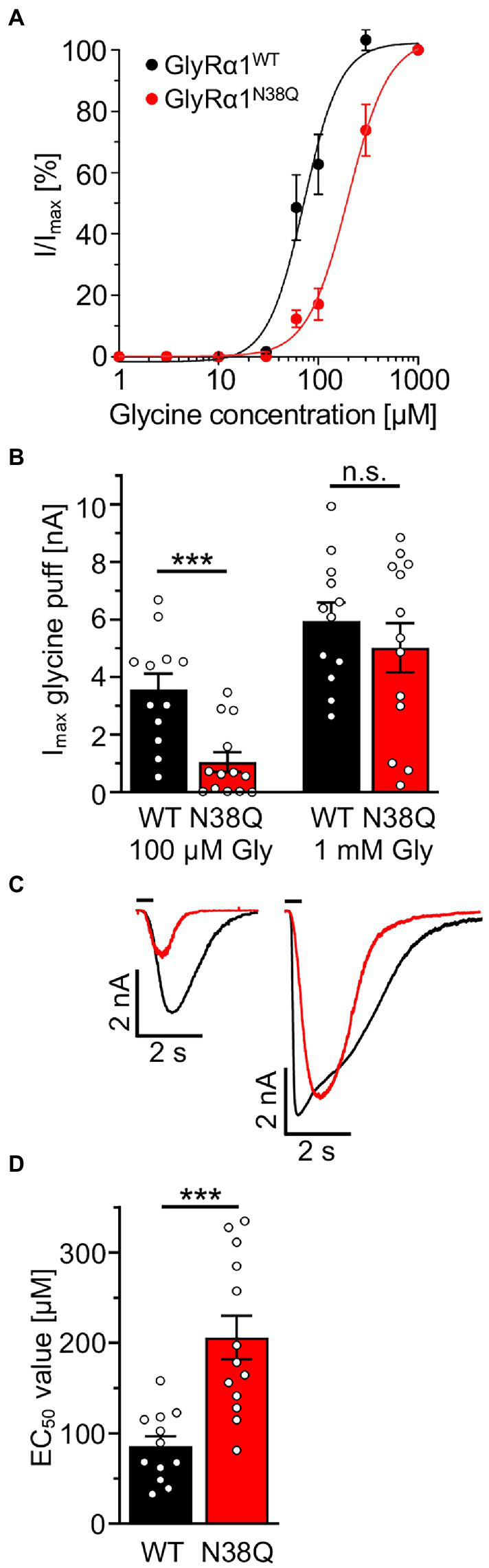
Glycine receptor (GlyR) autoantibodies are associated with stiff-person syndrome and the life-threatening progressive encephalomyelitis with rigidity and myoclonus in children and adults. Patient histories show variability in symptoms and responses to therapeutic treatments. A better understanding of the autoantibody pathology is required to develop improved therapeutic strategies. So far, the underlying molecular pathomechanisms include enhanced receptor internalization and direct receptor blocking altering GlyR function. A common epitope of autoantibodies against the GlyRα1 has been previously defined to residues 1A-33G at the N-terminus of the mature GlyR extracellular domain. However, if other autoantibody binding sites exist or additional GlyR residues are involved in autoantibody binding is yet unknown. The present study investigates the importance of receptor glycosylation for binding of anti-GlyR autoantibodies. The glycine receptor α1 harbors only one glycosylation site at the amino acid residue asparagine 38 localized in close vicinity to the identified common autoantibody epitope. First, non-glycosylated GlyRs were characterized using protein biochemical approaches as well as electrophysiological recordings and molecular modeling. Molecular modeling of non-glycosylated GlyRα1 did not show major structural alterations. Moreover, non-glycosylation of the GlyRα1N38Q did not prevent the receptor from surface expression. At the functional level, the non-glycosylated GlyR demonstrated reduced glycine potency, but patient GlyR autoantibodies still bound to the surface-expressed non-glycosylated receptor protein in living cells. Efficient adsorption of GlyR autoantibodies from patient samples was possible by binding to native glycosylated and non-glycosylated GlyRα1 expressed in living not fixed transfected HEK293 cells. Binding of patient-derived GlyR autoantibodies to the non-glycosylated GlyRα1 offered the possibility to use purified non-glycosylated GlyR extracellular domain constructs coated on ELISA plates and use them as a fast screening readout for the presence of GlyR autoantibodies in patient serum samples. Following successful adsorption of patient autoantibodies by GlyR ECDs, binding to primary motoneurons and transfected cells was absent. Our results indicate that the glycine receptor autoantibody binding is independent of the receptor's glycosylation state. Purified non-glycosylated receptor domains harbouring the autoantibody epitope thus provide, an additional reliable experimental tool besides binding to native receptors in cell-based assays for detection of autoantibody presence in patient sera.

Keywords: adsorption; autoantibodies; extracellular domain; glycine receptor; glycosylation.


Conflict of interest statement

The authors declare that the research was conducted in the absence of any commercial or financial relationships that could be construed as a potential conflict of interest.

Figures

Figure 1
Figure 1
Non-glycosylated GlyRs show no obvious structural alterations. (A) Alignment of GlyRα1WT, GlyRα1N38Q, GlyRα2WT, and GlyRα3WT. The position of the amino acid exchange of the non-glycosylation mutant (N38Q) is indicated in red. All GlyRα subunits share the asparagine at this position and with this a consensus site for glycosylation N-X-S/T. The MAb2b antibody epitope is depicted in purple underlined (Ala1-Ser9, numbers refer to mature protein) and the MAb4a antibody epitope is shown in cyan underlined (Pro96-Gly105). The autoantibody epitope (Ala29-Gly62) (Rauschenberger et al., 2020) is indicated by a blue box. Non-homologous amino acids between GlyRα1WT, α2WT and α3WT are highlighted as bold letters. (B) Crystal structure of human GlyRα3 homopentamer (PDB: 5CFB) (Huang et al., 2015) in side view and top view (C). The antagonist strychnine (orange) is shown in space-filling representation and glycans attached to N38 in ball and stick representation (marked by black box). (D) Enlarged views at position Asn38 in GlyRα3WT (left) and GlyRα3N38Q (right). The proline residue lost a hydrogen bond to asparagine Asn31, but no further obvious changes were detected.
Figure 2
Figure 2
Lack of glycosylation in GlyRα1 leads to slightly decreased whole cell and cell surface expression level. (A) Cell lysates of GlyRα1WT (left) and GlyRα1N38Q (right) undigested or treated with glycosidases PNGaseF and EndoH. Blots were stained with MAb4a and lysates of untransfected HEK293 cells served as negative controls. (B) Representative blots of whole cell (WC), intracellular (IC) and surface (SF) protein levels of pan-cadherin and glycine receptor (MAb4a) of transfected HEK293 cells with GlyRα1WT, GlyRα1N38Q compared to untransfected cells (UT). (C) Positive control blot of HEK293 cells transfected with gephyrin. (D) Quantification of whole cell, intracellular and surface expression of GlyRα1WT (black), and GlyRα1N38Q (red). Expression levels were normalized to pan-cadherin signal and GlyRα1WT signal was set to 1 afterwards. Number of independent experiments n = 5–6; n.s., non-significant.
Figure 3
Figure 3
Patient sera bind to GlyRα1 transfected but not untransfected cells and differ in GlyR autoantibody titres. (A) Patient sera were first tested on untransfected HEK293 cells and cells transfected with the empty pRK5 plasmid. As negative controls, serum from a patient suffering from multiple sclerosis (disease control, DC) and a mix of sera from healthy controls (HC) were used. As control that untransfected cells do not endogenously express the GlyR, MAb4a (binds to all GlyRα subunits) was used. Scale bar for left columns (untransfected and MOCK) represents 100 μm, right columns 10 μm. (B) Patient sera were investigated in a dilution series from 1:10 to 1:5000 for binding to transfected HEK293 cells with GlyRα1 and GFP as internal transfection efficiency control. Incubation with sera was performed for 1 h at living cells, followed by fixation. As secondary antibody an anti-human Cy3 antibody was used (magenta). Pat3 was excluded from analysis due to limited amount of serum available. Scale bar represents 10 μm. All stainings were performed three times (n = 3) and representative images are shown.
Figure 4
Figure 4
Patient anti-GlyR autoantibodies bind to the glycosylated and the non-glycosylated receptor. (A) HEK293 cells co-transfected with GlyRα1 and GFP (cyan, transfection control), followed by an immunocytochemical staining with patient sera (Pat1-5) or MAb2b antibody (magenta). As negative controls, serum from a patient suffering from multiple sclerosis (disease control, DC) and a mix of sera from healthy controls (HC) were used. (B) Immunocytochemical stainings of GlyRα1N38Q and GFP (cyan) co-transfected HEK293 cells that were incubated with patient sera 1–5 or MAb2b (magenta). Healthy control (HC) and disease control (DC) were used as negative controls. Scale bar refers to 10 μm. Immunocytochemical stainings were performed in three independent experiments (n = 3).
Figure 5
Figure 5
Non-glycosylated GlyRα1 differs in functionality to glycosylated receptors. (A) Dose–response curves resulting from electrophysiological recordings of transfected HEK293 cells with GlyRα1WT (black) and GlyRα1N38Q (red). Glycine was applied in a concentration series of 1, 3, 10, 30, 60, 100, 300, 1,000 μM glycine. (B) Maximal currents of transfected HEK293 cells upon glycine application of 100 μM (left) or 1 mM glycine (right). (C) Representative current traces of GlyRα1WT (black) and GlyRα1N38Q (red) upon application of 100 μM glycine (left traces) and saturating glycine concentration at 1 mM (right traces). (D) EC50 values determined from dose–response curves of GlyRα1WT (black) and GlyRα1N38Q (red). Data result from three independent experiments (n = 3) with 12–13 cells total recorded. Significance values reflect ***p < 0.001.
Figure 6
Figure 6
High expression level of GlyRα1 reduce GlyR autoantibodies with high efficiency. (A) GlyRα1WT and GFP (cyan) co-transfected HEK293 cells were incubated with patient sera (1:50 in MEM medium with supplements; magenta) and supernatant was transferred to the next dish with transfected cells (scheme at the top). Afterwards, immunocytochemical stainings were performed including co-stainings with MAb2b (left column, yellow). Note the decrease in autoantibody signal the more often the supernatant was transferred (from left to right). (B) Quantification of ratio of the mean signal intensity of patient serum signal to mean signal intensity of GFP signal. Calculations of cells incubated with patient sera and controls MAb2b, disease control (DC) and healthy control (HC) are shown. The GFP signal indicated the positively transfected HEK293 cells. Significance values were calculated by comparison of the values from coverslips 1, 2, 3, and 4. Data were derived from four independent experiments (n = 4) with a total of 10–15 images being analyzed. Scale bar refers to 20 μm. Significance values reflect *p < 0.05, **p < 0.01, ***p < 0.001, ****p < 0.0001.
Figure 7
Figure 7
Patient autoantibody binding to the non-glycosylated GlyRα1 ECD prevents subsequent binding to endogenous GlyRs expressed in spinal cord neurons. (A) Detection of GlyRα1 ECD construct using Western Blot analysis. Coomassie stain (left) as well as blot stained with MAb4a (middle) or anti-his-tag antibody (right). (B) CD spectrum of GlyR ECD after refolding. The spectrum is baseline corrected and represent the average of 10 individual scans. (C) Analysis of absorbance values recorded from GlyRα1 ECD coated ELISA plates that were incubated with patient sera (Pat1-5), MAb4a, healthy control (HC) and disease control (DC). Number of measured data were n = 6 from 2 independent experiments. All significance values were calculated with unpaired t tests followed by a Holms-Sidaks correction for multiple samples. (D) Immunocytochemical stainings of cultivated motoneurons (DIV14) with patient serum (magenta) which were performed before autoantibody binding to GlyR ECD. As control stainings, synapsin (cyan) and DAPI (blue) are included. (E) After patient autoantibodies were neutralized with the GlyRα1 ECD coated on ELISA plates, the suspension was transferred to cultivated motoneurons (magenta) and stained together with synapsin (cyan) and DAPI (blue). Significance values reflect *p < 0.05, **p < 0.01, ***p < 0.001.
Figure 8
Figure 8
Patient autoantibodies are also efficiently targeting GlyRα2 ECDs. (A) Absorbance values detected from samples of patient sera (Pat1, 2, 4, 5; serum of Pat3 was not tested due to limited amount of sample; value of p for Pat1 p = 0.0457; Pat2 p = 0.0005; Pat4 p = 0.0048, Pat5 p = 0.0457; MAb4a p = 0.0012 always compared to HC) incubated with GlyRα2 ECD coated ELISA plates. Data were obtained from n = 4–8 data points measured in two independent experiments. Significances were calculated with a t test with Holms-Šidáks correction for multiple samples. (B) GlyRα2 transfected HEK293 cells were stained using Pat1, 2, 4, 5 sera or supernatants following incubation on GlyRα2 ECD coated ELISA plates (α2 magenta; GFP cyan, and DAPI blue). Scale bar refers to 10 μm. Significance values reflect *p < 0.05, **p < 0.01, **p < 0.001.

Similar articles

Cited by

References

    1. Albahra S., Yates S. G., Joseph D., De Simone N., Burner J. D., Sarode R. (2019). Role of plasma exchange in stiff person syndrome. Transfus. Apher. Sci. 58, 310–312. doi: 10.1016/j.transci.2019.03.015, PMID: - DOI - PubMed
    1. Atak S., Langlhofer G., Schaefer N., Kessler D., Meiselbach H., Delto C., et al. . (2015). Disturbances of ligand potency and enhanced degradation of the human glycine receptor at affected positions G160 and T162 originally identified in patients suffering from Hyperekplexia. Front. Mol. Neurosci. 8:79. doi: 10.3389/fnmol.2015.00079 - DOI - PMC - PubMed
    1. Bormann J., Rundstrom N., Betz H., Langosch D. (1993). Residues within transmembrane segment M2 determine chloride conductance of glycine receptor homo- and hetero-oligomers. EMBO J. 12, 3729–3737. doi: 10.1002/j.1460-2075.1993.tb06050.x, PMID: - DOI - PMC - PubMed
    1. Breitinger U., Breitinger H. G., Bauer F., Fahmy K., Glockenhammer D., Becker C. M. (2004). Conserved high affinity ligand binding and membrane association in the native and refolded extracellular domain of the human glycine receptor alpha1-subunit. J. Biol. Chem. 279, 1627–1636. doi: 10.1074/jbc.M303811200, PMID: - DOI - PubMed
    1. Carvajal-Gonzalez A., Leite M. I., Waters P., Woodhall M., Coutinho E., Balint B., et al. . (2014). Glycine receptor antibodies in PERM and related syndromes: characteristics, clinical features and outcomes. Brain 137, 2178–2192. doi: 10.1093/brain/awu142, PMID: - DOI - PMC - PubMed

LinkOut - more resources