

Case Reports
doi: 10.1002/ccr3.6760.
eCollection 2023 Feb.
A severe case of PLOD1-related kyphoscoliotic Ehlers-Danlos syndrome associated with several arterial and venous complications: A case report
Affiliations
- PMID: 36860721
- PMCID: PMC9969762
- DOI: 10.1002/ccr3.6760
Item in Clipboard
Case Reports
A severe case of PLOD1-related kyphoscoliotic Ehlers-Danlos syndrome associated with several arterial and venous complications: A case report
Clin Case Rep.
.
Abstract
Kyphoscoliotic Ehlers-Danlos syndrome (kEDS) is a rare genetic disorder combining congenital hypotonia, congenital/early onset and progressive kyphoscoliosis, and generalized joint hypermobility. Vascular fragility is another characteristic of the disease rarely described. We report a severe case of kEDS-PLOD1 with several vascular complications leading to difficulties in disease management.
Keywords: PLOD1 gene; case report; kyphoscoliotic Ehlers–Danlos syndrome; vascular complications.
© 2023 The Authors. Clinical Case Reports published by John Wiley & Sons Ltd.
Conflict of interest statement
No conflict of interest to disclose.
Figures
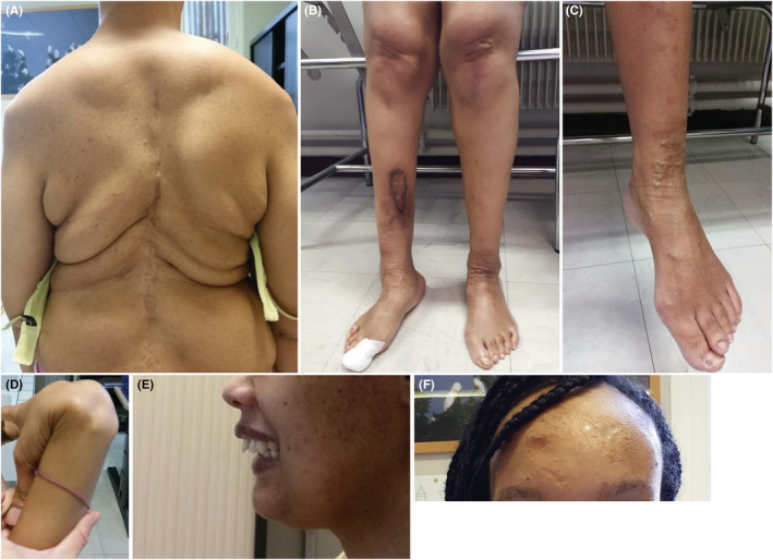
Clinical phenotype of the kEDS patient. (A) Patient at the age of 29 years after multiple surgical corrections of severe thoracolumbar scoliosis. (B) Pes planovarus and scar hyperpigmentation. (C) Varicose veins of the left lower limb; (D) Joint hyperlaxity: passive apposition of the thumb on the forearm. (E) Mandibular retrognathia. (F) Atrophic scars on the forehead
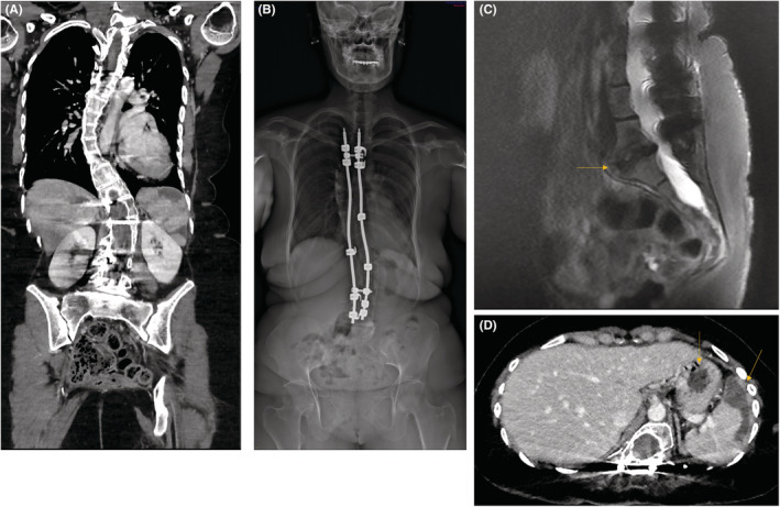
Radiological findings at the age of 29 and 30 years. (A and B) Left thoracolumbar kyphoscoliosis corrected by arthrodesis. (C) L5‐S1 disc herniation. (D) Splenic infarction (full arrow) and angiomyolipoma (dashed arrow)
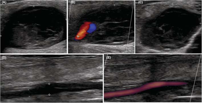
Arterial ultrasonography and Doppler of the false thrombosed occlusive aneurysm (proximal right anterior tibial artery). (A) and (B): Longitudinal view. (C): Transverse view. (D) Longitudinal view on ultrasonography and (E) Doppler ultrasonography of the dissection of the right posterior tibial artery
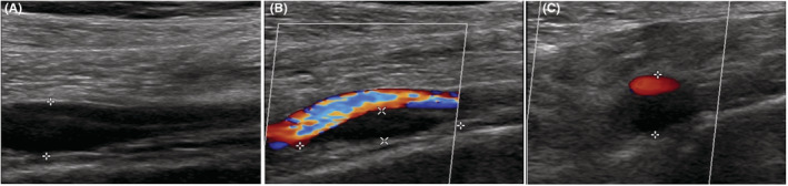
Arterial ultrasonography and Doppler of the dissecting aneurysm of the left tibial‐peroneal trunk. (A) and (B): Longitudinal view. (C): Transversal view
Similar articles
-
Spontaneous celiac artery aneurysms in 13-year-old and 10-year-old brothers with PLOD1-related kyphoscoliotic Ehlers-Danlos syndrome.J Vasc Surg Cases Innov Tech. 2024 Mar 21;10(3):101465. doi: 10.1016/j.jvscit.2024.101465. eCollection 2024 Jun. J Vasc Surg Cases Innov Tech. 2024. PMID: 38694482 Free PMC article.
-
The first case report of Kyphoscoliotic Ehlers-Danlos syndrome of chinese origin with a novel PLOD1 gene mutation.BMC Med Genet. 2020 Oct 31;21(1):214. doi: 10.1186/s12881-020-01154-3. BMC Med Genet. 2020. PMID: 33129265 Free PMC article.
-
Transcriptome Profiling of Primary Skin Fibroblasts Reveal Distinct Molecular Features Between PLOD1- and FKBP14-Kyphoscoliotic Ehlers-Danlos Syndrome.Genes (Basel). 2019 Jul 8;10(7):517. doi: 10.3390/genes10070517. Genes (Basel). 2019. PMID: 31288483 Free PMC article.
-
Nevo syndrome is allelic to the kyphoscoliotic type of the Ehlers-Danlos syndrome (EDS VIA).Am J Med Genet A. 2005 Mar 1;133A(2):158-64. doi: 10.1002/ajmg.a.30529. Am J Med Genet A. 2005. PMID: 15666309 Review.
-
Further delineation of FKBP14-related Ehlers-Danlos syndrome: A patient with early vascular complications and non-progressive kyphoscoliosis, and literature review.Am J Med Genet A. 2016 Aug;170(8):2031-8. doi: 10.1002/ajmg.a.37728. Epub 2016 May 5. Am J Med Genet A. 2016. PMID: 27149304 Review.
Cited by
-
Spontaneous celiac artery aneurysms in 13-year-old and 10-year-old brothers with PLOD1-related kyphoscoliotic Ehlers-Danlos syndrome.J Vasc Surg Cases Innov Tech. 2024 Mar 21;10(3):101465. doi: 10.1016/j.jvscit.2024.101465. eCollection 2024 Jun. J Vasc Surg Cases Innov Tech. 2024. PMID: 38694482 Free PMC article.
References
-
- Malfait F, Francomano C, Byers P, et al. The 2017 international classification of the Ehlers‐Danlos syndromes. Am J Med Genet Part C Semin Med Genet. 2017;175(1):8‐26. - PubMed
-
- Yeowell HN, Walker LC, Farmer B, Heikkinen J, Myllyla R. Mutational analysis of the lysyl hydroxylase 1 gene (PLOD) in six unrelated patients with Ehlers‐Danlos syndrome type VI: prenatal exclusion of this disorder in one family. Hum Mutat. 2000;16(1):90. - PubMed
-
- Hyland J, Ala‐Kokko L, Royce P, Steinmann B, Kivirikko KI, Myllylä R. A homozygous stop codon in the lysyl hydroxylase gene in two siblings with Ehlers–Danlos syndrome type VI. Nat Genet. 1992;2(3):228‐231. - PubMed
Publication types
LinkOut - more resources
Full Text Sources
Miscellaneous
