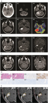
First case of posterior cranial fossa myopericytoma treated with a combined microsurgery and stereotactic radiosurgery approach: Case report and literature review
- PMID: 36861000
- PMCID: PMC9970739
First case of posterior cranial fossa myopericytoma treated with a combined microsurgery and stereotactic radiosurgery approach: Case report and literature review
Keywords: Gamma Knife radiosurgery; intracranial tumor; myopericytoma; posterior cranial fossa; stereotactic radiosurgery.
Figures

References
-
- Granter SR, Badizadegan K, Fletcher CD. Myofibromatosis in adults, glomangiopericytoma, and myopericytoma: a spectrum of tumors showing perivascular myoid differentiation Am J Surg Pathol 1998;22(5):513-525 - PubMed
-
- Rousseau A, Kujas M, van Effenterre R, Boch AL, Carpentier A, Leroy JP, Poirier J. Primary intracranial myopericytoma: report of three cases and review of the literature Neuropathol Appl Neurobiol 2005;31(6):641-648 - PubMed
-
- Calderaro J, Polivka M, Gallien S, Bertheau P, Thiebault JB, Molina JM, Gray F. Multifocal Epstein Barr virus (EBV)-associated myopericytoma in a patient with AIDS Neuropathol Appl Neurobiol 2008;34(1):115-117 - PubMed
-
- Lau PP, Wong OK, Lui PC, Cheung OY, Ho LC, Wong WC, To KF, Chan JK. Myopericytoma in patients with AIDS: a new class of Epstein-Barr virus-associated tumor Am J Surg Pathol 2009;33(11):1666-1672 - PubMed
-
- Zhang CH, Hasegawa H, Johns P, Martin AJ. Myopericytoma of the posterior cranial fossa Br J Neurosurg 2014;29(1):90-91 - PubMed
LinkOut - more resources
Full Text Sources
