. 2023 Aug;19(8):2338-2352.
doi: 10.1080/15548627.2023.2181615. Epub 2023 Mar 2.

N protein of PEDV plays chess game with host proteins by selective autophagy

Affiliations

N protein of PEDV plays chess game with host proteins by selective autophagy

Xueying Zhai et al. Autophagy. 2023 Aug.

Abstract

Macroautophagy/autophagy is a cellular degradation and recycling process that maintains the homeostasis of organisms. The protein degradation role of autophagy has been widely used to control viral infection at multiple levels. In the ongoing evolutionary arms race, viruses have developed various ways to hijack and subvert autophagy in favor of its replication. It is still unclear exactly how autophagy affects or inhibits viruses. In this study, we have found a novel host restriction factor, HNRNPA1, that could inhibit PEDV replication by degrading viral nucleocapsid (N) protein. The restriction factor activates the HNRNPA1-MARCHF8/MARCH8-CALCOCO2/NDP52-autophagosome pathway with the help of transcription factor EGR1 targeting the HNRNPA1 promoter. HNRNPA1 could also promote the expression of IFN to facilitate the host antiviral defense response for antagonizing PEDV infection through RIGI protein interaction. During viral replication, we found that PEDV can, in contrast, degrade the host antiviral proteins HNRNPA1 and others (FUBP3, HNRNPK, PTBP1, and TARDBP) through its N protein through the autophagy pathway. These results reveal the dual function of selective autophagy in PEDV N and host proteins, which could promote the ubiquitination of viral particles and host antiviral proteins and degradation both of the proteins to regulate the relationship between virus infection and host innate immunity.Abbreviations: 3-MA: 3-methyladenine; ATG: autophagy related; Baf A1: bafilomycin A1; CALCOCO2/NDP52: calcium binding and coiled-coil domain 2; ChIP: chromatin immunoprecipitation; Co-IP: co-immunoprecipitation; CQ: chloroquine; DAPI: 4',6-diamidino-2-phenylindole; GPI: glycosyl-phosphatidylinositol; hpi: hours post infection; MARCHF8/MARCH8: membrane-associated ring-CH-type finger 8; MOI: multiplicity of infection; N protein: nucleocapsid protein; PEDV: porcine epidemic diarrhea virus; siRNA: small interfering RNA; TCID50: 50% tissue culture infectious doses.

Keywords: HNRNPA1; IFN; PEDV; nucleocapsid protein; selective autophagy.


Conflict of interest statement

The authors declare that they have no potential conflicts of interest.

Figures
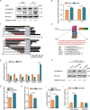
Figure 1.
Figure 1.
HNRNPA1 expression can be influenced in PEDV-infected cells through transcription factor EGR1. (A) PEDV infected or negatively infected LLC-PK1 cells (strain JS-2013) at MOI = 1 were collected at 20 and 24 hpi. Western blotting was applied to detect the expression levels of HNRNPA1 and PEDV N proteins with ACTB as a loading control. (B) qRT-PCR was employed to evaluate the HNRNPA1 relative mRNA expression levels. (C) HEK 293 T cells transfected with pGL3-Basic luciferase vector carrying truncated constructs (−1737 to – 1) of HNRNPA1 promoter were analyzed for the luciferase activity. (D) The TFBS of the HNRNPA1 promoter expected with JASPAR. (E) Relative mRNA levels of predicted genes explored using qRT-PCR in LLC-PK1 cells infected with PEDV 24 hpi. (F, G, and H) HNRNPA1 relative mRNA levels in the EGR1 over-expressed or EGR1 knockdown LLC-PK1 cells explored using western blotting and qRT-PCR. (I) The luciferase activity was analyzed in EGR1 over-expressing HEK 293 T cells. (J) Following transfection with Flag-EGR1 plasmid or blank vector, ChIP analysis for LLC-PK1 cells subjected to harvesting and treatment. Data presented are means ±SD from triplicate experiments. *, ** and ***, respectively indicate P < 0.05, P < 0.01 and P < 0.001 (two-tailed Student’s t-test).
Figure 2.
Figure 2.
Overexpression of HNRNPA1 Inhibits PEDV Infection. (A, B, and C) Vero cells transfected with HNRNPA1 plasmid and infected with PEDV at an MOI of 0.01 at 24 h post-transfection. The PEDV titers were explored with western blotting, qRT-PCR, and TCID50. (D and E) Gradient concentrations of HNRNPA1 plasmids transfected into Vero cells infected with PEDV at an MOI of 0.01 at 24 h post-transfection. (F, G, H, and I) HNRNPA1 plasmids or HNRNPA1 siRNA transfected into LLC-PK1 cells and infected with PEDV at 24 h post-transfection at the MOI of 1. The cell lysates were detected with western blotting and qRT-PCR. Data are presented as means ±SD of triplicate samples. * P < 0.05, ** P < 0.01, *** P < 0.001 (two-tailed Student’s t-test).
Figure 3.
Figure 3.
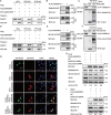
HNRNPA1 can degrade PEDV N protein via autophagy. (A) HEK 293 T cells transfected with the HA-N- and Flag-HNRNPA1-encoding plasmids or RNase and assayed for Co-IP with anti-Flag binding beads. The precipitated proteins were analyzed with western blotting. (B) Following negative infection or infection using PEDV at MOI = 0.01, the Vero cells gathered for the endogenous HNRNPA1 immunoprecipitation based on the antibody of N protein. (C) The pCold TF and pCold GST plasmids utilized for independent cloning of the HNRNPA1, PEDV N and subsequently denoted in the BL21 (DE3) bacterial strain for affinity isolation of GST. The eluted proteins were analyzed with western blotting. (D) HeLa cells co-transfected using HA-N- and FLAG-HNRNPA1-encoding plasmids and cellular labeled with specific antibodies. DAPI (4,6-diamidino-2-phenylindole) labeling for the cellular nuclei and fluorescent signals were monitored with a confocal immunofluorescent microscope (scale bars = 100 µm). (E) Gradient concentration of Flag-HNRNPA1 and HA-N-plasmids were co-transfected into HEK 293 T cells. 24 h later, the cellular lysates were detected by western blotting. (F) Flag-HNRNPA1 and HA-N-plasmids co-transfected into HEK 293 T cells, and then the cells treated with the MG132, CQ, Baf A1, and 3-MA. Western blotting was utilized to detect the cellular lysates. (G) Co-transfection of HEK 293 T cells with MYC-HNRNPA1 and Flag-N plasmids and immunoprecipitation of the ubiquitinated N proteins were with an anti-Flag antibody analyzed with western blotting.
Figure 4.
Figure 4.
PEDV replication can be hindered by HNRNPA1 through the HNRNPA1-MARCHF8-CALCOCO2-autophagosome pathway. (A) HEK 293 T cells treated with Flag-HNRNPA1- and MYC-MARCHF8-encoding plasmids, anti-Flag binding beads were utilized for the Co-IP procedure 24 h post-transfection. Western blotting analysis was adopted for analyzing the precipitated proteins. (B) Following transfected with Flag-HNRNPA1 plasmids or negative controls, the HEK 293 T cells were collected for the endogenous MARCHF8 immunoprecipitation. (C) The GST affinity-isolation assay detected the GST-MARCHF8 and HNRNPA1. (D) HEK 293 T cells treated with Flag-HNRNPA1- and MYC-CALCOCO2-encoding plasmids were precipitated, and proteins were analyzed using anti-Flag binding beads. (E) Following transfected with Flag-HNRNPA1 plasmids or negative controls, the Vero cells were collected for the endogenous CALCOCO2 immunoprecipitation. (F) The GST affinity-isolation assay detected the interaction of GST-CALCOCO2 and HNRNPA1. (G) Flag-HNRNPA1 and MYC-MARCHF8 or MYC-CALCOCO2 plasmids transfected into Hela cells were subsequently labeled with antibodies for confocal immunofluorescence microscopy. Scale bars: 100 µm. (H) HA-N, Flag-HNRNPA1 plasmids, and siRNA (MARCHF8 siRNA or CALCOCO2 siRNA) co-transfected into HEK 293 T cells. (I) Flag-HNRNPA1 plasmid and MARCHF8 siRNA transfected into the Vero cells and, after 24 h, infected with PEDV at MOI = 0.01. Western blotting analysis was applied to detect N protein.
Figure 5.
Figure 5.
HNRNPA1 promotes IFN expression by its interaction with RIGI. (A and B) HEK 293 T cells transfected with IFN-β or ISRE luciferase reporter along with the increasing amounts (wedge) of Flag-HNRNPA1, and dual luciferase activity was detected. (C) HNRNPA1, IFN-β luciferase reporter, plasmids encoding MDA5, RIGI, IKK, MAVS, MYD88, TRAF3, TRAF6, IRF3 or TBK1, co-transfected into HEK 293 T cells to detect dual luciferase activity. (D) HEK 293 T cell lysates were transfected with Flag-HNRNPA1, MDA5 siRNA, or RIGI siRNA to detect the dual luciferase activity. (E) HEK 293 T cells transfected with FLAG-HNRNPA1. The qRT-PCR was employed to value the RIGI relative mRNA expression. (F) HEK 293 T cells co-transfected with the Flag-RIGI and HA-HNRNPA1-encoding plasmids to conduct the Co-IP procedure. The precipitated proteins were analyzed with western blotting. (G) HEK 293 T cells were transfected with the Flag-RIGI for the endogenous HNRNPA1 immunoprecipitation. (H) HeLa cells were transfected with Flag-RIGI and HA-HNRNPA1 plasmids and labeled with specific antibodies. The confocal immunofluorescent microscope was utilized to monitor fluorescent signals (scale bars = 100 µm). (I and J) HEK 293 T cells co-transfected with increasing amounts of Flag-HNRNPA1 plasmids and RIGI siRNA.
Figure 6.
Figure 6.
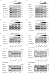
PEDV N protein can exploit the cellular autophagic machinery to degrade HNRNPA1 protein. (A) LLC-PK1 cells co-transfected with the increasing amounts of HA-N and over-expressed Flag-HNRNPA1. (B) LLC-PK1 cells transfected with increasing amounts of HA-N. (C) Flag-HNRNPA1 and HA-N plasmids co-transfected into HEK 293 T cells, and then the cells were treated with the MG132, CQ, Baf A1, and 3-MA. (D) LLC-PK1 cells infected with PEDV and treated with the protease inhibitor or autophagy inhibitor. (E) Co-transfection of HEK 293 T cells with FLAG-HNRNPA1 and MYC-N plasmids and cellular lysates collected following a 24 h post-transfection. The ubiquitinated N proteins were immunoprecipitated with an anti-Flag antibody. All samples were analyzed with western blotting.
Figure 7.
Figure 7.
PEDV N protein can degrade host antiviral proteins by selective autophagy. (A and B) LLC-PK1 cells and HEK 293 T cells transfected with increasing amounts of HA-N. Western blotting was used to detect and analyze the cell lysates. (C, D, E, and F) HEK 293 T cells co-transfected with increasing amounts of HA-N and over-expressed Flag-FUBP3, Flag-HNRNPK, Flag-PTBP1, or Flag-TARDBP. (G, H, I, and J) Flag-FUBP3, Flag-HNRNPK, Flag-PTBP1, or Flag-TARDBP plasmids and HA-N-plasmids were co-transfected into HEK 293 T cells; then the cells were treated with the Baf A1, 3-MA and MG132. All samples were analyzed with western blotting.
Figure 8.
Figure 8.
N protein of PEDV plays a chess game with HNRNPA1. During PEDV infection, the host antiviral protein HNRNPA1 triggered the MARCHF8-CALCOCO2-autophagosome pathway to thwart viral replication. Moreover, HNRNPA1 promoted IFN expression to stimulate the host antiviral defense response to antagonize PEDV infection by RIGI protein interaction. Meanwhile, PEDV also utilized viral N protein to degrade the host antiviral protein HNRNPA1, FUBP3, HNRNPK, PTBP1, and Flag-TARDBP through the host autophagy process to facilitate virus replication. All samples were analyzed with western blotting.

References

    1. Jung K, LJ Saif, Wang Q.. Porcine epidemic diarrhea virus (PEDV): an update on etiology, transmission, pathogenesis, and prevention and control. Virus Res. 2020;286:198045. - PMC - PubMed
    1. EN Wood. An apparently new syndrome of porcine epidemic diarrhoea. Vet Rec. 1977;100:243–244. - PubMed
    1. PR Davies. The dilemma of rare events: porcine epidemic diarrhea virus in North America. Prev Vet Med. 2015;122:235–241. - PMC - PubMed
    1. Jung K, LJ Saif. Porcine epidemic diarrhea virus infection: etiology, epidemiology, pathogenesis and immunoprophylaxis. Vet J. 2015;204:134–143. - PMC - PubMed
    1. Wang D, Fang L, Xiao S. Porcine epidemic diarrhea in China. Virus Res. 2016;226:7–13. - PMC - PubMed

Publication types

LinkOut - more resources