. 2023 Apr 17;133(8):e161156.
doi: 10.1172/JCI161156.

CEP162 deficiency causes human retinal degeneration and reveals a dual role in ciliogenesis and neurogenesis

Affiliations

CEP162 deficiency causes human retinal degeneration and reveals a dual role in ciliogenesis and neurogenesis

Nafisa Nuzhat et al. J Clin Invest. .

Abstract

Defects in primary or motile cilia result in a variety of human pathologies, and retinal degeneration is frequently associated with these so-called ciliopathies. We found that homozygosity for a truncating variant in CEP162, a centrosome and microtubule-associated protein required for transition zone assembly during ciliogenesis and neuronal differentiation in the retina, caused late-onset retinitis pigmentosa in 2 unrelated families. The mutant CEP162-E646R*5 protein was expressed and properly localized to the mitotic spindle, but it was missing from the basal body in primary and photoreceptor cilia. This impaired recruitment of transition zone components to the basal body and corresponded to complete loss of CEP162 function at the ciliary compartment, reflected by delayed formation of dysmorphic cilia. In contrast, shRNA knockdown of Cep162 in the developing mouse retina increased cell death, which was rescued by expression of CEP162-E646R*5, indicating that the mutant retains its role for retinal neurogenesis. Human retinal degeneration thus resulted from specific loss of the ciliary function of CEP162.

Keywords: Cell Biology; Genetic diseases; Genetics; Molecular genetics; Retinopathy.


Figures

Figure 1
Figure 1. Ophthalmological data of patients 1 and 2 with RP.
Ophthalmological data of patient 1 (AF) and patient 2 (GN) with RP are presented throughout (A) Right eye (OD) and (B) left eye (OS) with pale optic disc, narrow vessels, and bone spicule pigmentation on color fundus photography. (C and D) Granular decreased autofluorescence throughout the posterior pole on blue light autofluorescence. (E and F) Spectral domain optical coherence tomography (SDOCT): Cystoid spaces in the inner and outer nuclear layer and thinning of the outer nuclear layer with sparing of the central fovea. (G) Color fundus photography of the left eye: Pale optic disc, narrow vessels with pronounced sheathing giving a white, pseudothrombotic, aspect, bone spicule pigmentation. (H) Fluorescein angiography of the right eye: Strong atrophy of the outer retina and few intraretinal pigment migrations. Autofluorescence from (I) right eye and (J) left eye: bull’s eye aspect of the macula. Goldmann visual fields for (K) left eye and (L) right eye: constriction of < 10°. (M) Pattern ERG of left eye: Reduced macular activity (visual acuity 3/10). No responses for the right eye. (N) Optical coherence tomography: Absence of the outer nuclear layer beyond the macula.
Figure 2
Figure 2. Homozygous CEP162 frameshift variant causes RP in 2 unrelated Moroccan families.
(A) Pedigrees with individuals who were available for genotyping of the c.1935dupA [p.(E646R*5)] in CEP162. M, mutation. (B) Electropherograms of an individual with WT sequence (upper panel), patient 1 (middle; homozygous 1-bp insertion with frameshift and premature termination codon), and a heterozygous carrier (bottom; all children of the patients). (C) ROH on chromosome 6, comprising CEP162, shared by patient 1 and patient 2. (D) Scheme of CEP162 gene (to scale). Vertical bars: exons. The pathogenic variant resides in exon 15.
Figure 3
Figure 3. Effect of E646R*5 mutation on CEP162 protein expression and localization.
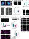
(A) Scheme of human CEP162 protein with 3 C-terminal CC domains and the truncated CEP162-E646R*5 mutant protein. The aa residues are given for each scheme. (B) Western blot of 239T cell lysates from untransfected control, transfected FLAG-CEP162 (approximately 165 kDa, red arrow) and FLAG-CEP162-E646R*5 (approximately 80 kDa, red asterisk). Blots were probed for CEP162 to detect expressed protein and α-tubulin was used as a loading control. (C) Microtubule binding assay. Purified FLAG-CEP162 or FLAG-CEP16-E646R*5 was copelleted with taxol-stabilized microtubules. CEP162 was probed by anti-FLAG antibodies and pelleted microtubules were detected by anti–α-Tubulin antibodies. S, supernatant; P, pellet. (D) Serum-starved IMCD3 cells coexpressing Htr6-GFP and FLAG-CEP162 or FLAG-CEP162-E646R*5. Transfected cells were identified by GFP (green) fluorescence in the cilium. FLAG (magenta) was coimmunostained with either γ-tubulin (cyan, basal bodies) or CEP250 (cyan, proximal-end centriolar protein). Scale bars: 5 μm and 2 μm. (E) FLAG IP from mock, FLAG-CEP162 (approximately 165 kDa, red arrow), or FLAG-CEP162-E646R*5 (approximately 80 kDa, red asterisk) transfected 293T cells lysates. Eluates shown on top and inputs below. Blots probed with anti-CEP290 and anti-FLAG antibodies. (F) CEP290 (magenta) immunostaining with acetylated/γ-tubulin (Ac/γTub, green) in control and patient fibroblasts, with (+) and without (–) cilia. Scale bars: 2 μm.
Figure 4
Figure 4. CEP162-E646R*5 localizes to the mitotic spindle in patient fibroblasts, but its absence from the basal body delays ciliation.
(A) Control and patient fibroblasts costained with CEP162 (magenta) and α-tubulin (green, mitotic spindle). (B) Control and patient fibroblasts costained with CEP162 (magenta) and Ac/γTub (green). Scale bars: 2 μm. (C) Percent ciliation in control and patient fibroblasts 24, 48, or 72 hours after serum withdrawal. A Welch’s 2-tailed t test performed at each time point: ***P = 0.00012 (24 hours), **P = 0.0396 (48 hours), P = 0.4083 (72 hours; NS). (D) CLSEM performed in control and patient fibroblasts by staining cilia for Arl13b (green). Top: merged images. Bottom: magnified SEM. Scale bars: 4 μm and 2 μm. (E) Cilia length measurements. A Welch’s 2-tailed t test performed at each timepoint: P = 1 (24 hours; NS), P = 1 (48 hours; NS), ***P < 0.001 (72 hours). (F) Control and patient fibroblast cilia stained with Arl13b (green) and γ-tubulin (cyan). Scale bar: 5 μm. (G) Immunostaining of CP110 (magenta) in control and patient fibroblasts, ± cilia (Percent observed indicated above). Bar graph quantifying CP110 dots. 2-way ANOVA, P = 0.1701 (0 dots; NS), **P = 0.0068 (1 dot), *P = 0.0329 (2 dots). (H) Western blot from control and patient fibroblast lysates after 24 hours ± serum were probed for CP110 and α-tubulin. Bar graph quantifying levels of CP110 normalized to control + serum, below. Ordinary 1-way ANOVA, **P = 0.0096 (control + serum versus control – serum), P = 0.1929 (control + serum versus patient + serum; NS), P = 0.3042 (control + serum versus patient – serum; NS). (I) Immunostaining of TZ proteins TMEM67, RPGRIP1L, NPHP1, and TCTN1 (magenta) costained with Ac/γTub (green) in control and patient fibroblasts, ± cilia. Cyan arrows mark TZ. Scale bars: 2 μm. (J) Htr6-GFP cilia (green) in control or CEP162 shRNA targeted cells (mCherry, magenta) and coexpressing either FLAG-CEP162 or FLAG-CEP162-E646R*5. Scale bars: 10 μm. Bar graph shows percent ciliation in control or CEP162 shRNA expressing IMCD3 cells, right. Ordinary 1-way ANOVA, *P = 0.0012 (control versus CEP162 shRNA), ***P < 0.0001 (CEP162 versus FLAG-CEP162 shRNA), **P = 0.0002 (FLAG-CEP162 versus FLAG-CEP162-E646R*5 shRNA), P = 0.6186 (CEP162 versus FLAG-CEP162-E646R*5 shRNA; NS). For all graphs, error bars represent SD.
Figure 5
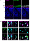
Figure 5. Endogenous CEP162 is localized to the distal-end of centrioles at the base of the photoreceptor outer segment in adult WT mouse retina.
(A) Cetn2-GFP transgenic mouse retinal sections stained with an anti-CEP162 (magenta) antibody. CEP162 is localized at the photoreceptor connecting cilium, which is marked by GFP fluorescence (green). Scale bar: 10 μm. (B) High-resolution Airyscan images: CEP162 (magenta) staining decorates the distal ends of each centriole of the basal body at the base of the connecting cilium (Cetn2-GFP, green). Additional Airyscan images of CEP162 counterstained with multiple ciliary markers: Acetylated Tubulin (AcTub), CEP290, CEP164, and CP110 (cyan). Scale bar: 1 μm.
Figure 6
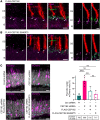
Figure 6. CEP162-E646R*5 mutant protein does not localize to centrioles in adult mouse rod photoreceptors but does participate in retinal neurogenesis.
(A and B) Adult (P21) WT mouse retinal sections expressing either FLAG-CEP162 (A) or FLAG-CEP162-E646R*5 (B, magenta) and rhodopsin-mCherry (Rho-mCherry, red) as a transfection marker. Centrin1 (green) immunostaining was conducted to label the connecting cilium. In the transfected rods, overexpressed FLAG-CEP162 is predominantly localized to the basal body (arrows) but is also be seen within the connecting cilium. Scale bars: 5 μm and 2 μm. (C) Developing mouse retina (P0–P1) were electroporated with control or CEP162 shRNA plasmids coexpressing a soluble mCherry (magenta), and CEP162 knockdown was rescued with either FLAG-CEP162 or FLAG-CEP162-E646R*5. Retinal sections were collected at P14 for analysis. Scale bar: 5 μm. Within a mCherry-positive retinal section, cell death was assessed by counting pycnotic nuclei labeled with DAPI (grey, cyan asterisk) and normalizing to mCherry cells to account for variations in electroporation efficiency. Ordinary 1-way ANOVA, ***P = 0.0001 (control versus CEP162 shRNA), **P = 0.01 (CEP162 shRNA versus FLAG-CEP162), P = 0.3389 (control versus FLAG-CEP162; NS), P = 0.9217 (control versus FLAG-CEP162-E646R*5; NS), P = 0.746 (FLAG-CEP162 versus FLAG-CEP162-E646R*5; NS).

References

    1. Vasquez SSV, et al. An updated SYSCILIA gold standard (SCGSv2) of known ciliary genes, revealing the vast progress that has been made in the cilia research field. Mol Biol Cell. 2021;32(22):br13. doi: 10.1091/mbc.E21-05-0226. - DOI - PMC - PubMed
    1. Morleo M, Franco B. OFD Type I syndrome: lessons learned from a rare ciliopathy. Biochem Soc Trans. 2020;48(5):1929–1939. doi: 10.1042/BST20191029. - DOI - PubMed
    1. Pezzella N, et al. OFD1: One gene, several disorders. Am J Med Genet C Semin Med Genet. 2022;190(1):57–71. doi: 10.1002/ajmg.c.31962. - DOI - PMC - PubMed
    1. Abramowicz I, et al. Oral-facial-digital syndrome type I cells exhibit impaired DNA repair; unanticipated consequences of defective OFD1 outside of the cilia network. Hum Mol Genet. 2017;26(1):19–32. - PubMed
    1. Alfieri M, et al. The centrosomal/basal body protein OFD1 is required for microtubule organization and cell cycle progression. Tissue Cell. 2020;64:101369. doi: 10.1016/j.tice.2020.101369. - DOI - PubMed

Publication types

Substances