. 2023 Mar:89:104492.
doi: 10.1016/j.ebiom.2023.104492. Epub 2023 Feb 28.

PPM1K-regulated impaired catabolism of branched-chain amino acids orchestrates polycystic ovary syndrome

Affiliations

PPM1K-regulated impaired catabolism of branched-chain amino acids orchestrates polycystic ovary syndrome

Liangshan Mu et al. EBioMedicine. 2023 Mar.

Abstract

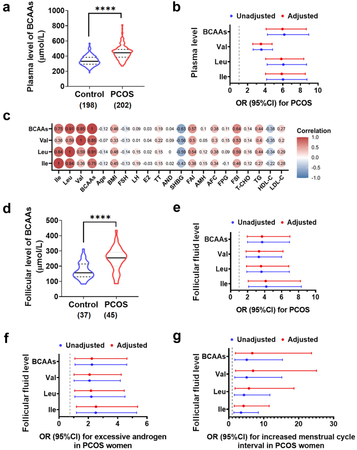
Background: Polycystic ovary syndrome (PCOS) is one of the most common diseases with the coexistence of reproductive malfunction and metabolic disorders. Previous studies have found increased branched chain amino acid (BCAA) levels in women with PCOS. However, it remains unclear whether BCAA metabolism is causally associated with the risk of PCOS.

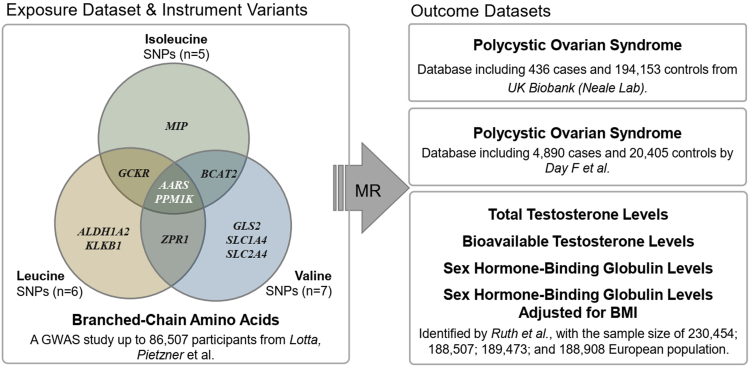
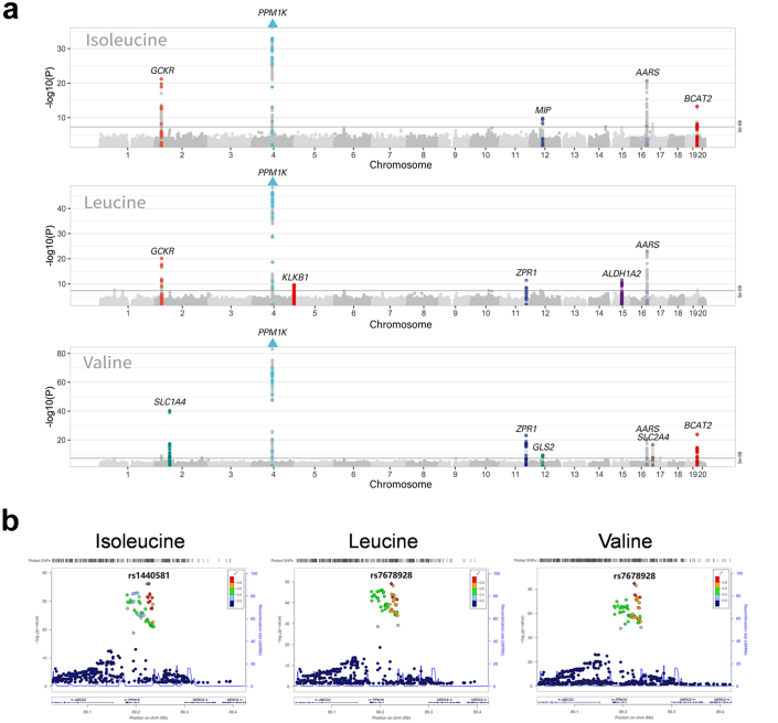
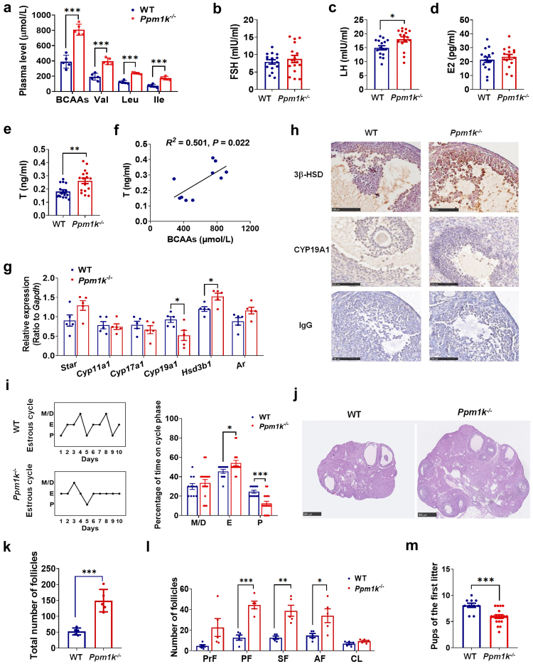
Methods: The changes of BCAA levels in the plasma and follicular fluids of PCOS women were detected. Mendelian randomization (MR) approaches were used to explore the potential causal association between BCAA levels and the risk of PCOS. The function of the gene coding the protein phosphatase Mg2+/Mn2+-dependent 1K (PPM1K) was further explored by using Ppm1k-deficient mouse model and PPM1K down-regulated human ovarian granulosa cells.

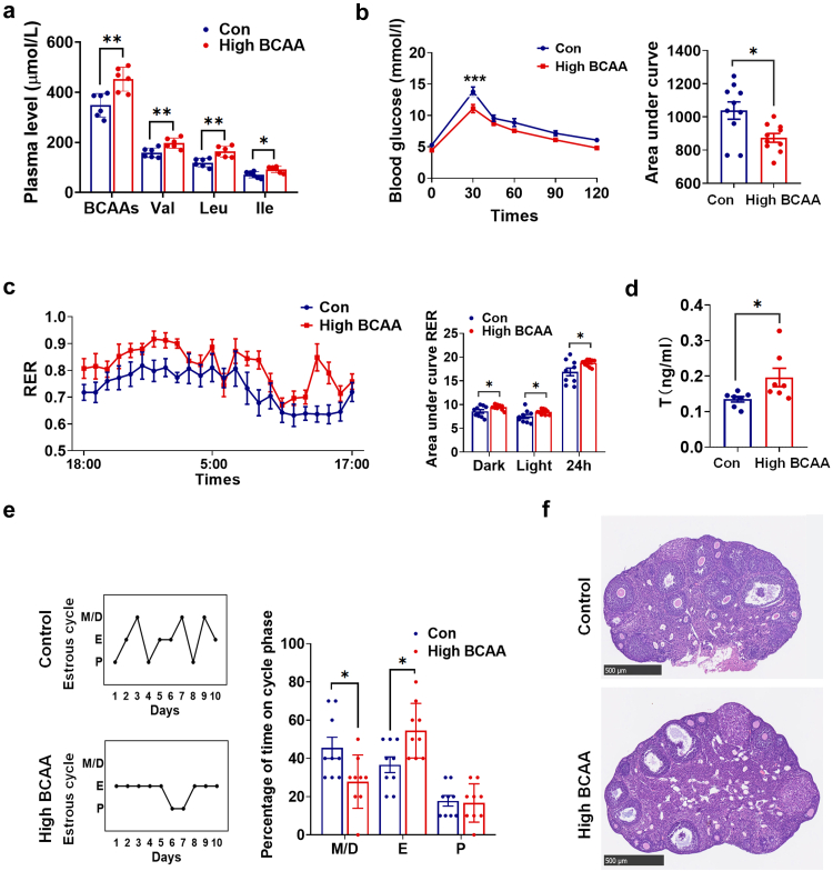
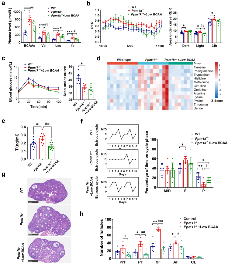
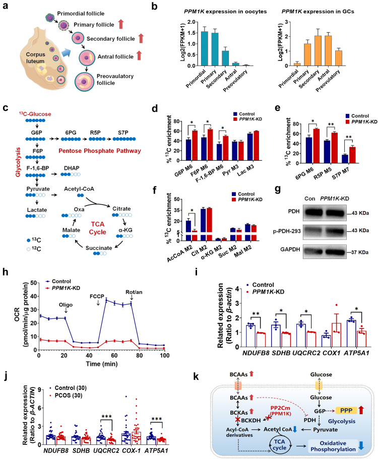
Findings: BCAA levels were significantly elevated in both plasma and follicular fluids of PCOS women. Based on MR, a potential direct, causal role for BCAA metabolism was revealed in the pathogenesis of PCOS, and PPM1K was detected as a vital driver. Ppm1k-deficient female mice had increased BCAA levels and exhibited PCOS-like traits, including hyperandrogenemia and abnormal follicle development. A reduction in dietary BCAA intake significantly improved the endocrine and ovarian dysfunction of Ppm1k-/- female mice. Knockdown of PPM1K promoted the conversion of glycolysis to pentose phosphate pathway and inhibited mitochondrial oxidative phosphorylation in human granulosa cells.

Interpretation: Ppm1k deficiency-impaired BCAA catabolism causes the occurrence and development of PCOS. PPM1K suppression disturbed energy metabolism homeostasis in the follicular microenvironment, which provided an underlying mechanism of abnormal follicle development.

Funding: This study was supported by the National Key Research and Development Program of China (2021YFC2700402, 2019YFA0802503), the National Natural Science Foundation of China (81871139, 82001503, 92057107), the CAMS Innovation Fund for Medical Sciences (2019-I2M-5-001), Key Clinical Projects of Peking University Third Hospital (BYSY2022043), the China Postdoctoral Science Foundation (2021T140600), and the Collaborative Innovation Program of Shanghai Municipal Health Commission (2020CXJQ01).

Keywords: BCAA Catabolism; Mendelian randomization; PCOS; PPM1K.


Conflict of interest statement

Declaration of interests The authors declare that they have no competing interests.

Figures

Fig. 1
Fig. 1
BCAA levels associate with clinical features of PCOS women. (a) The plasma levels of BCAAs in PCOS (n = 202) and control (n = 198) groups. (b) The odds ratios (95% CIs) for PCOS per 1-SD increase in plasma abundance of BCAAs with and without age and BMI adjustment. (c) The correlations between the plasma levels of BCAAs and the endocrine and metabolic parameters of PCOS. (d) The follicular fluid levels of BCAAs in PCOS (n = 45) and control (n = 37) groups. (e) The odds ratios (95% CIs) for PCOS per 1-SD increase in BCAA levels in follicular fluids with and without age and BMI adjustment. (f and g) The odds ratios (95% CIs) for excessive androgen level (f) or increased menstrual cycle interval (g) in PCOS women per 1-SD increase in BCAA levels in follicular fluids with and without age and BMI adjustment. P values were determined by two-tailed Mann–Whitney U-test. ∗∗∗∗, p < 0.0001.
Fig. 2
Fig. 2
Flow diagram of Mendelian randomization analysis. We used two-sample MR with summary statistics from 3 GWAS genetic datasets to determine the causal relationship between BCAAs and PCOS (with related traits). 18 robust variants were included as instruments, AARS and PPM1K were the shared gene in all BCAAs. “TwoSampleMR” R package was used in the analysis.
Fig. 3
Fig. 3
The Manhattan plot of BCAAs and Locus Zoom plots for PPM1K. (a) Genes associated with exposure are annotated the MR analysis annotates the genes associated with exposure. SNPs above the threshold (in black line) were considered significant (p < 5 × 10−8). Where 11 genes are highlighted (AARS, ALDH1A2, BCAT2, GCKR, GLS2, KLKB1, MIP, PPM1K, SLC1A4, SLC2A4, ZPR1). The triangle indicated the −log10(P) value of SNPs in the corresponding gene has exceeded the drawn region. (b) The Locus Zoom plots for PPM1K. Abbreviations: PPM1K, the protein phosphatase Mg2+/Mn2+ dependent 1K.
Fig. 4
Fig. 4
Ppm1k-deficient female mice have a PCOS-like phenotype. (a) The plasma levels of BCAAs in wild-type (WT) and Ppm1k−/− female mice (n = 5 mice per group). (b–e) The changes of serum levels of follicle-stimulating hormone (FSH), luteinizing hormone (LH), estradiol (E2) and testosterone (T) in WT and Ppm1k−/− female mice (n = 16 mice per group). (f) The correlation analysis between serum BCAAs and total testosterone levels (n = 10 mice per group). (g) The mRNA expression of androgen synthesis-related genes in the ovarian tissue of WT and Ppm1k−/− female mice (n = 5 mice per group). (h) Immunohistochemical analysis of the protein expression levels of CYP19A1 and 3β-HSD in ovarian tissues of WT and Ppm1k−/− mice, scale bar = 100 μm. (i) Representative and quantitative analysis of estrous cycles (WT, n = 11; Ppm1k−/−female mice, n = 17). (j) Representative ovarian morphology of WT and Ppm1k−/− mice by hematoxylin and eosin staining, scale bar = 500 μm, images are representative of three independent experiments with similar results. (k) The total number of follicles in the entire ovary of WT and Ppm1k−/− mice (n = 5 mice per group). (l) Differential follicle counts of primordial (PrF), primary (PF), secondary follicles (SF), antral follicles (AF) and corpus luteum (CL) (n = 5 mice per group). (m) Fertility test of female mice (WT, n = 6; Ppm1k−/−female mice, n = 18). Data are represented as means ± SEM. P values were determined by Student's t-test. ∗, p < 0.05; ∗∗, p < 0.01; ∗∗∗, p < 0.001.
Fig. 5
Fig. 5
The effects of high dietary BCAAs on the reproductive and metabolic phenotypes of female mice. (a) The plasma levels of BCAAs (n = 6 mice per group). (b) The glucose tolerance test (n = 10 mice per group). (c) Respiratory exchange ratios (RER) and the area under curve of RER during light and dark cycles (n = 10 mice per group). (d) The serum levels of testosterone (n = 7 mice per group). (e) Representative and quantitative analysis of estrous cycles (n = 9 mice per group). (f) Representative ovarian morphology by hematoxylin and eosin staining, scale bar = 500 μm. Data are presented as means ± SEM. P values were determined by Student's t-test.∗, p < 0.05; ∗∗, p < 0.01; ∗∗∗, p < 0.001.
Fig. 6
Fig. 6
Reduction of dietary BCAAs improves the abnormal reproductive and metabolic phenotypes of Ppm1k-deficient female mice. (a) The plasma levels of BCAAs (wild-type, n = 10; Ppm1k−/− female mice, n = 8; Ppm1k−/− mice with Low-BCAA diet, n = 7). (b) Respiratory exchange ratios (RER) and the area under curve of RER during light and dark cycles (wild-type, n = 10; Ppm1k−/− female mice, n = 5; Ppm1k−/− mice with Low BCAA diet, n = 8). (c) The glucose tolerance test (wild-type, n = 5; Ppm1k−/− female mice, n = 5; Ppm1k−/− mice with Low-BCAA diet, n = 6). (d) The plasma levels of other amino acids (wild-type, n = 10; Ppm1k−/− female mice, n = 10; Ppm1k−/− mice with Low-BCAA diet, n = 8) (e) The serum levels of testosterone (wild-type, n = 9; Ppm1k−/− female mice, n = 10; Ppm1k−/− mice with Low BCAA diet, n = 8). (f) Representative and quantitative analysis of estrous cycles (wild-type, n = 8; Ppm1k−/− female mice, n = 5; Ppm1k−/− mice with Low-BCAA diet, n = 8). (g) Representative ovarian morphology by hematoxylin and eosin staining, scale bar = 500 μm. (h) Differential follicle counts of primordial (PrF), primary (PF), secondary follicles (SF), antral follicles (AF) and corpus luteum (CL) (wild-type, n = 5; Ppm1k−/− female mice, n = 4; Ppm1k−/− mice with Low-BCAA diet, n = 8). Data are presented as means ± SEM. P values were determined by one-way ANOVA with Tukey's multiple comparison post-hoc test. Wild-type versus Ppm1k−/− female mice, ∗p < 0.05, ∗∗∗p < 0.001; Ppm1k−/− versus Ppm1k−/− mice with Low-BCAA diet, #p < 0.05, ##p < 0.01, ###p < 0.001.
Fig. 7
Fig. 7
Effects of PPM1K knockdown on the energy metabolism homeostasis of human granulosa cells. (a) Schematic diagram of follicle development (By Figdraw). Red arrows indicate abnormal follicle development in Ppm1k−/− female mice. (b) The changes of PPM1K gene expression in oocytes and granulosa cells in human follicles at different developmental stages. (c) Schematic map of 13C-glucose incorporation from glucose into TCA cycle in human ovarian granulosa cells. G6P, glucose 6-phosphate; F6P, fructose 6-phosphate; F-1,6-BP, fructose 1,6-bisphosphate; DHAP, dihydroxyacetone phosphate; 6 PG, glucose-6-phosphate; R5P, ribose 5-phosphate; S7P, sedoheptulose 7-phosphate; α-KG, α-Ketoglutarate; Oxa, oxaloacetic acid. (df) Relative levels of glycolysis (d), pentose phosphate pathway (e) and TCA cycle (f) determined by 13C-labeled metabolites in control and PPM1K knocked-down (PPM1K-KD) groups (n = 3). (g) Analysis of phosphorylation levels changes of pyruvate dehydrogenase (PDH) protein. (h) Real-time changes of the O2 consumption rate (OCR) in control and PPM1K-KD human ovarian granulosa cells. Cells were treated with 2 μM oligomycin (Oligo), 1 μM carbonyl cyanide-ptrifluoromethoxyphenylhydrazone (FCCP) and 1 μM rotenone and antimycin A (Rot/an), as indicated by the three black arrows. (i) The expressions of mitochondrial oxidative phosphorylation-related genes in control and PPM1K-KD granulosa cells (n = 3). (j) The expressions of mitochondrial oxidative phosphorylation-related genes in the primary granulosa cells of PCOS and non-PCOS control individuals (n = 30). (k) Summary of PPM1K knockdown inducing the conversion of glycolysis to pentose phosphate pathway and the suppression of oxidative phosphorylation in human ovarian GCs. Data are represented as means ± SEM. For d, e, f and i, p values were determined by Student's t-test. For j, p values were determined by two-tailed Mann–Whitney U-test. ∗, p < 0.05; ∗∗, p < 0.01; ∗∗∗, p < 0.001.

References

    1. Skiba M.A., Islam R.M., Bell R.J., Davis S.R. Understanding variation in prevalence estimates of polycystic ovary syndrome: a systematic review and meta-analysis. Hum Reprod Update. 2018;24(6):694–709. - PubMed
    1. Escobar-Morreale H.F. Polycystic ovary syndrome: definition, aetiology, diagnosis and treatment. Nat Rev Endocrinol. 2018;14(5):270–284. - PubMed
    1. Dumesic D.A., Oberfield S.E., Stener-Victorin E., Marshall J.C., Laven J.S., Legro R.S. Scientific statement on the diagnostic criteria, epidemiology, pathophysiology, and molecular genetics of polycystic ovary syndrome. Endocr Rev. 2015;36(5):487–525. - PMC - PubMed
    1. Lynch C.J., Adams S.H. Branched-chain amino acids in metabolic signalling and insulin resistance. Nat Rev Endocrinol. 2014;10(12):723–736. - PMC - PubMed
    1. Newgard C.B., An J., Bain J.R., et al. A branched-chain amino acid-related metabolic signature that differentiates obese and lean humans and contributes to insulin resistance. Cell Metab. 2009;9(4):311–326. - PMC - PubMed

Substances