. 2023 Mar 21;4(3):100959.
doi: 10.1016/j.xcrm.2023.100959. Epub 2023 Mar 1.

Bioengineered omental transplant site promotes pancreatic islet allografts survival in non-human primates

Affiliations

Bioengineered omental transplant site promotes pancreatic islet allografts survival in non-human primates

Hongping Deng et al. Cell Rep Med. .

Abstract

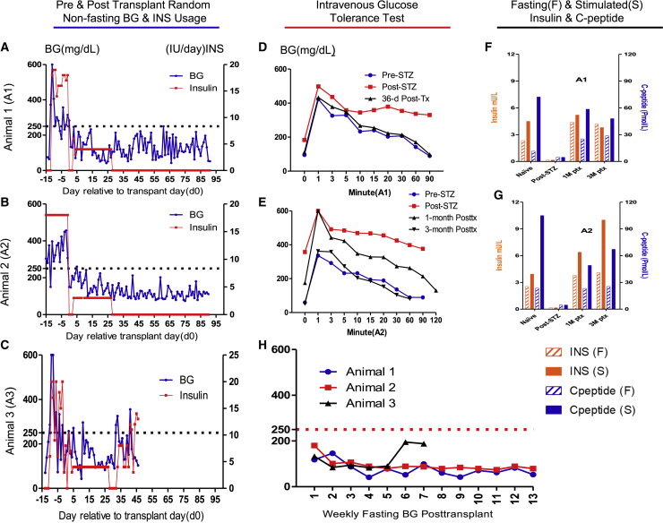
The transplanting islets to the liver approach suffers from an immediate posttransplant loss of islets of more than 50%, progressive graft dysfunction over time, and precludes recovery of grafts should there be serious complications such as the development of teratomas with grafts that are stem cell-derived islets (SC-islets). The omentum features an attractive extrahepatic alternative site for clinical islet transplantation. We explore an approach in which allogeneic islets are transplanted onto the omentum, which is bioengineered with a plasma-thrombin biodegradable matrix in three diabetic non-human primates (NHPs). Within 1 week posttransplant, each transplanted NHP achieves normoglycemia and insulin independence and remains stable until termination of the experiment. Success was achieved in each case with islets recovered from a single NHP donor. Histology demonstrates robust revascularization and reinnervation of the graft. This preclinical study can inform the development of strategies for β cell replacement including the use of SC-islets or other types of novel cells in clinical settings.

Keywords: allogeneic islet transplantation; bioengineering the omentum; euglycemia; graft survival; non-human primate; plasma-thrombin matrix; revascularization and reinnervation; single donor; stem cell-derived islets.


Conflict of interest statement

Declaration of interests The authors declare no competing interests.

Figures

None
Graphical abstract
Figure 1
Figure 1
Characteristics of transplanted islets and recipients, immunosuppression treatment regimen, and the process to engineer the omentum for islet implantation (A) Table 1: characteristics of transplanted islets and recipients. Three streptozotocin (STZ)-induced diabetic NHPs (5.6–6.5 kg body weight) were entered into this study, and each received allogeneic islet transplantation in a one-donor-one-recipient fashion in the omentum. Recipient animals 1, 2, and 3 received 17,571, 15,554, and 17,213 islet equivalents (IEQ)/kg, respectively. (B) Immunosuppression treatment regimen: the recipient cynomolgus macaque was given: (1) thymoglobulin (rATG) intravenously (i.v.) 5.0 mg/kg on days −3, 0, +5, and +10, (2) anti-CD20 antibody i.v. 375 mg/m2 on days 0 and +5, and (3) rapamycin (Rapa) 0.1 mg/kg intramuscularly, daily for 3 months adjusted for a target blood trough level of 10–20 ng/mL. The day of allogeneic islet transplantation was defined as day 0. (C) Schema describing the process to engineer the omentum for islet implantation. The procedure of implantation of high purity islets (C1) onto the omentum (C2) consists of (1) suspension of islets with recipient’s autologous plasma was dripped onto the omentum surface (C3), (2) then islets were immobilized onto the omentum by topical recombinant thrombin (Recothrom) layered over the islet slurry (C4), (3) followed by another layer of autologous plasma to create a degradable biologic fibrin matrix (C5), (4) the omentum was then folded onto itself and held in place by the thrombin-induced adherent fibrin glue to form a pouch (C6), a microenvironment to contain islets and increase graft contact surface with the surrounding microvasculature bed to promote engraftment. Scale bar, 150 μm (C1).
Figure 2
Figure 2
Islet transplant onto the bioengineered omentum restores euglycemic control in diabetic NHPs (A–C) Daily pre- and posttransplant random non-fasting blood glucose (BG) reading and insulin (INS) usage for animals 1–3, respectively. Daily BG readings (blue line, left axis) and daily total external INS requirement (red line, right axis) are indicated. Animals exhibited high BG levels (averaged ∼300 mg/dL) and 16–20 units of external insulin demand after STZ induction but before transplant (defined as post-STZ). After islet transplantation (Tx), all animals rapidly became normoglycemic with non-fasting BG readings ranging from 42 to 250 mg/dL with most measures below 100 mg/dL throughout the 90-day study duration for animals 1 (A) and 2 (B), and 32 days for animal 3 (C), before contracting an infection and being sacrificed at day 46 posttransplant. Exogenous insulin was not administered except for 1–3 units of administered daily for the first 28 days posttransplant to encourage islet “rest” and to prevent post-prandial blood glucose fluctuation as per our standard protocol and as is done clinically, except that animal 3 required INS from day 32 to the end of the study. (D and E) Intravenous glucose tolerance test (IVGTT) for animals 1 and 2, respectively. BG disposal dynamics during IVGTT performed at naive (pre-STZ), post-STZ, 1- and 3-month (only animal 2 data available) posttransplant time points showed excellent glucose disposal, mimicking the glucose dynamics when each animal was naive. (F and G) Fasting (F) and stimulated (S) insulin (n = 2 technical replicates) and C-peptide (n = 2 technical replicates) for animals 1 (F) and 2 (G), respectively. Insulin (left axis) and C-peptide (right axis) levels in serum for recipients under fasting and post-stimulation at naive (pre-STZ), Post STZ, 1- and 3-month posttransplantation revealed that both animals restored stable insulin and C-peptide levels that were comparable with the pre-STZ (naive) levels. Both animals manifest no detectable insulin or C-peptide levels post-STZ. (H) Posttransplant weekly fasting BG readings for animals 1–3. The weekly 12-h fasting BG readings demonstrated that animals 1 and 2 consistently achieved fasting BG levels within the normal range (30–110 mg/dL) of naive healthy cynomolgus monkeys during the entire study posttransplant period, and for the first 32 days posttransplant for animal 3.
Figure 3
Figure 3
Body weight dynamics and islet graft histology demonstrate viable graft and its revascularization and reinnervation (A–C) Body weight dynamics pre- and posttransplant for animals 1–3, respectively. All animals suffered mild weight loss immediately posttransplant due to surgery and possibly the side effects of Rapa, but continual stable weight gain followed over time for animals 1 and 2 (A and B) indicating that posttransplant euglycemia was not due to malnutrition except for animal 3 (C) suffered continued weight loss after day 32 due to ill health. (D–I) Representative images of necropsy graft samples of animal 1. Graft tissue samples were collected, and sections were stained with H&E to reveal islets that were stained with insulin (brown), anti-CD3 (brown), and anti-CD20 (brown). Histology revealed well-preserved islets (D) with strong insulin staining (E), minimal CD3+ (F), as well as minimal CD20+ (G) cell infiltration. Representative images of animal 1 graft revascularization and reinnervation. Graft at 91 days after omental implantation shows CD31+ cells (red, white arrow) are richly present (H), indicating that revascularization has well established in islets (circled by white dotted line); and β-III-tubulin-positive cells (red, white arrow) are well present (I), indicating that reinnervation has established in islets (circled by white dotted line). Sections were also stained for cell nuclei (DAPI, blue) and for insulin (green). Scale bars, 150 μm (D, E, F, and G) and 50 μm (H and I). (J and K) Representative images of necropsy native pancreas of animal 1 showed that the native pancreas was devoid of islet structures (J) and insulin staining (K). Scale bars, 150 μm (J and K).
Figure 4
Figure 4
Systemic immunosuppression effectively diminishes B and T cell populations Immune population dynamics in peripheral blood were assessed. (A–C) White blood cells (blue line, left axis) and lymphocyte absolute counts/μL (red line, left axis) and lymphocyte percentage of WBC (black line, right axis) flow cytometry showed that posttransplant lymphocytes remained mostly depleted during the first 1–2 months, followed by a gradual and partial recovery toward the end of this study for animals 1 (A) and 2 (B). For animal 3, the number of lymphocytes stayed low during the study, but the lymphocyte percentage increased and reached peak level on day 32 when the animal became ill (C). (D–F) Flow cytometry posttransplant showed similar trends for CD3+CD4+ cells (red line) and CD3+CD8+ cells (black line) populations for all animals. (G–I) Flow cytometry posttransplant showed that CD3CD20+ cells were almost completely depleted and remained so during the first 2 months, followed by a gradual and partial recovery toward the end of the study.
Figure 5
Figure 5
Recipients display suppressed changes in pre- to posttransplant IFN-γ-secreting cell numbers, anti-donor MHC antibodies, and CD4+ and CD8+ T cell MLR to donor and third-party stimulators (A–C) ELISpot IFN-γ counts per one million PBMCs for the donor (red bar) and third-party (blue bar) stimulation for all subjects (n = 2 technical replicates). Animal 1 showed a decrease in the frequency of IFN-γ-secreting cells in circulation between pre- and posttransplant time points (A). Animals 2 (B) and 3 (C) showed a slight increase of IFN-γ-secreting cells posttransplant, but all within the normal range of naive or tolerated animals, which can be up to 1,000 spots per million PBMCs. (D–F) Serum samples were incubated with donor PBMCs, and the degree of IgG binding to either MHC I or MHC II was analyzed by flow cytometry to detect alloantibody response (n = 2 technical replicates). The MFI readings when the animal is naive is defined as 1 for each animal. MHC I (blue bar) and MHC II (red bar) at different time points demonstrated no donor-specific activation for animals 1 (D) and 2 (E) during the study except for animal 2, which had an increase of MHC II at 1 month posttransplant, possibly due to the residual anti-CD20 administered. For animal 3, anti-donor-specific antibodies were detected at high titer at 1 month posttransplant, especially IgG-MHC II was elevated at about 50 times relative to naive (F) when the animal was ill. (G–J) Mixed lymphocyte reaction to CD4+ (G and I) and CD8+ (H and J) to the donor (red bar) and third-party (blue bar) stimulators demonstrated marked decreases in both CD4+ and CD8+ compartments compared with pretransplant levels when exposed to the irradiated donor or third-party PBMCs (n = 2 technical replicates).

References

    1. CDC National Diabetes Statistics Report: Estimates of Diabetes and its Burden in the United States. https://www.cdc.gov/diabetes/pdfs/data/statistics/national-diabetes-stat...
    1. Fiorina P., Shapiro A.M.J., Ricordi C., Secchi A. The clinical impact of islet transplantation. Am. J. Transplant. 2008;8:1990–1997. doi: 10.1111/j.1600-6143.2008.02353.x. - DOI - PubMed
    1. Vaithilingam V., Sundaram G., Tuch B.E. Islet cell transplantation. Curr. Opin. Organ Transplant. 2008;13:633–638. doi: 10.1097/MOT.0b013e328317a48b. - DOI - PubMed
    1. Hering B.J., Clarke W.R., Bridges N.D., Eggerman T.L., Alejandro R., Bellin M.D., Chaloner K., Czarniecki C.W., Goldstein J.S., Hunsicker L.G., et al. Phase 3 trial of transplantation of human islets in type 1 diabetes complicated by severe hypoglycemia. Diabetes Care. 2016;39:1230–1240. doi: 10.2337/dc15-1988. - DOI - PMC - PubMed
    1. Markmann J.F., Rickels M.R., Eggerman T.L., Bridges N.D., Lafontant D.E., Qidwai J., Foster E., Clarke W.R., Kamoun M., Alejandro R., et al. Phase 3 trial of human islet-after-kidney transplantation in type 1 diabetes. Am. J. Transplant. 2021;21:1477–1492. doi: 10.1111/ajt.16174. - DOI - PMC - PubMed

Publication types

MeSH terms