. 2023 May;23(5):619-628.
doi: 10.1016/j.ajt.2023.02.021. Epub 2023 Mar 1.

Engineering pancreatic islets with a novel form of thrombomodulin protein to overcome early graft loss triggered by instant blood-mediated inflammatory reaction

Affiliations

Engineering pancreatic islets with a novel form of thrombomodulin protein to overcome early graft loss triggered by instant blood-mediated inflammatory reaction

Ali Turan et al. Am J Transplant. 2023 May.

Abstract

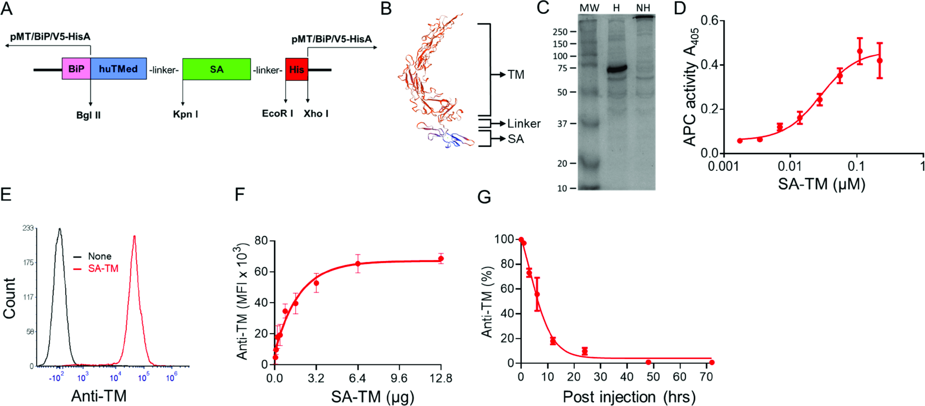
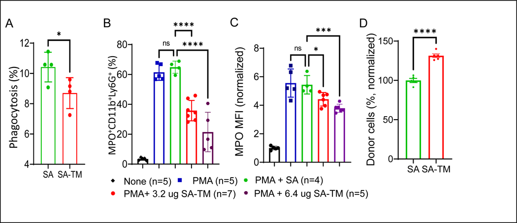
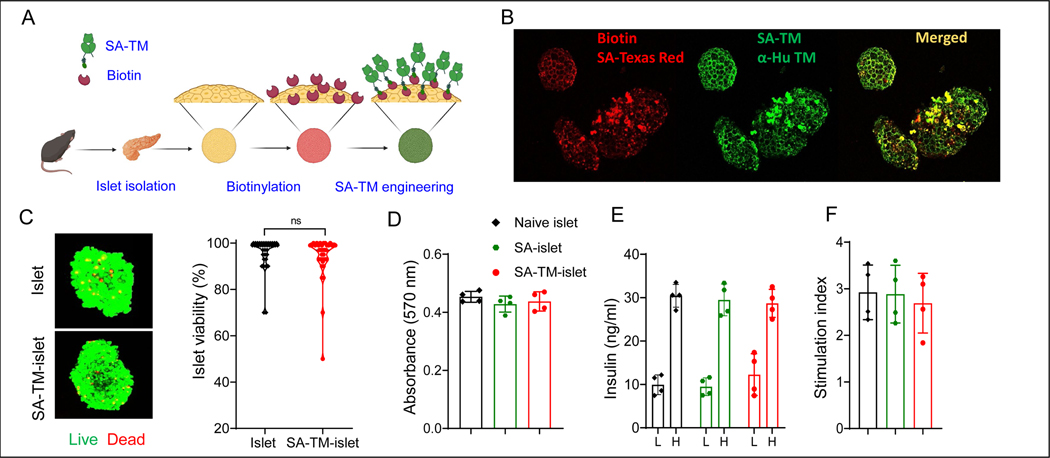
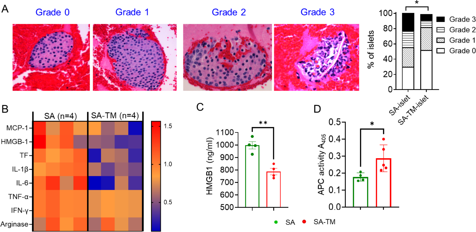
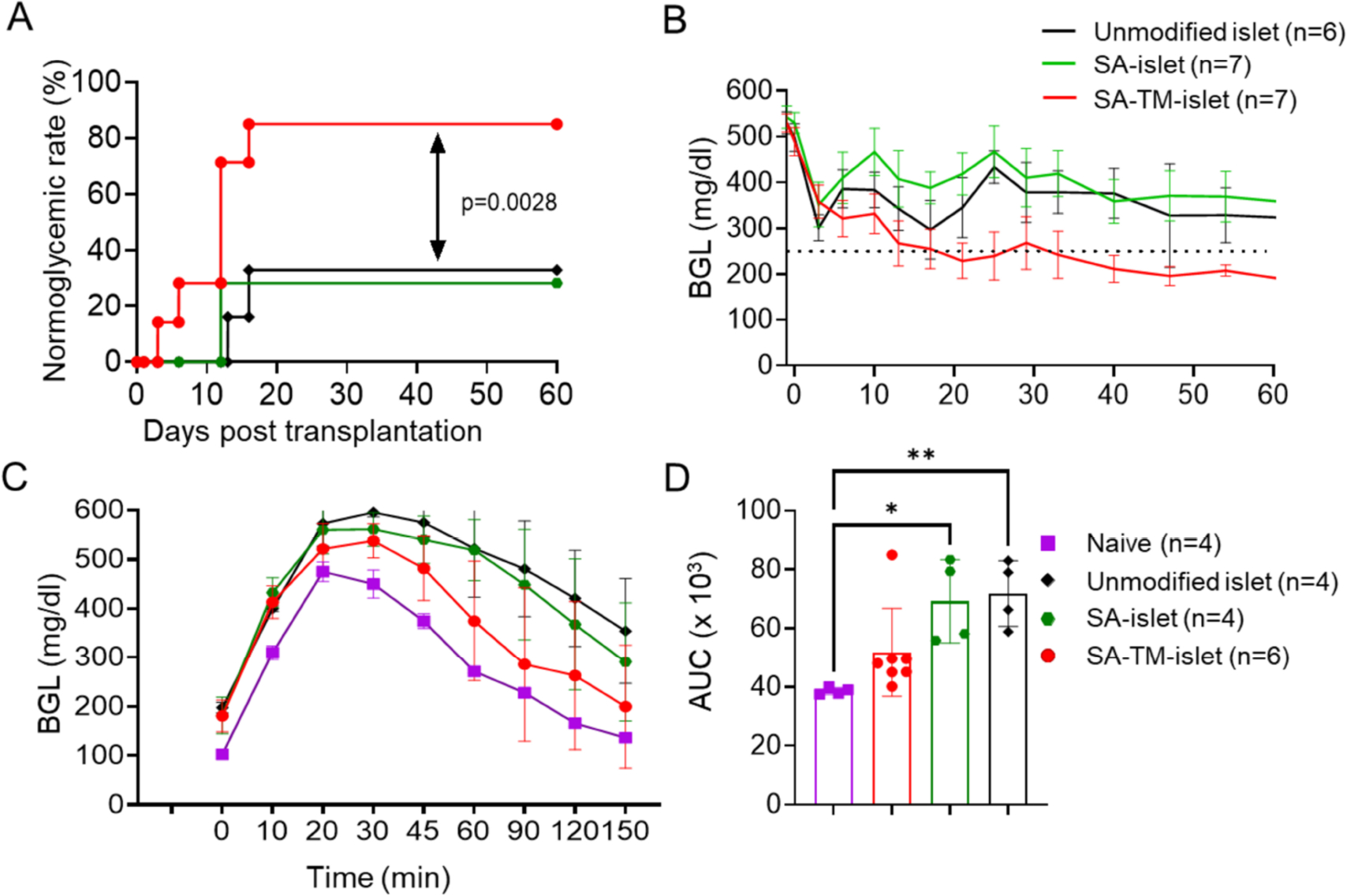
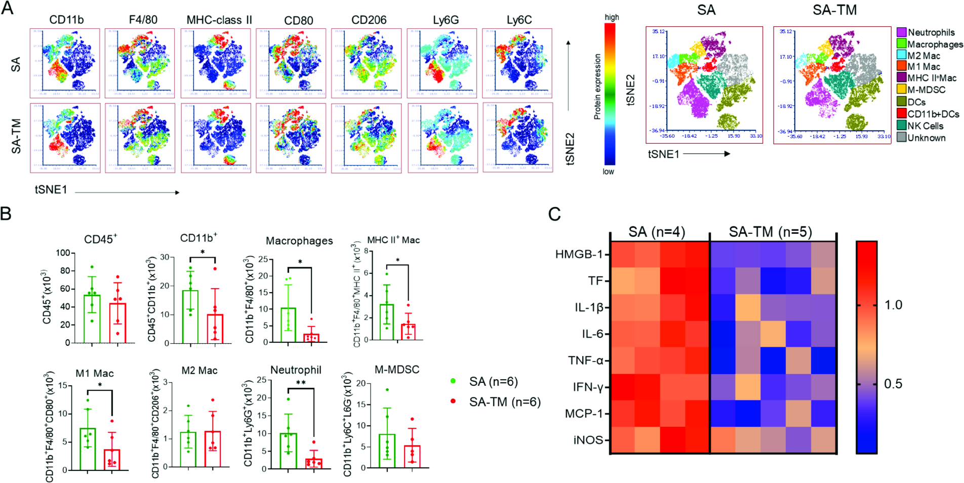
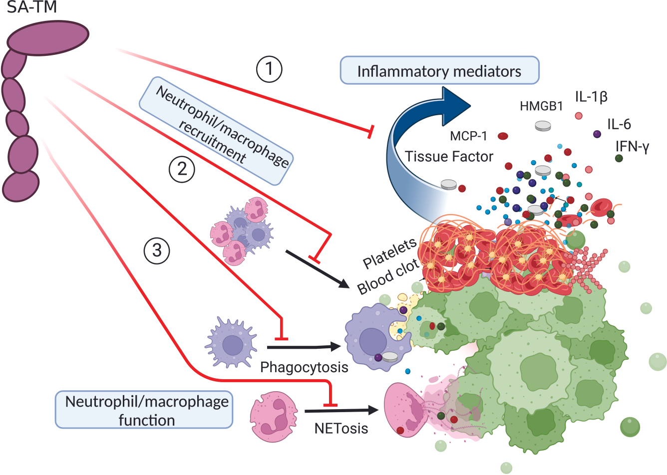
The instant blood-mediated inflammatory reaction (IBMIR) is initiated by innate immune responses that cause substantial islet loss after intraportal transplantation. Thrombomodulin (TM) is a multifaceted innate immune modulator. In this study, we report the generation of a chimeric form of thrombomodulin with streptavidin (SA-TM) for transient display on the surface of islets modified with biotin to mitigate IBMIR. SA-TM protein expressed in insect cells showed the expected structural and functional features. SA-TM converted protein C into activated protein C, blocked phagocytosis of xenogeneic cells by mouse macrophages and inhibited neutrophil activation. SA-TM was effectively displayed on the surface of biotinylated islets without a negative effect on their viability or function. Islets engineered with SA-TM showed improved engraftment and established euglycemia in 83% of diabetic recipients when compared with 29% of recipients transplanted with SA-engineered islets as control in a syngeneic minimal mass intraportal transplantation model. Enhanced engraftment and function of SA-TM-engineered islets were associated with the inhibition of intragraft proinflammatory innate cellular and soluble mediators of IBMIR, such as macrophages, neutrophils, high-mobility group box 1, tissue factor, macrophage chemoattractant protein-1, interleukin-1β, interleukin-6, tumor necrosis factor-α, interferon-γ. Transient display of SA-TM protein on the islet surface to modulate innate immune responses causing islet graft destruction has clinical potential for autologous and allogeneic islet transplantation.

Keywords: immunomodulation; instant blood-mediated inflammatory reaction; intraportal transplantation; islet transplantation; thrombomodulin; thrombomodulin chimeric with streptavidin innate immunity.


Conflict of interest statement

DISCLOSURE

The authors of this manuscript have no conflicts of interest to disclose as described by the American Journal of Transplantation.

Declaration of interests

The authors declare that they have no known competing financial interests or personal relationships that could have appeared to influence the work reported in this paper.

Figures

FIGURE 1
FIGURE 1
Generation of SA-TM protein and structural and functional characterization. (A) Schematic representation of the SA-TM construct. A synthetic chimeric gene containing the coding sequences for the extracellular domain of human TM and core streptavidin (SA) with flexible linkers and a 6xHis tag to facilitate protein purification was constructed and subcloned into the CuSO4-inducible pMT-Bip-V5-HisA S2 insect cell expression vector. (B) The anticipated three-dimensional structure of SA-TM protein using the SWISS-MODEL. (C) SDS-PAGE profile of SA-TM. The protein was produced in S2 cells, isolated from cell supernatant using metal affinity chromatography, left unheated (NH) or heated at 100°C, and analyzed using denaturing SDS-PAGE for structure and purity. (D) SA-TM converts protein C to activated protein C (APC). Lightly heparinized mouse blood was incubated with the indicated doses of soluble SA-TM for 30 min and the activity of APC was measured at OD405. (E) Splenocytes engineered with SA-TM protein. Mouse splenocytes were biotinylated (15 μM), engineered with SA-TM protein (3.2 μg/106 cells), stained with an Ab to human TM (red line), and analyzed in flow cytometry with unmodified splenocytes (green line) serving as control. (F) Dose-dependent binding of SA-TM protein to biotinylated splenocytes. Data expressed as mean ± SD and representative of 3 independent experiments. (F) In vivo kinetics of SA-TM on the surface of splenocytes. C57BL/6.SJL (CD45.1) splenocytes were engineered with SA-TM protein as in (E) and injected i.v. into C57BL/6 (CD45.2) mice. Spleens were harvested at indicated time points and analyzed for the frequency of SA-TM positive cells in flow cytometry by gating on donor cells (CD45.1). Data from 3 separate experiments expressed as mean ± SD.
FIGURE 2
FIGURE 2
SA-TM mitigates effector function of macrophages and neutrophils and prevents clearance of allogeneic cells in vivo. (A) SA-TM on xenogeneic rat splenocytes inhibits phagocytosis by macrophages in vitro. CFSE labeled and SA-TM-engineered rat splenocytes (3.2 μg/106 cells) were cocultured with mouse RAW 264.7 macrophage cells at 1:5 ratio for 18 hrs. Phagocytosis of rat cells (CD11b+F4/80+CFSE+) was assessed using flow cytometry. Data are shown as mean ± SD pooled from 3 separate experiments; Student’s t test. (B) SA-TM mitigates PMA-induced NETosis. C57BL/6 bone marrow neutrophils were stimulated with 200 nM PMA in the presence of the indicated doses of SA-TM and SA as control protein for 3 hrs. NETosis was assessed using an Ab to myeloperoxidase (MPO) in flow cytometry gating on CD11b+Gr-1+MPO+ cells. (C) MFI of MPO in panel B are plotted as fold values over unstimulated neutrophils. Data for B and C are shown as mean ± SD of 3 independent experiments; ANOVA with Bonferroni’s post hoc test. (D) SA-TM inhibits in vivo clearance of allogeneic bone marrow cells. Bone marrow from NSG mice deficient for MHC class I and II were engineered with SA (0.8 μg/106 cells) or SA-TM (3.2 μg/106 cells) proteins followed by labeling with CTV and CFSE, respectively. Cells were then mixed at 1:1 ratio and injected i.v. into allogeneic C57BL/6 recipients (in total 20×106 cells/animal). Splenocytes were harvested 24 hrs post-transplantation and analyzed using flow cytometry by gating on CFSE+ or CTV+ donor cells. Data are shown as mean ± SD of 4 independent experiments; Student’s t test. *P < 0.05, **P < 0.01, ***P < 0.001, ****P < 0.0001.
FIGURE 3
FIGURE 3
Engineering islets with the SA-TM protein does not impact islet viability and function. (A) Schematic drawing of islet engineering with the SA-TM protein. (B) A representative image of SA-TM-engineered islets (15 μM biotin and 3.2 μg/500 islets). Biotinylation and engineering were assessed using fluorescence labeled streptavidin (red) and an antibody to human TM (green) in confocal microscopy. (C) Turnover kinetics of SA-TM on islet surface. SA-TM engineered islets were cultured in vitro and stained with anti-SA-DyLight 488 Ab at the indicated time points. Islet images were acquired using fluorescence microscopy. MFI values over the background were quantified using ImageJ software and time zero normalized values were plotted. Each time point corresponds to mean ± SD MFI from at least 10 islets pooled from two independent experiments. (D) Engineering with SA-TM does not impact the viability of islets. Islets were engineered with SA-TM followed by staining with FDA and PI to assess viability. A minimum of 50 islets from two independent experiments were scored under confocal microscopy for the percentage of dead (red) and live (green) cells in each islet. Data are shown as mean ± SD; Student’s t test, ns = not significant. (E) Islet metabolic activity. Islets were left unmodified or engineered with SA-TM or an equimolar of SA proteins and metabolic function was assessed using AlamarBlue cell viability assay. (F) Insulin stimulated glucose secretion assay performed using unmodified and SA-TM or SA-engineered islets. Islets were cultured in low (3.5 mM) and high (16.5 mM) doses of glucose and secreted insulin levels in the supernatant were measured using ELISA. (G) Stimulation index (mean secreted insulin in high glucose/mean secreted insulin in low glucose) of data shown in (F). For panel D, data are shown as mean ± SD pooled from a minimum of 2 separate experiments; Student’s 2-tailed unpaired t test. For panel E and G, data are shown as mean ± SD pooled from a minimum of 4 separate experiments; ANOVA with Bonferroni’s post hoc test.
FIGURE 4
FIGURE 4
SA-TM on islets inhibits various innate immune inflammatory responses. An in vitro blood loop assay mimicking IBMIR was performed by culturing 100 SA-TM- or SA-engineered islets in fresh autologous blood at 37 °C for 3 hrs. Islet thrombus was used for H&E staining and qPCR, while serum was analyzed using ELISA. (A) Histological assessment of islet thrombus. SA-TM maintains islet structure and protects from IBMIR as compared to SA. Data from at least 3 independent experiments. Statistical differences were assessed using Chi-square test. (B) Heatmap of proinflammatory factors. TaqMan qPCR were performed on total RNA extracted from islet thrombus. Data from 4 independent experiments. (C) HMGB1 levels in the serum of islet thrombus using ELISA. (D) APC activity in the serum of islet thrombus. Data shown as mean ± SD of at least 3 independent experiments. Statistical differences were assessed using Student’s 2-tailed unpaired t test with *P < 0.05, **P < 0.01.
FIGURE 5
FIGURE 5
SA-TM improves islet engraftment and function in a syngeneic marginal mass intraportal transplantation model. Islets from C57BL/6 mice were left unmodified or engineered with SA-TM or SA as control protein and 200 IEQ were transplanted intraportally into streptozotocin diabetic syngeneic recipients. (A) Rate of euglycemia over a 60-day observation period. Log-rank (Mantel-Cox) was used to assess statistical differences among groups. (B) Nonfasting blood glucose levels of transplant recipients in (A). (C) Intraperitoneal glucose tolerance test (IPGTT) on long-term (> 60 days) euglycemic mice in the indicated groups. Naïve C57BL/6 mice were used as controls. (D) Area under cure (AUC) analysis for (C). Data expressed as mean ± SD. Statistical differences were assessed using a one-way ANOVA with ***P < 0.001, ****P < 0.0001.
FIGURE 6
FIGURE 6
Engraftment and sustained function of SA-TM-engineered islets are associated with decreased intragraft proinflammatory innate immune mediators of IBMIR. Chemically diabetic C57BL/6 mice were transplanted with 200 IEQ engineered with control SA and SA-TM proteins. Intrahepatic islet infiltrates were harvested 3 hrs post-transplantation and analyzed in flow cytometry using antibodies to various cell surface markers demarking the indicated innate immune cells. (A) viSNE display of flow cytometry analysis by individual lineage identification markers of immune infiltrates and cell type annotation by different colors depiction (B) Absolute number of the indicated immune cell type plotted per gram of liver per animal. Data shown as mean ± SD of 2 independent experiments. (C) Heatmap of proinflammatory intrahepatic transcripts. Total RNA was harvested from the indicated transplant groups 3 hrs post-transplantation and analyzed for quantitative assessment of the transcript levels for the indicated proinflammatory mediators using the TaqMan Gene Expression Assay. Data shown as mean ± SD for a total of 4 animals for SA and 5 animals for SA-TM groups pooled from 2 independent experiments. Statistical differences for B and C were assessed using student t-test (unpaired, one tailed) with *P < 0.05, **P < 0.01.
FIGURE 7
FIGURE 7
Prevention of islet damage by SA-TM. SA-TM blocks islets damage throughout 3 distinct pathways. 1. SA-TM/thrombin complex converts protein C into its activated form APC that inhibits the induction and secretion of proinflammatory cytokines and chemokines by blocking NF-κB signaling. SA-TM and APC also degrade DAMPs released by damaged islets, further preventing coagulation and secretion of proinflammatory mediators. 2. SA-TM prevents the recruitment of macrophages and neutrophils into islet grafts and inhibits their effector function (3), culminating into enhanced engraftment and sustained survival and function.

References

    1. Naziruddin B, Iwahashi S, Kanak MA, Takita M, Itoh T, Levy MF. Evidence for instant blood-mediated inflammatory reaction in clinical autologous islet transplantation. Am J Transplant 2014;14(2):428–437. - PubMed
    1. Kanak MA, Takita M, Itoh T, SoRelle JA, Murali S, Kunnathodi F et al. Alleviation of instant blood-mediated inflammatory reaction in autologous conditions through treatment of human islets with NF-kappaB inhibitors. Transplantation 2014;98(5):578–584. - PMC - PubMed
    1. Hering BJ, Clarke WR, Bridges ND, Eggerman TL, Alejandro R, Bellin MD et al. Phase 3 trial of transplantation of human Islets in Type 1 diabetes complicated by severe hypoglycemia. Diabetes Care 2016;39(7):1230–1240. - PMC - PubMed
    1. Bennet W, Sundberg B, Groth CG, Brendel MD, Brandhorst D, Brandhorst H et al. Incompatibility between human blood and isolated islets of Langerhans: a finding with implications for clinical intraportal islet transplantation? Diabetes 1999;48(10):1907–1914. - PubMed
    1. Moberg L, Johansson H, Lukinius A, Berne C, Foss A, Kallen R et al. Production of tissue factor by pancreatic islet cells as a trigger of detrimental thrombotic reactions in clinical islet transplantation. Lancet 2002;360(9350):2039–2045. - PubMed

Publication types