

nanoGold and µGold inhibit autoimmune inflammation: a review
- PMID: 36864314
- PMCID: PMC10006034
- DOI: 10.1007/s00418-023-02182-9
nanoGold and µGold inhibit autoimmune inflammation: a review
Abstract
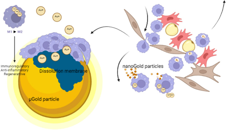
The newest data on metallic gold have placed the noble metal central in the fight for the safe treatment of autoimmune inflammation. There are two different ways to use gold for the treatment of inflammation: gold microparticles > 20 µm and gold nanoparticles. The injection of gold microparticles (µGold) is a purely local therapy. µGold particles stay put where injected, and gold ions released from them are relatively few and taken up by cells within a sphere of only a few millimeters in diameter from their origin particles. The macrophage-induced release of gold ions may continue for years. Injection of gold nanoparticles (nanoGold), on the other hand, is spread throughout the whole body, and the bio-released gold ions, therefore, affect multitudes of cells all over the body, as when using gold-containing drugs such as Myocrisin. Since macrophages and other phagocytotic cells take up and transport nanoGold and remove it after a short period, repeated treatment is necessary. This review describes the details of the cellular mechanisms that lead to the bio-release of gold ions in µGold and nanoGold.
Keywords: Gold microparticles; Gold nanoparticles; Inflammation; Macrophages; Mast cells.
© 2023. The Author(s).
Conflict of interest statement
The authors declare no competing interests.
Both authors are involved in companies working with gold and inflammation, Berlock ApS and ReGold ApS.
Figures


Similar articles
-
[Optical Analysis of the Interaction of Mercaptan Derivatives of Nanogold Particles with Carcinoembryonic Antigen].Guang Pu Xue Yu Guang Pu Fen Xi. 2016 Feb;36(2):478-81. Guang Pu Xue Yu Guang Pu Fen Xi. 2016. PMID: 27209753 Chinese.
-
Gold and thiol compounds in the treatment of rheumatoid arthritis: excretory fate and tissue distribution of thiomalate in relation to gold after administration of myocrisin (auro-thiomalate).Scand J Rheumatol Suppl. 1979;(28):28-36. Scand J Rheumatol Suppl. 1979. PMID: 109914
-
[Nanogold - Biological effects and occupational exposure levels].Med Pr. 2017 Jun 27;68(4):545-556. doi: 10.13075/mp.5893.00538. Epub 2017 May 9. Med Pr. 2017. PMID: 28584334 Review. Polish.
-
Amelioration of collagen-induced arthritis in rats by nanogold.Arthritis Rheum. 2007 Feb;56(2):544-54. doi: 10.1002/art.22401. Arthritis Rheum. 2007. PMID: 17265489
-
Gold Nanoparticles in Cancer Treatment.Mol Pharm. 2019 Jan 7;16(1):1-23. doi: 10.1021/acs.molpharmaceut.8b00810. Epub 2018 Nov 30. Mol Pharm. 2019. PMID: 30452861 Review.
Cited by
-
In focus in HCB.Histochem Cell Biol. 2023 Mar;159(3):221-224. doi: 10.1007/s00418-023-02184-7. Histochem Cell Biol. 2023. PMID: 36877266 No abstract available.
-
Intraarticular gold microparticles using hyaluronic acid as the carrier for hip osteoarthritis. A 2-year follow-up pilot study.Sci Rep. 2024 Nov 1;14(1):26249. doi: 10.1038/s41598-024-77760-5. Sci Rep. 2024. PMID: 39482349 Free PMC article.
-
Impacts of loading thymoquinone to gold or silver nanoparticles on the efficacy of anti-tumor treatments in breast cancer with or without chemotherapeutic cisplatin.BMC Biotechnol. 2025 Apr 10;25(1):26. doi: 10.1186/s12896-025-00958-6. BMC Biotechnol. 2025. PMID: 40211258 Free PMC article.
-
Exploring the Therapeutic Potential of Green-Synthesized Gold Nanoparticles and Ericaria selaginoides Extract for Inflammatory Bowel Disease.Antioxidants (Basel). 2024 Jul 23;13(8):884. doi: 10.3390/antiox13080884. Antioxidants (Basel). 2024. PMID: 39199130 Free PMC article.
-
Intra-articular injection of gold micro-particles with hyaluronic acid for painful knee osteoarthritis.BMC Musculoskelet Disord. 2024 Mar 12;25(1):211. doi: 10.1186/s12891-024-07321-4. BMC Musculoskelet Disord. 2024. PMID: 38475764 Free PMC article. Clinical Trial.
References
-
- Brown C, Bushell G, Whitehouse M, Agrawal DS, Tuoe SG, Paknikar KM, Tiekink E. Nanogold-pharmaceutics. Gold Bull. 2007;40:245–250. doi: 10.1007/BF03215588. - DOI
-
- Cardoso E, Rezin GT, Zanoni ET, de Souza Notoya F, Leffa DD, Damiani AP, Daumann F, Rodriguez JC, Benavides R, da Silva L, Andrade VM, da Silva Paula MM. Acute and chronic administration of gold nanoparticles cause DNA damage in the cerebral cortex of adult rats. Mutat Res. 2014 doi: 10.1016/j.mrfmmm.2014.05.009. - DOI - PubMed
Publication types
MeSH terms
Substances
LinkOut - more resources
Full Text Sources
Medical
