. 2023 Mar 2;11(1):33.
doi: 10.1186/s40478-023-01532-x.

An integrated genetic analysis of epileptogenic brain malformed lesions

Affiliations

An integrated genetic analysis of epileptogenic brain malformed lesions

Atsushi Fujita et al. Acta Neuropathol Commun. .

Abstract

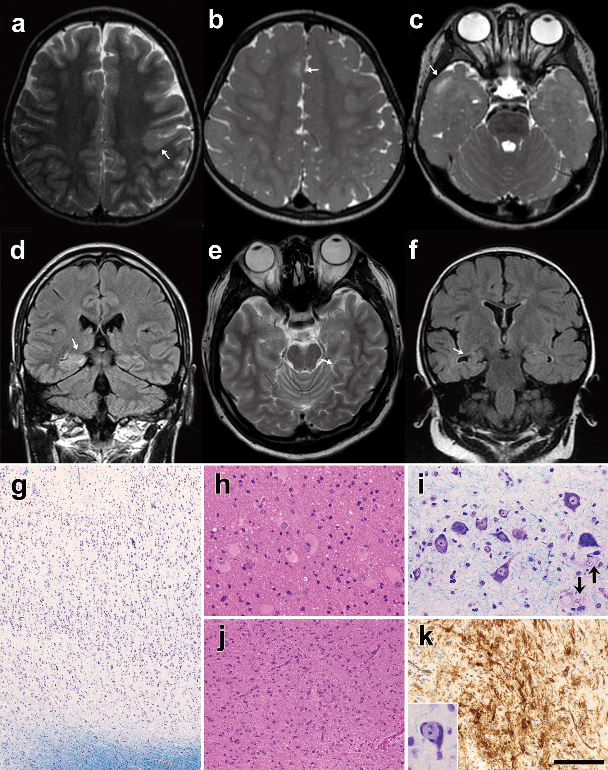
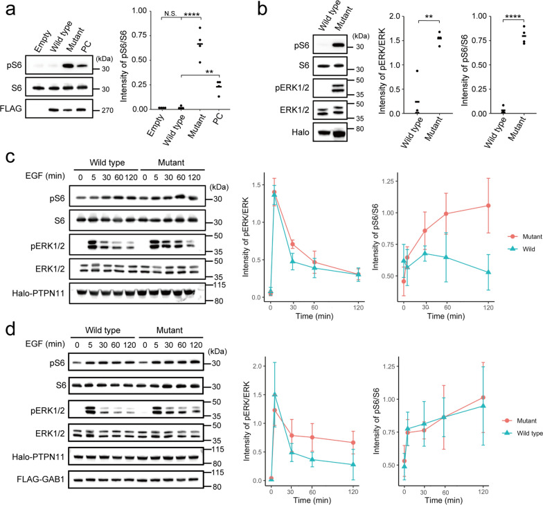
Focal cortical dysplasia is the most common malformation during cortical development, sometimes excised by epilepsy surgery and often caused by somatic variants of the mTOR pathway genes. In this study, we performed a genetic analysis of epileptogenic brain malformed lesions from 64 patients with focal cortical dysplasia, hemimegalencephy, brain tumors, or hippocampal sclerosis. Targeted sequencing, whole-exome sequencing, and single nucleotide polymorphism microarray detected four germline and 35 somatic variants, comprising three copy number variants and 36 single nucleotide variants and indels in 37 patients. One of the somatic variants in focal cortical dysplasia type IIB was an in-frame deletion in MTOR, in which only gain-of-function missense variants have been reported. In focal cortical dysplasia type I, somatic variants of MAP2K1 and PTPN11 involved in the RAS/MAPK pathway were detected. The in-frame deletions of MTOR and MAP2K1 in this study resulted in the activation of the mTOR pathway in transiently transfected cells. In addition, the PTPN11 missense variant tended to elongate activation of the mTOR or RAS/MAPK pathway, depending on culture conditions. We demonstrate that epileptogenic brain malformed lesions except for focal cortical dysplasia type II arose from somatic variants of diverse genes but were eventually linked to the mTOR pathway.

Keywords: Focal cortical dysplasia; LEATs; RAS/MAPK; Somatic variants; mTOR.


Conflict of interest statement

The authors have no competing interests to declare that are relevant to the content of this article.

Figures

Fig. 1
Fig. 1
Brain MRI and histopathologic features of patients with somatic variants a Patient F08 at 3 years (FCD type IIB, an MTOR in-frame variant), b, c patient F61 at 2 years (FCD type IIB, germline and somatic variants of TSC2), d patient F48 at 23 years (FCD type IB, a MAP2K1 in-frame deletion), e patient F70 at 21 years (ganglioglioma, deletion of entire chromosome 9), and f patient F30 at 2 years and 7 months (hippocampal sclerosis, 19p13.3p12 deletion). a, b, c, e are T2-weighted axial brain MRIs, and d, f are fluid-attenuated inversion recovery coronal MRIs. Brain MRI showing focal irregular gyri (arrows) with blurred junctions between the cortex and white matter (a) or hyperintensity of the subcortical white matter (be). g Patient F67 (FCD type I, a PTPN11 missense variant). Cytoarchitectural abnormality of the cortex. h Patient F08 with FCD type IIB showing several balloon cells in the white matter. i Patient F61 with FCD type IIB exhibiting dysmorphic neurons and balloon cells (arrows) in the cortex. j, k Patient F70 with ganglioglioma. j Astrocytic cells showing a wavy, fascicular arrangement with mild hypercellularity. A dysmorphic ganglion cell (inset in k). k CD34-immunopositive cells with fine processes. g, i and inset in k: Klüver–Barrera stain. h, j hematoxylin and eosin stain. k immunostained and counterstained with hematoxylin. Scale bar = 350 μm for g, 90 μm for h and i, 180 μm for j, 50 μm for inset in k, and 140 μm for k
Fig. 2
Fig. 2
Immunoblotting analysis of HEK293T cells transfected with mutant MTOR, MAP2K1, and PTPN11 a An in-frame (15-bp) MTOR deletion, c.4339_4353del p.(Ala1447_Glu1451del), detected in our study resulted in significantly higher phospho-S6 expression compared to wild-type (One-way ANOVA followed by Dunnett's post-hoc test; Empty: P = 0.99; Mutant: P =  < 0.0001; PC: P = 0.0079). Positive control (PC), c.4379T > C p.(Leu1460Pro). b The MAP2K1 variant, c.173_187del p.(Gln58_Glu62del), resulted in significantly higher phospho-S6 and phospho-ERK levels compared to the wild-type (Two-tailed paired t-test; pERK/ERK: P = 0.0048; pS6/S6: P ≤ 0.0001). c, d Time course of relative expression of phospho-ERK and phospho-S6 under stimulation with EGF in cells with PTPN11 transfection (c) and co-transfection with PTPN11 and GAB1 (d). Two-way repeated measurement ANOVA in c; pERK/ERK: P = 0.35; pS6/S6: P = 0.0028. Sidak’s post hoc test in pS6/S6 of c; 0 min: P = 0.63; 5 min: P = 0.96; 30 min: P = 0.58; 60 min: P = 0.34; 120 min: P = 0.14. Two-way repeated measurement ANOVA in d; pERK/ERK: P = 0.38; pS6/S6: P = 0.96. Bars in a, b represent the mean, and those in c, d represent SD. **P < 0.01; ****P < 0.0001. N.S. not significant

References

    1. Araki T, Mohi MG, Ismat FA, Bronson RT, Williams IR, Kutok JL, Yang W, Pao LI, Gilliland DG, Epstein JA, et al. Mouse model of Noonan syndrome reveals cell type- and gene dosage-dependent effects of Ptpn11 mutation. Nat Med. 2004;10:849–857. doi: 10.1038/nm1084. - DOI - PubMed
    1. Baldassari S, Picard F, Verbeek NE, van Kempen M, Brilstra EH, Lesca G, Conti V, Guerrini R, Bisulli F, Licchetta L, et al. The landscape of epilepsy-related GATOR1 variants. Genet Med. 2019;21:398–408. doi: 10.1038/s41436-018-0060-2. - DOI - PMC - PubMed
    1. Baldassari S, Ribierre T, Marsan E, Adle-Biassette H, Ferrand-Sorbets S, Bulteau C, Dorison N, Fohlen M, Polivka M, Weckhuysen S, et al. Dissecting the genetic basis of focal cortical dysplasia: a large cohort study. Acta Neuropathol. 2019;138:885–900. doi: 10.1007/s00401-019-02061-5. - DOI - PMC - PubMed
    1. Bedrosian TA, Miller KE, Grischow OE, Schieffer KM, LaHaye S, Yoon H, Miller AR, Navarro J, Westfall J, Leraas K, et al. Detection of brain somatic variation in epilepsy-associated developmental lesions. Epilepsia. 2022 doi: 10.1111/epi.17323. - DOI - PubMed
    1. Blumcke I, Spreafico R, Haaker G, Coras R, Kobow K, Bien CG, Pfäfflin M, Elger C, Widman G, Schramm J, et al. Histopathological findings in brain tissue obtained during epilepsy surgery. N Engl J Med. 2017;377:1648–1656. doi: 10.1056/NEJMoa1703784. - DOI - PubMed

Publication types

Supplementary concepts