

Image-guided tunneled peritoneal dialysis catheter placement
- PMID: 36864963
- PMCID: PMC9971291
- DOI: 10.21037/cdt-21-579
Image-guided tunneled peritoneal dialysis catheter placement
Abstract
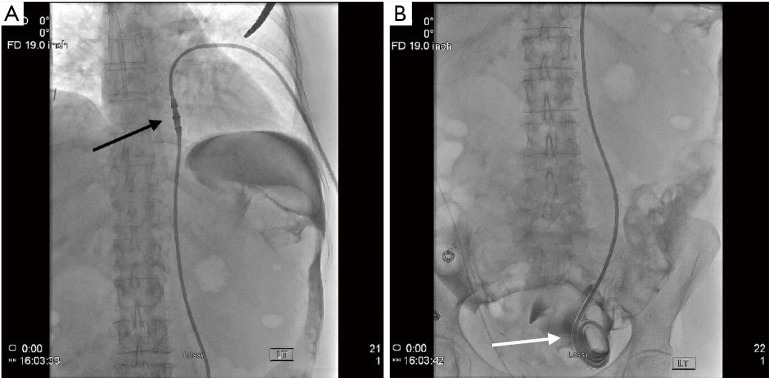
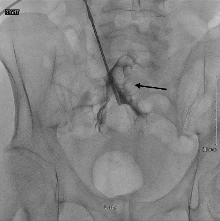
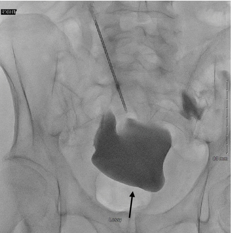
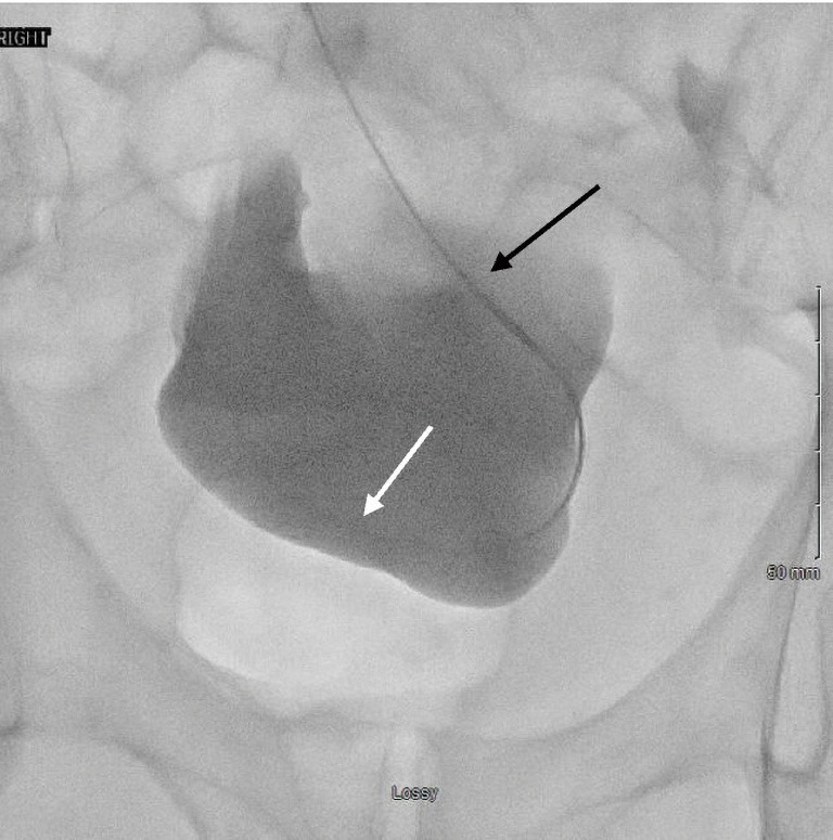
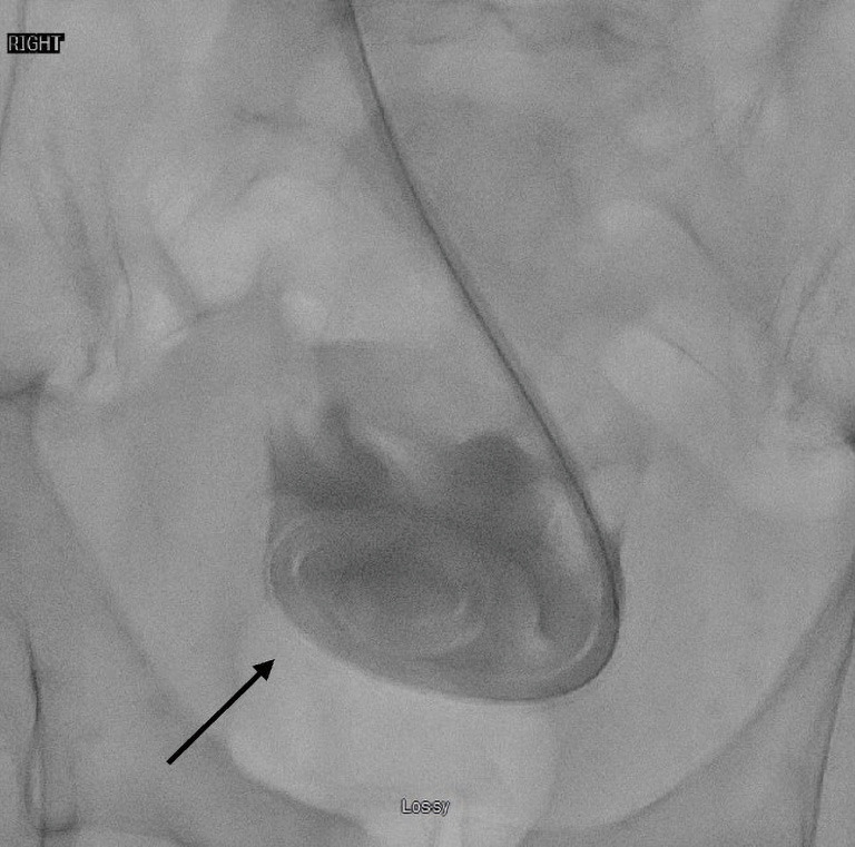
Patients with end-stage renal disease (ESRD) often will ultimately require dialysis to survive. One type of dialysis is peritoneal dialysis (PD), which utilizes the vessel-rich peritoneum as a semi-permeable membrane to filter blood. In order to perform PD, a tunneled catheter must be placed through the abdominal wall and into the peritoneal space, with ideal positioning of the catheter within the most dependent portion of the pelvis, represented by the rectouterine or rectovesical space in women and men, respectively. There are several approaches to PD catheter insertion, including open surgical, laparoscopic surgical, blind percutaneous, and image-guided with the use of fluoroscopy techniques. Interventional radiology (through the use of image-guided percutaneous techniques) is an infrequently utilized resource to place PD catheters, and offers real-time imaging confirmation of catheter positioning with similar outcomes to more invasive surgical catheter insertion approaches. Although the vast majority of dialysis patients receive hemodialysis instead of peritoneal dialysis in the United States, some countries have moved towards a "Peritoneal Dialysis First" initiative, prioritizing initial PD, as it is less burdensome on healthcare facilities as it can be performed at home. In addition, the outbreak of the COVID-19 pandemic has produced shortages of medical supplies and delays in care delivery worldwide, while simultaneously generating a shift away from in-person medical visits and appointments. This shift may be met with more frequent utilization of imaged-guided PD catheter placement, reserving surgical and laparoscopic placement for complex patients who may require omental periprocedural revisions. This literature review outlines a brief history of PD, the various techniques of PD catheter insertion, patient selection criteria, and new COVID-19 considerations, in anticipation for the increased demand for PD in the United States.
Keywords: End-stage renal disease (ESRD); interventional radiology; peritoneal dialysis; review.
2023 Cardiovascular Diagnosis and Therapy. All rights reserved.
Conflict of interest statement
Conflicts of Interest: All authors have completed the ICMJE uniform disclosure form (available at https://cdt.amegroups.com/article/view/10.21037/cdt-21-579/coif). The series “Endovascular and Surgical Interventions in the End Stage Renal Disease Population” was commissioned by the editorial office without any funding or sponsorship. GJ reports UVMMC Radiology funding received for travel for poster presentation at ECTRIMS conference in 2019, and will be funding his travel for RSNA in Chicago in 11/2021. CM reports Patent pending: Peritoneal dialysis catheter weighted anchor modification. This is a modification of existing peritoneal dialysis catheters to increase efficacy and decrease complications. No payment, external grant or funding, royalty, consulting fee, or any financial interest has occurred related to this activity in the past 36 months. Christopher Morris did receive an internal grant to fund a study related to this medical device. The authors have no other conflicts of interest to declare.
Figures


References
-
- Berger A, Edelsberg J, Inglese GW, et al. Cost comparison of peritoneal dialysis versus hemodialysis in end-stage renal disease. Am J Manag Care 2009;15:509-18. - PubMed
Publication types
LinkOut - more resources
Full Text Sources
Miscellaneous