Case Reports
. 2023 Feb 15:30:101818.
doi: 10.1016/j.ajoc.2023.101818. eCollection 2023 Jun.

An unusual case of a pigment epithelial cyst masquerading as a uveal melanoma after zoster ophthalmicus-related iris atrophy

Affiliations
Case Reports

An unusual case of a pigment epithelial cyst masquerading as a uveal melanoma after zoster ophthalmicus-related iris atrophy

Jordan Finley et al. Am J Ophthalmol Case Rep. .

Abstract

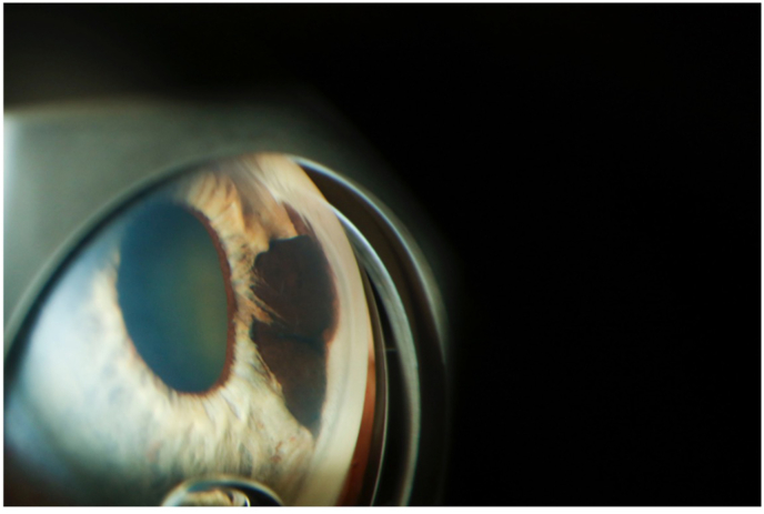
Purpose: To report the case of a 69-year-old male who was referred for a previously unidentified pigmented iris lesion with surrounding iris atrophy masquerading as an iris melanoma.

Observations: A sharply demarcated pigmented lesion extending from the trabecular meshwork to the pupillary margin was identified in the left eye. There was adjacent iris stromal atrophy. Testing was consistent with a cyst-like lesion. The patient later described a prior episode of ipsilateral herpes zoster involving the ophthalmic division of cranial nerve five.

Conclusions and importance: Iris cysts present an uncommon iris tumor, often going unrecognized especially if located on the posterior iris surface. When they present acutely, as in this case where a previously unidentified cyst was revealed following zoster-induced sectoral iris atrophy, these pigmented lesions can be concerning for malignancy. Accurately identifying iris melanomas and differentiating them from benign iris lesions is imperative.

Keywords: Cancer; Herpes zoster; Iris atrophy; Iris melanoma; Iris nevus; Iris pigmented lesion.


Conflict of interest statement

None for all authors. There is no funding or grant support for this case report. All authors attest that they meet the current ICMJE criteria for Authorship. Internal Review Board (IRB) approval was deemed not necessary by the IRB as the research is not generalizable.

Figures

Fig. 1
Fig. 1
Pigmented iris lesion. Slit lamp photo of darkly pigmented, bilobed epithelial iris lesion with sharply demarcated borders.
Fig. 2
Fig. 2
Lesion Transillumination. A slit lamp view of the lesion under retro-illumination revealing transillumination defects.
Fig. 3
Fig. 3
Obscuration of the angle. Gonioscopic view of pigmented lesion extending to and obscuring the angle.
Fig. 4
Fig. 4
4a & 4b Ultrasonography studies. 4a: High frequency anterior segment ultrasound biomicroscopy with 1.42 × 4.53 (basal dimensions), irregularly shaped, echolucent lesion of the iris with angle involvement. 4b: High frequency anterior segment ultrasound biomicroscopy with 1.40 × 4.07 (basal dimensions), semicircular shaped, echolucent lesion of the iris with no angle involvement.

References

    1. Shields C.L., Shields J.A. Ocular melanoma: relatively rare but requiring respect. Clin Dermatol. 2009;27(1):122–133. doi: 10.1016/J.CLINDERMATOL.2008.09.010. - DOI - PubMed
    1. Rao A., Gupta V., Bhadange Y., Sharma R., Shields J.A. Iris cysts: a review. Semin Ophthalmol. 2011;26(1):11–22. doi: 10.3109/08820538.2010.541319. - DOI - PubMed
    1. Georgalas I., Petrou P., Papaconstantinou D., Brouzas D., Koutsandrea C., Kanakis M. Iris cysts: a comprehensive review on diagnosis and treatment. Surv Ophthalmol. 2018;63(3):347–364. doi: 10.1016/J.SURVOPHTHAL.2017.08.009. - DOI - PubMed
    1. Mor J.M., Koch K.R., Heindl L.M. Diagnosis and therapy of Iris lesions. Klin Monbl Augenheilkd. 2017;234(12):1541–1554. doi: 10.1055/S-0043-120093. - DOI - PubMed
    1. Conway R.M., Chua W.C.T., Qureshi C., Billson F.A. Primary iris melanoma: diagnostic features and outcome of conservative surgical treatment. Br J Ophthalmol. 2001;85(7):848–854. doi: 10.1136/BJO.85.7.848. - DOI - PMC - PubMed

Publication types

LinkOut - more resources