[Preprint]. 2023 Feb 24:2023.02.23.529575.
doi: 10.1101/2023.02.23.529575.

Multi-omic screening of invasive GBM cells in engineered biomaterials and patient biopsies reveals targetable transsulfuration pathway alterations

Affiliations

Multi-omic screening of invasive GBM cells in engineered biomaterials and patient biopsies reveals targetable transsulfuration pathway alterations

Joseph H Garcia et al. bioRxiv. .

Update in

Abstract

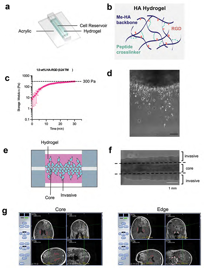
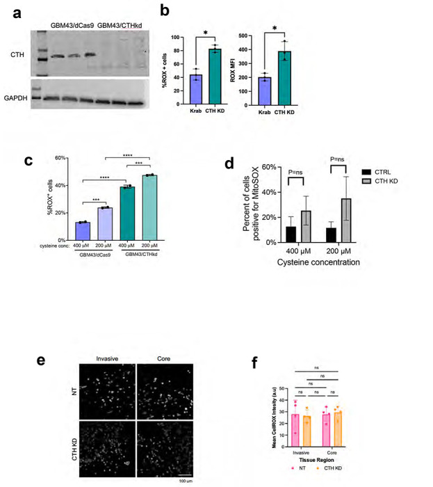
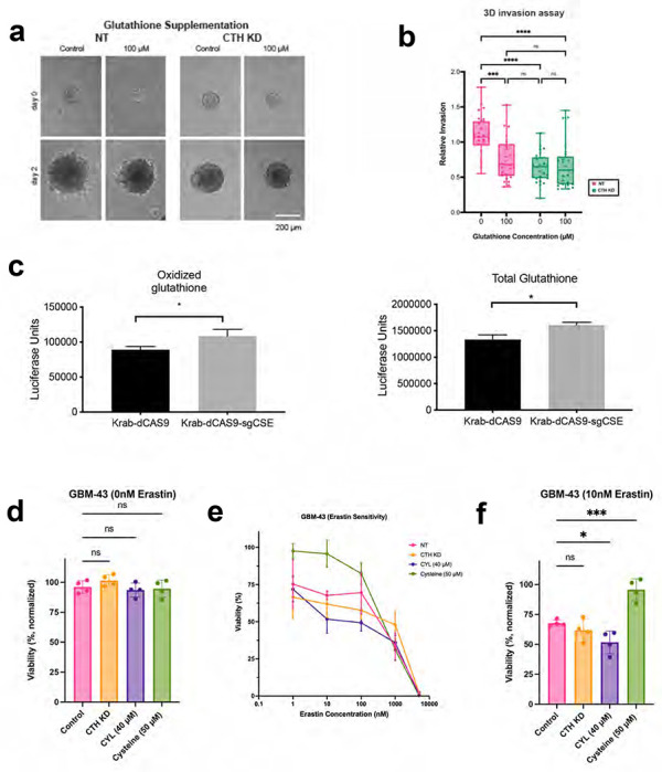
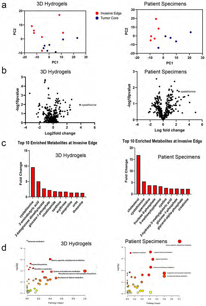
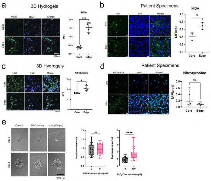
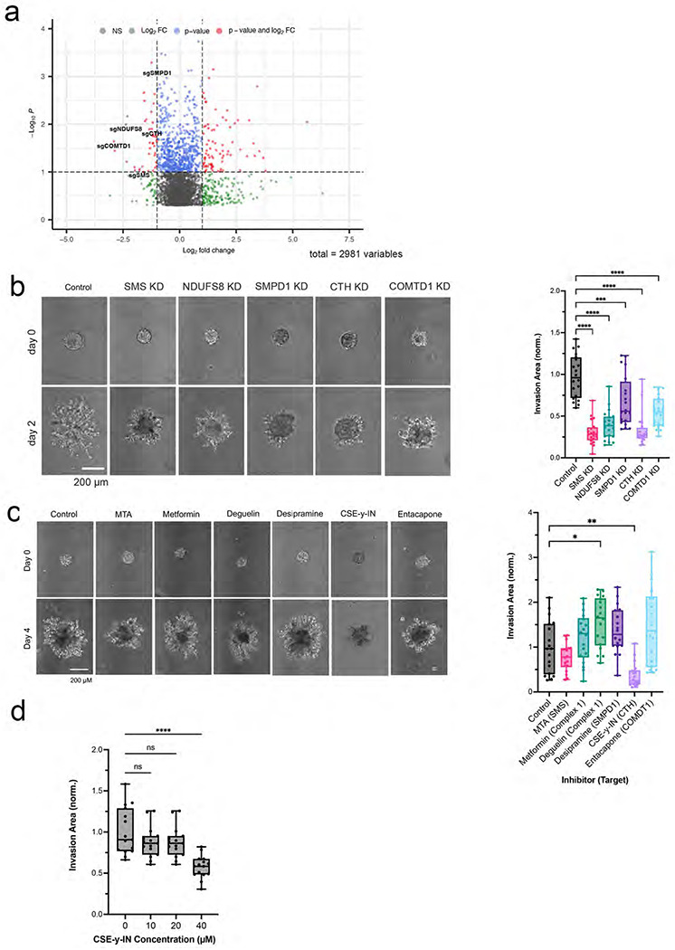
While the poor prognosis of glioblastoma arises from the invasion of a subset of tumor cells, little is known of the metabolic alterations within these cells that fuel invasion. We integrated spatially addressable hydrogel biomaterial platforms, patient site-directed biopsies, and multi-omics analyses to define metabolic drivers of invasive glioblastoma cells. Metabolomics and lipidomics revealed elevations in the redox buffers cystathionine, hexosylceramides, and glucosyl ceramides in the invasive front of both hydrogel-cultured tumors and patient site-directed biopsies, with immunofluorescence indicating elevated reactive oxygen species (ROS) markers in invasive cells. Transcriptomics confirmed upregulation of ROS-producing and response genes at the invasive front in both hydrogel models and patient tumors. Amongst oncologic ROS, hydrogen peroxide specifically promoted glioblastoma invasion in 3D hydrogel spheroid cultures. A CRISPR metabolic gene screen revealed cystathionine gamma lyase (CTH), which converts cystathionine to the non-essential amino acid cysteine in the transsulfuration pathway, to be essential for glioblastoma invasion. Correspondingly, supplementing CTH knockdown cells with exogenous cysteine rescued invasion. Pharmacologic CTH inhibition suppressed glioblastoma invasion, while CTH knockdown slowed glioblastoma invasion in vivo. Our studies highlight the importance of ROS metabolism in invasive glioblastoma cells and support further exploration of the transsulfuration pathway as a mechanistic and therapeutic target.

Keywords: CRISPR; Glioblastoma; Hydrogels; Invasion; Metabolism.


Conflict of interest statement

The authors have declared that no conflict of interest exists.

Figures

Extended Data Figure 1.
Extended Data Figure 1.. Setup for 3D hydrogel devices and patient site-directed biopsies.
(a) Shown is an illustration of the invasion device which consists of a cell reservoir within a hyaluronic acid hydrogel on an acrylic slide. (b) HA-methacrylate (Me-HA) was functionalized with an integrin binding (RGD) peptide using the Michael-type addition reaction between the methacrylate groups on the polymer and the cysteine thiol groups on the peptide. The same addition reaction with the methacrylate groups was used to induce crosslinking with protease-sensitive peptide crosslinkers to form HA hydrogels. (c) Hydrogel storage modulus (G’) (in Pascals) versus time after the gel is casted. Time=0 represents the intersection of storage modulus (G’) and loss modulus (G’’) (n=3 hydrogels). (d) Phase image (10x magnification) of single cells of invasive fraction in HA hydrogel invasion device; scale bar=100 μm. (e) Illustration of invasion device (f) Phase image of invasion device with dotted lines indicating microdissection boundaries to isolate core and invasive cells. (g) Shown are representative screen shots taken from the neuro-navigation (BrainLAB system) illustrating locations of site-directed biopsies taken from the central core vs. enhancing edge of a newly diagnosed glioblastoma. The red voxels posterior to the gadolinium enhancement illustrate the bilateral locations of corticospinal tracts.
Extended Data Figure 2.
Extended Data Figure 2.. Metabolomic and transcriptomic analysis of invasive versus core GBM cells in 3D hydrogels and patient specimens.
(a) Shown are levels of individual metabolites in the invasive fraction vs. core of 3D hydrogels after long-term culture (n=7 paired samples; left) and site-directed biopsies from patient tumor cores vs. invasive front (n=5 paired samples; right). (b-e) After GBM43 cells invaded 3D hydrogels during long-term culture, tumor cells in the invasive vs. core fractions were profiled (n=3 devices) using a 770 gene multiplex to analyze expression of metabolism genes, revealing: (b) an MA plot, in which the y-axis represents log2(fold change), the x-axis represents normalized RNA read counts of a particular gene, and each dot represents a gene, with < 0 indicating an increase in log2(fold change) in the Invasive fraction as compared to the Core fraction; (c) principal component analysis (PCA) of the data, showing that cells in the invasive front clustered together, but separate from cells in the tumor core; (d) a heatmap of the top 85 differentially expressed genes; (e) a Volcano plot illustrating differentially expressed genes in the invasive (right) and core (left) fractions; and (f) upregulated genes in the invasive fraction in the ROS response pathway. (g-h) Paired specimens from the invasive front vs. core of patient GBMs (n=3 pairs) were profiled using a 770 gene multiplex to analyze expression of metabolism genes, revealing (g) a Volcano plot illustrating differentially expressed genes in the invasive (right) and core (left) fractions; and (h) upregulated genes in the invasive fraction in the glutamine metabolism and mitochondrial respiration pathways.
Extended Data Figure 3.
Extended Data Figure 3.. Validation of metabolic genes upregulated in the invasive front of patient GBM.
Shown is the expression of 8 metabolic genes involved in the adaptive response to ROS that were found to be upregulated via RNA-seq in the invasive front versus core of patient GBMs. Validation was done using analysis of the Ivy Glioblastoma Atlas Project which sampled GBMs from 10 patients. *P< 0.05; **P< 0.01; ***P < 0.001.
Extended Data Figure 4.
Extended Data Figure 4.. Hydrogen peroxide increases GBM invasion in 3D hydrogels.
(a) Tumorsphere invasion assay schematic. (b) GBM43 spheroid invasion assay with various NAC and H2O2 concentrations (n=24 spheres, collected from 3 independent experiments). (c) Treating cultured GBM43 cells with 30 μM SOD mimetic MnTBAP lowered superoxide levels as assessed with the MitoSOX dye by 80% within 3 hours. (d) 30 μM SOD mimetic MnTBAP increased invasion of GBM43 cells in tumorsphere invasion assay. (e) Quantification of data from (d). (f) NAC did not alter superoxide levels in GBM43 cells assessed by MitoSOX (n=30 spheres, collected from 3 independent experiments). *P< 0.05; **P< 0.01; ***P<0.001; ****P<0.0001; (NAC=N-acetylcysteine).
Extended Data Figure 5.
Extended Data Figure 5.. CRISPR knockdown screen of metabolic genes reveals a role for CTH in GBM invasion.
(a) CRISPR Screening schematic. (b) Volcano plot displaying the enrichment of sgRNAs for metabolic genes in the core (left) and invasive front (right) of GBM 3D tumor models. (c) Results of qPCRs validating CRISPRi knockdown of five metabolic genes emerging as playing a role for GBM43 invasion into 3D hydrogels based on a CRISPRi screen of 3000 metabolic genes. (d) Results from tumorsphere invasion assays in U251 GBM cells treated with vehicle or 40 μM CSE-y-IN, a CTH small molecule inhibitor. (n=30 spheres, collected from 3 independent experiments *** P < 0.001). (e) Dose response curve for GBM43 (left) and U251 (right) GBM cells grown in varying concentrations of CSE-γ-IN for 48 hours after which cell viability was assessed (n=3 biological replicates). Based on these results, 40 μM of CSE-γ-IN was used for subsequent experiments. (f) Expression of pyridoxal kinase (PDXK), the enzyme that converts pyridoxine and other vitamin B6 precursors into pyridoxal-5’-phosphate (PLP), the bioactive form of CTH cofactor vitamin B6, in the invasive front versus core of patient GBMs was assessed using the Ivy Glioblastoma Atlas Project which sampled GBMs from 10 patients. *P< 0.05; **P< 0.01; ***P < 0.001; ****P<0.0001; CSE-γ- IN=Cystathioinine-γ-lyase inihibitor.
Extended Data Figure 6.
Extended Data Figure 6.. Effect of CTH knockdown on GBM cellular ROS levels during invasion.
(a) Western blot validating CRISPRi knockdown of CTH in GBM43 cells (three replicates of GBM43/dCas9 in the left most lanes, three replicates of GBM43/CTHkd in the right most lanes). (b) CTH knockdown increases ROS levels in GBM43 cells as determined using the CellROX reagent and measuring percent of cells that are CellROX positive (left; P=0.01) or mean fluorescence intensity (MFI; P=0.03; right). (c) Cysteine deprivation increased ROS accumulation in GBM43 cells expressing dCas9-KRAB with or without sgRNAs targeting CTH. (d) CTH knockdown did not affect superoxide levels in GBM43 cells in 400 or 200 μM cysteine, as determined using the MitoSOX reagent and measuring percent of cells that are MitoSOX positive. (e-f) Cellular ROS levels were measured by the CellROX reagent in GBM43 control and CTH KD cells isolated from 3D invasion devices, by (e) imaging the cells incubated with CellROX after completing the invasion assay and (f) measuring mean fluorescence intensity (P=ns, n=4 devices) . *P< 0.05; **P< 0.01; ***P < 0.001; ****P<0.0001
Extended Data Figure 7.
Extended Data Figure 7.. CTH drives GBM invasion due to cysteine not glutathione biosynthesis.
(a-b) The impact of glutathione supplementation on invasiveness of GBM43 cells with CTH knockdown was assessed through (a) images of GBM43 control and CTH KD spheroid invasion assay in 3D Hydrogels with glutathione supplementation, from which (b) invasion was quantified and normalized relative to control cells without glutathione (n=26 spheres, collected from 3 independent experiments). (c) When GBM43 cells expressing Krab-dCAS9 and sgRNA targeting CTH were compared to GBM43 cells expressing just Krab-dCAS9, oxidized (left; P=0.03) and total (right; P=0.01) glutathione levels were slightly elevated. (d-e) The effect of CTH knockdown, treatment with 40 μM CSE-y-IN, or 50 μM additional cysteine (total cysteine=450 μM) was assessed on the response of GBM43 cells to ferroptosis inducer erastin (n=4 biological replicates). (d) Baseline cell viability was not affected by CTH knockdown, treatment with 40 μM CSE-y-IN, or 50 μM cysteine (P=ns). (e) Cell viability in 10 nM erastin was reduced by 40 μM CSE-y-IN (P<0.05) and increased by 50 μM cysteine (P<0.001). *P< 0.05; **P< 0.01; ***P < 0.001; ****P<0.0001.
Extended Data Figure 8.
Extended Data Figure 8.. CTH drives GBM invasion due to cysteine not glutathione biosynthesis.
(a) KI-67 staining of dCas9 and CTHkd GBM43 cells in hydrogel invasion devices revealed no differences in proliferation between core vs. edge fractions or between control and knockdown cells (n=4 devices). (b-f) GBM43 cells with CRISPRi targeting of CTH were seeded into invasion devices, after which cells isolated from core and invasive fractions were transcriptomically assessed using the NanoString nCounter platform and the 770 metabolic gene multiplex, revealing (b) an MA plot, in which the y-axis represents log2(fold change) and the x-axis represents normalized RNA read counts of a particular gene, with each dot representing a gene, and < 0 indicating an increase in log2(fold change) in the Invasive fraction as compared to the Core fraction; (c) PCA plots in which cells in the invasive front clustered together and apart from cells in the core; (d) Volcano plot from CTHkd core (log2fold change<0 ) versus CTHkd invasive (log2fold change>0) comparison; (e) heatmap identified enriched metabolic genes in the invasive fractions relative to the core fractions of the hydrogels; and (f) GSEA identified several upregulated pathways in CTHkd cells in the invasive fraction versus the core fraction, including the DNA repair genes listed here. (g) Upregulated genes from the 770 metabolic gene multiplex in GBM43 cells with CTH knockdown in the invasive fraction were similar to genes upregulated in control GBM43 cells in the invasive fraction, creating a Volcano plot with log2fold change>0 representing genes upregulated in invasive CTH kd cells relative to invasive control GBM43 cells and log2 fold change<0 representing genes upregulated in invasive control GBM43 cells relative to invasive CTH kd cells, revealing few differentially expressed genes between the invasive fractions of the two cell types. (h) Bulk RNA-seq was used to compare invasive versus core CTH KD cells, creating a Volcano plot, with log2fold change>0 representing genes upregulated in invasive cells and log2 fold change<0 representing genes upregulated in core cells.
Extended Data Figure 9.
Extended Data Figure 9.. CTH knockdown slows GBM43 invasion in vivo.
(a) After mice carrying intracranial GBM43 xenografts with CRISPRi targeting of CTH reached endpoint, the tumors maintained their knockdown of CTH, as assessed by qPCR of explanted tumors. (b-c) Compared to intracranial control GBM43 xenografts, GBM43 xenografts with CRISPRi targeting of CTH (b) had a larger maximal cross-sectional area when assessed just before endpoint (P=0.02), but were (c) less invasive, as measured in Fig. 6f and exemplified here by brainstem invasion seen in 25% of intracranial GBM43/dCas9 xenografts, but not seen in GBM43/CTHkd xenografts.
Figure 1.
Figure 1.. Invasive GBM cells display a distinct metabolic profile in which cystathionine and other oxidative stress metabolites are upregulated.
Shown are results from unbiased metabolomic analysis of cells from the invasive front and tumor core of GBM43 cells in 3D hydrogels (left) and site directed biopsies (right) of patient GBMs. (a) Principal Component Analysis (PCA) from 3D hydrogels (left; n=7/group) and site directed patient biopsies (right, n=5/group). (b) Volcano plots displaying fold-change for metabolites in the invasive front of 3D hydrogels (left) and patient tumors (right) compared with the tumor core. (c) Bar graphs displaying 10 most enriched metabolites at the invasive tumor front of 3D hydrogels (left) and patient tumors (right). (d) Pathway analysis was performed using MetaboAnalyst to identify pathways upregulated at the invasive tumor front of 3D hydrogels (left) and patient tumors (right). Pathways are plotted according to significance (y-axis) and pathway impact value (x-axis). The node color is based on its p value (darker colors represent more significance) and the node radius is determined based on their pathway impact values (larger circles represent greater pathway enrichment). Most contributing pathways are in the top right corner.
Figure 2.
Figure 2.. Lipidomic profiling indicates increased oxidative stress, lipid peroxidation, and apoptotic signaling at the invasive GBM front.
Shown are results from unbiased lipidomic analysis of cells from the invasive front and tumor core of GBM43 cells in 3D hydrogels and site directed biopsies of patient GBMs. (a) Volcano plots displaying relative fold-change for individual lipid abundance at the invasive front of 3D hydrogels (top) and patient specimens (bottom) compared with the tumor core. (b) Heat maps displaying relative abundance of lipid species in 3D hydrogels (top) and patient specimens (bottom) organized by lipid classification. (c) KEGG pathway enrichment analysis of untargeted lipidomics displaying lipid pathways preferentially upregulated at the invasive tumor front of 3D hydrogels (top) and patient tumors (bottom) using bubble plots. (d) Relative fold change of hexosylceramide and glucosylceramide species at the invasive tumor front in 3D hydrogels (left) and patient tumors (right). (e) Illustration depicting lipidomic pathways enabling hexosylceramide and glucosylceramide species to protect against apoptosis in invasive GBM cells exposed to oxidative stress.
Figure 3.
Figure 3.. Gene expression profiling demonstrates upregulation of pathways involved in adapting to oxidative stress in invasive GBM cells.
(a-b) RNA extracted from invasive and core (a) GBM43 cells from hydrogel invasion devices or (b) site-directed biopsies of patient GBMs were assessed using the NanoString nCounter panel consisting of a multiplex to analyze the expression of 770 genes across 34 annotated metabolic pathways, with GSEA revealing enriched metabolic pathways, including five shared between GBM43 cells in hydrogels and patient specimens (shown in green). Volcano plots (P and FC=probability of significance and fold change invasive vs. core) are shown for genes in two of these pathways - mitochondrial respiration (left) and ROS response genes (right), highlighting genes in the invasive (log2FC>0) and core (log2FC<0) samples. (c-f) We performed bulk RNA sequencing on invasive and core GBM43 cells isolated from our hydrogel invasion devices, revealing that (c) of the 250 genes most enriched in the invasive edge relative to the core of hydrogels, 57 (23%) were involved in cellular metabolism; (d) among the metabolic genes overrepresented at the invasive edge of the tumors, most were involved in responding to reactive oxidative species (ROS), including PRDX4, PRDX3, GPX8, ABCC3, NNMT, SLC14A1, NOX4, and HSD11B1; and (e) KEGG pathway analysis implicated pathways involved in the production of and response to ROS rather than pathways related to invasive cellular behavior; and (f) gene expression changes overlaid on a schematic of oxidative phosphorylation revealed particular upregulation of genes encoding mitochondrial complex I proteins, a major producer of cellular ROS, in invasive GBM43 cells relative to those in the core.
Figure 4.
Figure 4.. Invasive GBM cells exhibit increased ROS.
(a-b) Malondialdehyde (MDA) staining of (a) hydrogel devices and (b) patient specimens revealed increasing MDA in the invasive edge relative to the core of hydrogels (P<0.001; n=4 pairs) and patient specimens (P=0.03; n=3 pairs). (c-d) Nitrotyrosine staining of (c) 3D hydrogels and (d) patient specimens revealed increased staining in the edge relative to the core in the hydrogels (P<0.05; n=4 pairs) but not in the patient specimens (P=0.4; n=3 pairs). (e) While hydrogen peroxide increased invasion of GBM43 cells in HA hydrogels (P<0.0001), ROS scavenger N-acetylcysteine did not affect invasion (P=ns) of GBM spheroids in HA hydrogel invasion assays (n=24 spheres, collected across 3 independent experiments).
Figure 5.
Figure 5.. A focused metabolic CRISPR screen to identify metabolic genes essential to GBM invasion reveals that ROS response genes including CTH are necessary for tumor cell invasion.
(a) Shown is a volcano plot displaying the enrichment of sgRNAs for metabolic genes in the core (log2fold change < 0) and invasive front (log2fold change > 0) of GBM 3D invasion devices, with labeling of the five genes (COMTD1, SMS, CTH, SMPD1, and NDUFS8) selected for further evaluation. (b) Quantification and representative images of spheroid invasion assays of five knockdown GBM43 cell lines selected from CRISPR screen hits compared with control cells expressing dCas9 (n=20 spheres, collected across 3 independent experiments). (c) Spheroid invasion assays of GBM43 cells treated with inhibitors of the five metabolic enzymes encoded by the genes chosen for further evaluation from the CRISPR screen (n=18 spheres, collected across 3 independent experiments). (d) CSE-γ-IN, an inhibitor of CTH, slowed GBM43 tumorsphere invasion at 40 μM (P<0.0001; n=15 spheres, collected across 3 independent experiments).
Figure 6.
Figure 6.. Targeting of cystathionine gamma lyase (CTH) inhibits GBM invasion.
(a) GBM43 cells with CRISPRi targeting of CTH were validated to be less invasive in 3D hydrogel invasion devices as determined by bulk invasive area (left; P<0.05; n=6 regions of interest, collected across 3 devices) and number of detached invasive cells (right; P<0.001), with morphology of invasive cells not affected by CTH knockdown (right; P>0.05; n=16 regions of interest, collected across 3 devices). (b) Spheroid invasion assays revealed that additional cysteine supplementation (increasing from 400 μM in normal media to 450 μM) reversed the slowed invasion caused by CTH knockdown (n=24 spheres, collected across 3 independent experiments). (c-e) GBM43 cells with CRISPRi knockdown of CTH were seeded into invasion devices, after which cells isolated from core and invasive fractions were transcriptomically assessed using the NanoString nCounter platform and the 770 metabolic gene multiplex, yielding (c) a GSEA in which 6 of the 13 upregulated pathways were shared with control cells invading hydrogel devices (shown in green); (d) a heatmap depicting normalized gene expression (NGE) of cells in the invasive fraction relative to core fraction from same hydrogel for CTHkd (red bars) and control GBM43 cells (black bars) (n=3 per group), with uniform gene expression changes across control GBM43 vs. CTHkd cells suggesting similar transcriptional profile in this platform amongst invasive GBM cells regardless of CTH expression; and (e) scatter plot depicting the fold-change in gene expression for the same genes, in invasive compared to core fractions for GBM43 control cells (x-axis) and GBM43 CTHkd (y-axis). The high correlation between fold-change values in invasive control GBM43 vs. CTHkd cells (P<0.001) illustrates that transcriptional patterns related to metabolism change during invasion similarly regardless of CTH expression. Purple dots indicate genes with discordant expression changes in control GBM43 vs. CTHkd cells, which are scant (20 of 322 total genes=6.2%). (f-h) Intracranial PDXs derived from GBM43 cells expressing mCherry along with dCas9 or dCas9 with sgRNAs targeting CTH (f-g) were less invasive with CTH knockdown (P=0.03; n=5-7/group) based on fractal analysis of images of tumors and their surrounding brain, which yields the fractal dimension, a numeric description of invasive tumor growth pattern as a continuous number between 1 and 2, with higher numbers representing greater invasiveness (10x and 20x magnification with 1000 μm and 25 μm scale bars); and (h) exhibited prolonged survival with CTH knockdown (P=0.008; n=4/group).

References

    1. Wen P. Y. et al. Glioblastoma in adults: a Society for Neuro-Oncology (SNO) and European Society of Neuro-Oncology (EANO) consensus review on current management and future directions. Neuro Oncol 22, 1073–1113, doi:10.1093/neuonc/noaa106 (2020). - DOI - PMC - PubMed
    1. Nakada M. et al. Molecular targets of glioma invasion. Cell Mol Life Sci 64, 458–478, doi:10.1007/s00018-007-6342-5 (2007). - DOI - PMC - PubMed
    1. de Gooijer M. C., Guillén Navarro M., Bernards R., Wurdinger T. & van Tellingen O. An Experimenter’s Guide to Glioblastoma Invasion Pathways. Trends Mol Med 24, 763–780, doi:10.1016/j.molmed.2018.07.003 (2018). - DOI - PubMed
    1. Perry J. R. et al. Short-Course Radiation plus Temozolomide in Elderly Patients with Glioblastoma. N Engl J Med 376, 1027–1037, doi:10.1056/NEJMoa1611977 (2017). - DOI - PubMed
    1. Davis M. E. Glioblastoma: Overview of Disease and Treatment. Clin J Oncol Nurs 20, S2–8, doi:10.1188/16.Cjon.S1.2-8 (2016). - DOI - PMC - PubMed

Publication types