

This is a preprint.
Synthesis and preclinical evaluation of a novel fluorine-18 labeled small-molecule PET radiotracer for imaging of CXCR3 receptor in mouse models of atherosclerosis
- PMID: 36865232
- PMCID: PMC9980197
- DOI: 10.21203/rs.3.rs-2539952/v1
Synthesis and preclinical evaluation of a novel fluorine-18 labeled small-molecule PET radiotracer for imaging of CXCR3 receptor in mouse models of atherosclerosis
Update in
-
Synthesis and preclinical evaluation of a novel fluorine-18 labeled small-molecule PET radiotracer for imaging of CXCR3 receptor in mouse models of atherosclerosis.EJNMMI Res. 2023 Jul 13;13(1):67. doi: 10.1186/s13550-023-01017-x. EJNMMI Res. 2023. PMID: 37438543 Free PMC article.
Abstract
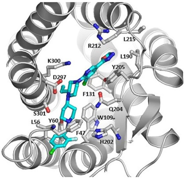
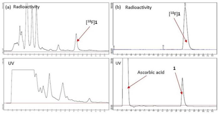
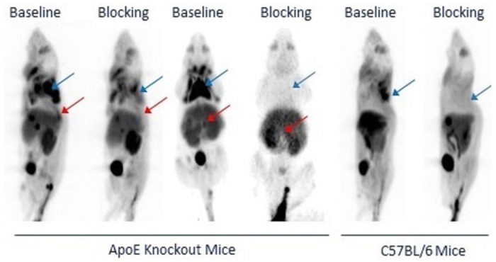
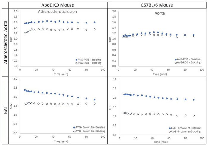
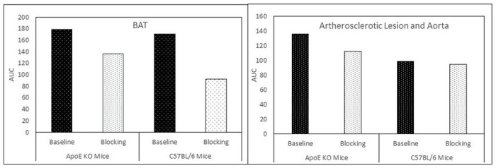
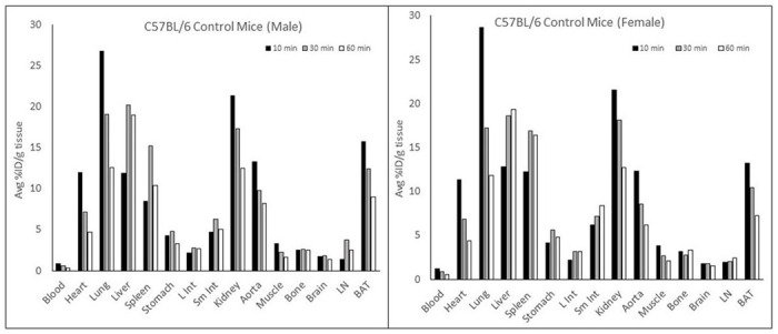
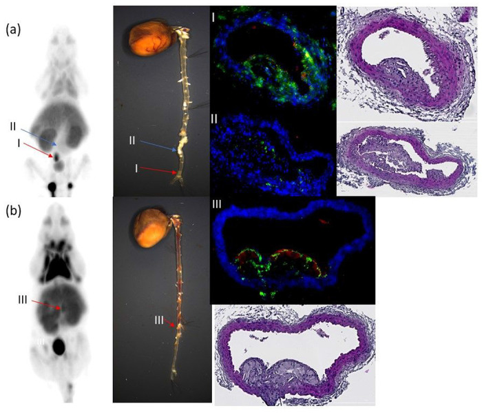
Background: CXCR3 is a chemokine receptor and is expressed on innate and adaptive immune cells. It promotes the recruitment of T-lymphocytes and other immune cells to the inflammatory site in response to the binding of cognate chemokines. Upregulation of CXCR3 and its chemokines has been found during atherosclerotic lesion formation. Therefore, the detection of CXCR3 by positron emission tomography (PET) radiotracer may be a useful tool to detect atherosclerosis development noninvasively. Herein, we report the synthesis, radiosynthesis, and characterization of a novel fluorine-18 (F-18, 18 F) labeled small-molecule radiotracer for the imaging of the CXCR3 receptor in mouse models of atherosclerosis. Methods: The reference standard ( S )-2-(5-chloro-6-(4-(1-(4-chloro-2-fluorobenzyl)piperidin-4-yl)-3-ethylpiperazin-1-yl)pyridin-3-yl)-1,3,4-oxadiazole ( 1 ) and its corresponding precursor 9 were synthesized using organic syntheses. The radiotracer [ 18 F] 1 was prepared in one-pot, two-step synthesis via aromatic 18 F-substitution followed by reductive amination. Cell binding assays were conducted using 1 , [ 125 I]CXCL10, and CXCR3A- and CXCR3B-transfected human embryonic kidney (HEK) 293 cells. Dynamic PET imaging studies over 90 min were performed on C57BL/6 and apolipoprotein E (ApoE) knockout (KO) mice that were subjected to a normal and high-fat diet for 12 weeks, respectively. Blocking studies were conducted with preadministration of the hydrochloride salt of 1 (5 mg/kg) to assess the binding specificity. Time-activity curves (TACs) for [ 18 F] 1 in both mice were used to extract standard uptake values (SUVs). Biodistribution studies were performed on C57BL/6 mice, and the distribution of CXCR3 in the abdominal aorta of ApoE KO mice was assessed by immunohistochemistry (IHC). Results: The reference standard 1 and its precursor 9 were synthesized over 5 steps from starting materials in good to moderate yields. The measured K i values of CXCR3A and CXCR3B were 0.81 ± 0.02 nM and 0.31 ± 0.02 nM, respectively. [ 18 F] 1 was prepared with decay-corrected radiochemical yield (RCY) of 13 ± 2%, radiochemical purity (RCP) >99%, and specific activity of 44.4 ± 3.7 GBq/µmol at the end of synthesis (EOS) ( n =6). The baseline studies showed that [ 18 F] 1 displayed high uptake in the atherosclerotic aorta and brown adipose tissue (BAT) in ApoE KO mice. The uptake of [ 18 F] 1 in these regions was reduced significantly in self-blocking studies, demonstrating CXCR3 binding specificity. Contrary to this, no significant differences in uptake of [ 18 F] 1 in the abdominal aorta of C57BL/6 mice were observed in both baseline and blocking studies, indicating increased CXCR3 expression in atherosclerotic lesions. IHC studies demonstrated that [ 18 F] 1 -positive regions were correlated with CXCR3 expression, but some atherosclerotic plaques with significant size were not detected by [ 18 F] 1 , and their CXCR3 expressions were minimal. Conclusion: The novel radiotracer, [ 18 F] 1 was synthesized with good RCY and high RCP. In PET imaging studies, [ 18 F] 1 displayed CXCR3-specific uptake in the atherosclerotic aorta in ApoE KO mice. [ 18 F] 1 visualized CXCR3 expression in different regions in mice is in line with the tissue histology studies. Taken together, [ 18 F] 1 is a potential PET radiotracer for the imaging of CXCR3 in atherosclerosis.
Figures
References
Publication types
Grants and funding
LinkOut - more resources
Full Text Sources
Research Materials
Miscellaneous
