[Preprint]. 2023 Feb 27:2023.02.25.530020.
doi: 10.1101/2023.02.25.530020.

Cerebellar Granule Cells Develop Non-neuronal 3D Genome Architecture over the Lifespan

Affiliations

Cerebellar Granule Cells Develop Non-neuronal 3D Genome Architecture over the Lifespan

Longzhi Tan et al. bioRxiv. .

Update in

Abstract

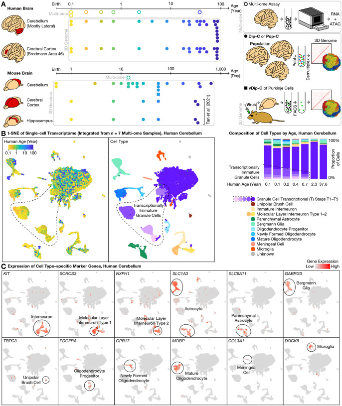
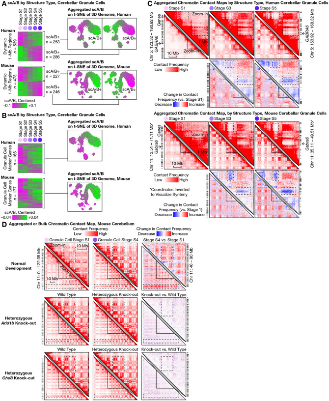
The cerebellum contains most of the neurons in the human brain, and exhibits unique modes of development, malformation, and aging. For example, granule cells-the most abundant neuron type-develop unusually late and exhibit unique nuclear morphology. Here, by developing our high-resolution single-cell 3D genome assay Dip-C into population-scale (Pop-C) and virus-enriched (vDip-C) modes, we were able to resolve the first 3D genome structures of single cerebellar cells, create life-spanning 3D genome atlases for both human and mouse, and jointly measure transcriptome and chromatin accessibility during development. We found that while the transcriptome and chromatin accessibility of human granule cells exhibit a characteristic maturation pattern within the first year of postnatal life, 3D genome architecture gradually remodels throughout life into a non-neuronal state with ultra-long-range intra-chromosomal contacts and specific inter-chromosomal contacts. This 3D genome remodeling is conserved in mice, and robust to heterozygous deletion of chromatin remodeling disease-associated genes (Chd8 or Arid1b). Together these results reveal unexpected and evolutionarily-conserved molecular processes underlying the unique development and aging of the mammalian cerebellum.


Conflict of interest statement

Competing interests: LT is an inventor on a patent application US16/615,872 filed by Harvard University that covers Dip-C.

Figures

Fig. 1.
Fig. 1.. Single-cell 3D genome atlas across lifespan for human and mouse cerebellum with single-cell multi-ome atlas of postnatal cerebellar development.
(A) Schematic of overall study design. To understand the genomic underpinning for cerebellar development and aging, we created a 3D genome atlas (filled dots and squares) of 13,161 cells from the cerebellum and cerebral cortex (and additionally, hippocampus in mouse) across the human (0.1–86 years) and mouse (0–2 years) lifespan, using our diploid chromosome conformation capture (Dip-C) (2), population-scale Dip-C (Pop-C), and virus-enriched Dip-C (vDip-C) methods (lower right). We additionally created a multi-ome (simultaneous transcriptome and chromatin accessibility) atlas (circles) of 63,768 cells from the developing cerebellum (human: 0.1–2.3 years and 1 adult; mouse: postnatal day (P) 14) (upper right; Methods). Data were compared to mouse cerebral cortex and hippocampus 3D genome data from previous work (5). (B) Integrative transcriptome analysis of the 7 human multi-ome samples revealed transcriptionally immature granule cells (dashed outlines) in the newborn cerebellum. We visualized the transcriptome portion of our human multi-ome atlas with t-distributed stochastic neighbor embedding (t-SNE) after cross-sample integration (21) (left: each dot represents a single cell). Granule cells existed in both mature form, which we termed transcriptional (T) stage 5 (T5; darkest purple), and a variety of immature forms, which we termed T1–T4 (dashed outlines; lighter shades of purple). Transcriptionally immature granule cells were abundant (14–34%) in the first postnatal year, but vanished (<1%) in the 2.3- and 37.6-year-old donors (right). (C) Representative gene expression profiles of cell type–specific marker genes (see Fig. 2A for granule cells).
Fig. 2.
Fig. 2.. Simultaneous transcriptome and chromatin accessibility profiling revealed continuous maturation of cerebellar granule cells over the first postnatal year.
(A) From our multi-ome atlas of the developing human cerebellum (Fig. 1B), we identified marker genes with expression enriched in each of the 5 transcriptional (T) stages (T1–T5; shades of purple) of granule cell maturation. Marker genes were ranked by specificity (area under the receiver operating characteristic (AUROC)). Expression of the top marker gene for each T stage was visualized on the integrated transcriptome t-SNE plot, while the top 2–10 genes were listed below. Enriched pathways (gene ontology (GO) terms) were summarized for the top 100 genes at each T stage. Note that overlap between marker genes at adjacent T stages suggested a continuous spectrum of transcriptional maturation. (B) We individually visualized each multi-ome sample with joint uniform manifold approximation and projection (UMAP) of transcriptome and chromatin accessibility (22), colored by cell types jointly defined by transcriptome and chromatin accessibility (top row). In each newborn sample (bottom: 0.1-year-old donor shown as example), granule cells (purple) exhibited a continuous maturation pseudotime (dashed arrows), suggesting gradual changes in both transcriptome and chromatin accessibility rather than discrete jumps. We identified dynamically expressed genes, dynamically accessible chromatin regions (peaks), and dynamically accessible transcription factor binding site (TFBS) motifs during granule cell maturation. Inset showed maturation pseudotime of molecular layer interneurons (MLIs).
Fig. 3.
Fig. 3.. High-throughput, high-precision 3D genome profiling uncovered progressive lifelong genome remodeling in the human and mouse cerebellum.
(A) Traditional 3C/Hi-C methods such as Dip-C measure one sample at a time (left); in contrast, the new population-scale Dip-C (Pop-C) method simultaneously profiles many samples at once, by pooling samples and computationally demultiplexing single cells based on linear DNA sequences (genotypes), thereby eliminating batch effects and substantially reducing labor and cost (right). This approach was found to be highly reproducible; note consistency between the same sample (Donor 4) assayed both individually (left) and by Pop-C (right), visualized with t-SNE of the single-cell 3D genome (based on single-cell chromatin A/B compartment values; scA/B) (5); each dot represents a single cell). (B) Rare cell types, such as adult Purkinje cells (<0.5% in mouse), were often missed by conventional Dip-C (left); in contrast, virus-enriched Dip-C (vDip-C) enabled efficient isolation of adult Purkinje cells with minimally invasive, fixation-robust fluorescent labeling in wild-type mouse tissue (right). (CD) With Pop-C and vDip-C, we created a high-resolution, cross-species 3D genome atlas for the developing and aging cerebellum (with cerebral cortex as counterpoint), and resolved the first 3D genome structures of single cerebellar cells (bottom (D)). In both species (human (C); mouse (D)), all samples were visualized together with t-SNE of scA/B; 3D genome structure types were identified with hierarchical clustering of scA/B (top). Cerebellar granule cells (shades of purple) exhibited by far the most dramatic structural transformation—born with an immature structure type, which we termed structural (S) stage S1 (lightest purple), that closely resembled forebrain neurons (shades of brown), and evolving progressively into new structure types, which we termed stages S2–S5 (darker shades of purple), that drastically differed from all other neurons as the cerebellum developed and aged. Abundances of the various S stages peaked around ages 0.2, 1, 10, 30, and 80 yr in human, and around P3, P14, P21, P56 (~2 months), and P365 (~12 months) in mouse (bottom, right).
Fig. 4.
Fig. 4.. Large-scale 3D genome remodeling of cerebellar granule cells formed ultra-long-range (10–100 Mb) intra-chromosomal contacts and specific inter-chromosomal contacts during development and aging.
(A) The most prominent architectural changes in granule cells (first 5 columns) were emergence of ultra-long-range (10–100 Mb) intra-chromosomal contacts (dashed boxes) thought to be exclusive to non-neuronal cells (16, 34) such as microglia (second to last column). Distribution of genomic distances of chromatin contacts (in base pairs (bp); on logarithmic scale) quantified by histogram for each 3D genome structure type (top) in human (middle) and mouse (bottom). (B) Emergent ultra-long-range contacts in granule cells formed prominent checkerboard patterns on contact maps—suggesting strong phase separation between the newly formed chromatin A/B compartments; this effect was generally stronger in the gene-poor, heterochromatic compartment B. For each species, an aggregated contact map of each structural (S) stage (S1–S5) of granule cell maturation was shown for an example chromosome (lower left triangles) and for an example zoomed-in (50-Mb) genomic region (upper right triangles). Contact maps (matrices of contact frequencies) were visualized with Juicebox (47), both as absolute values (first and third rows) and as relative changes compared to stage S1 (second and fourth rows). Zoomed-in regions are homologous between human and mouse; mouse coordinates were inverted for synteny. Dashed boxes highlight prominent changes during granule cell maturation. Bin size: 250 kb. (C) Granule cells formed specific inter-chromosomal contacts during development and aging; note increasing interactions between certain chromosomes—most prominently within a multi-chromosome hub of Chr 1/9/11/14/15/16/17/21/22, and between chromosome pairs such as Chr 2/9, Chr 4/14, Chr 8/11, Chr 13/20 in human. In each species, aggregated contact maps are shown for 2 example chromosome pairs. Dashed boxes highlight prominent changes during granule cell maturation. Bin sizes: 6 Mb (human genome-wide); 5 Mb (mouse genome-wide); 500 kb (zoom-in).
Fig. 5.
Fig. 5.. Continuous lifelong maturation of single-cell chromatin A/B compartment value (scA/B) was associated with cerebellar granule cell–specific marker genes and resistant to modulation of Arid1b or Chd8.
(A) Relationship between transcriptional and architectural changes in granule cells. We calculated the mean scA/B of each 1-Mb genomic region at each structural (S) stage (S1–S5; columns) of granule cell maturation, and identified the top 20% dynamic regions (rows) based on between-stage variance. Dynamic regions (rows) are shown in a heatmap (left) ordered by hierarchical clustering of scA/B correlation, and clustered into 2 temporal modes: continuous up- or down-regulation across the lifespan. Each mode was additionally visualized with aggregated scA/B of all its regions on the 3D genome t-SNE plot (right). (B) Continuously-progressing scA/B changes correlated with expression of mature granule cell–specific genes—suggesting continued 3D genome rewiring well after initial transcriptional up-regulation. Mean scA/B of each 1-Mb genomic region harboring conserved, granule cell–specific marker genes (Supplementary Table 5 of (18)) at each S stage reveals that the majority of such regions continually increased scA/B across the lifespan (left). Shown: aggregated scA/B of all such regions on the 3D genome t-SNE plot (right). (C) The granule cell–specific marker gene GABRA6 (arrows) steadily increased scA/B by losing contacts with two nearby gene-poor regions over the lifespan (dashed boxes; note the gene-poor regions formed strong contacts with each other over time) in both species. Contact maps were visualized with Juicebox (47), both as absolute values (first and third rows) and as relative changes compared to stage S1 (second and fourth rows). Zoom-in regions homologous between human and mouse; mouse coordinates inverted for synteny. Bin size: 250 kb. (D) Functional perturbation by disrupting chromatin remodelers. Bulk Dip-C on whole adult cerebellum (chiefly granule cells) of mice with clinically-relevant heterozygous deletion of autism-implicated genes Arid1b (middle) or Chd8 (bottom) had little effect on 3D genome (top; stage S4 chosen to match ages). Contact maps visualized both as absolute values (left and middle) and as relative changes (right). Dashed boxes highlighted prominent changes during granule cell maturation. Bin size: 250 kb.

References

    1. Chi Y., Shi J., Xing D., Tan L., Every gene everywhere all at once: High-precision measurement of 3D chromosome architecture with single-cell Hi-C. Frontiers in Molecular Biosciences. 9 (2022) (available at https://www.frontiersin.org/articles/10.3389/fmolb.2022.959688). - DOI - PMC - PubMed
    1. Tan L., Xing D., Chang C.-H., Li H., Xie X. S., Three-dimensional genome structures of single diploid human cells. Science. 361, 924–928 (2018). - PMC - PubMed
    1. Tan L., Xing D., Daley N., Xie X. S., Three-dimensional genome structures of single sensory neurons in mouse visual and olfactory systems. Nat Struct Mol Biol. 26, 297–307 (2019). - PubMed
    1. Tan L., Determining the 3D genome structure of a single mammalian cell with Dip-C. STAR Protocols. 2, 100622 (2021). - PMC - PubMed
    1. Tan L., Ma W., Wu H., Zheng Y., Xing D., Chen R., Li X., Daley N., Deisseroth K., Xie X. S., Changes in genome architecture and transcriptional dynamics progress independently of sensory experience during post-natal brain development. Cell. 184, 741–758.e17 (2021). - PubMed

Publication types