

This is a preprint.
Hippocampal Synaptic Alterations Associated with Tau Pathology in Primary Age-Related Tauopathy
- PMID: 36865237
- PMCID: PMC9980270
- DOI: 10.1101/2023.02.22.23286323
Hippocampal Synaptic Alterations Associated with Tau Pathology in Primary Age-Related Tauopathy
Update in
-
Hippocampal synaptic alterations associated with tau pathology in primary age-related tauopathy.J Neuropathol Exp Neurol. 2023 Sep 20;82(10):836-844. doi: 10.1093/jnen/nlad064. J Neuropathol Exp Neurol. 2023. PMID: 37595576 Free PMC article.
Abstract
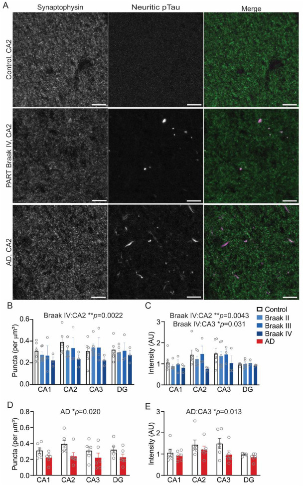
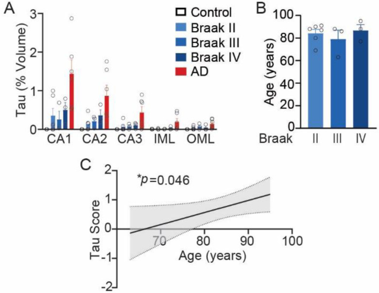
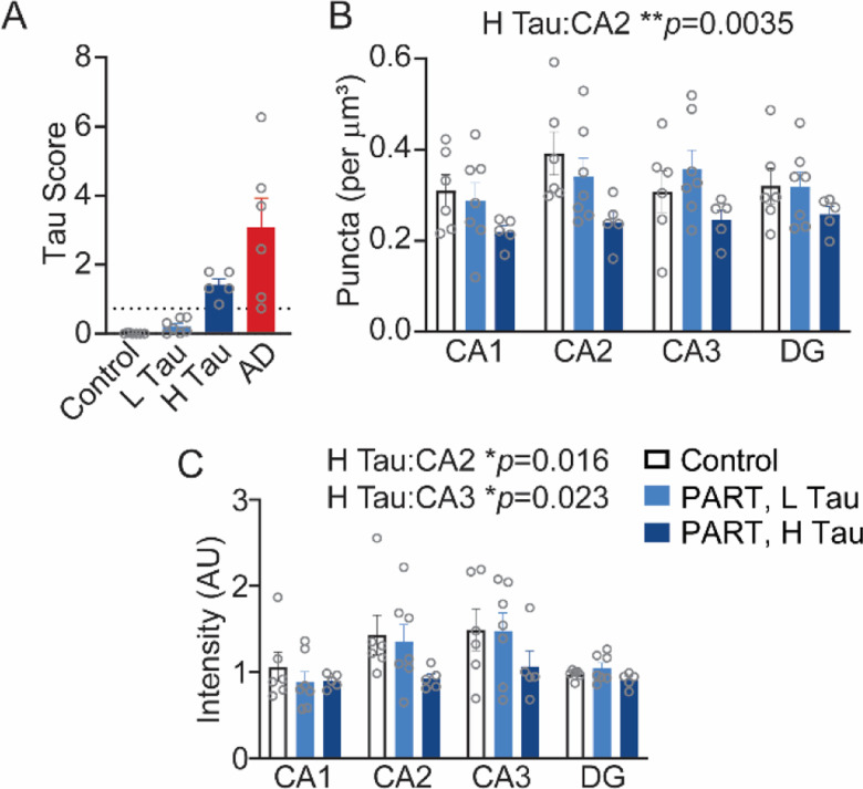
Primary Age-Related Tauopathy (PART) is characterized by the aggregation of tau in the mesial temporal lobe in older individuals. High pathologic tau stage (Braak stage) or a high burden of hippocampal tau pathology have been associated with cognitive impairment in PART. However, the underlying mechanisms of cognitive impairment in PART are not well understood. Cognitive impairment in many neurodegenerative diseases correlates with synaptic loss, raising the question of whether synaptic loss occurs in PART. To address this, we investigated synaptic changes associated with tau Braak stage and a high tau pathology burden in PART using synaptophysin and phospho-tau immunofluorescence. We compared twelve cases of definite PART with six young controls and six Alzheimer's disease cases. In this study, we identified loss of synaptophysin puncta and intensity in the CA2 region of the hippocampus in cases of PART with either a high stage (Braak IV) or a high burden of neuritic tau pathology. There was also loss of synaptophysin intensity in CA3 associated with a high stage or high burden of tau pathology. Loss of synaptophysin signal was present in AD, but the pattern was distinct from that seen in PART. These novel findings suggest the presence of synaptic loss in PART associated with either a high hippocampal tau burden or a Braak stage IV. These synaptic changes raise the possibility that synaptic loss in PART could contribute to cognitive impairment, though future studies including cognitive assessments are needed to address this question.
Conflict of interest statement
Competing Interests: All authors have no conflicts of interest to declare.
Figures
References
-
- Bates D, Mächler M., Bolker B., & Walker S. (2015) Fitting linear mixed-effects models Usinglme4. Journal of Statistical Software 67: Doi 10.18637/jss.v067.i01 - DOI
-
- Bell WR, An Y, Kageyama Y, English C, Rudow GL, Pletnikova O, Thambisetty M, O’Brien R, Moghekar AR, Albert MS et al. (2019) Neuropathologic, genetic, and longitudinal cognitive profiles in primary age-related tauopathy (PART) and Alzheimer’s disease. Alzheimers Dement 15: 8–16 Doi 10.1016/j.jalz.2018.07.215 - DOI - PMC - PubMed
Publication types
Grants and funding
LinkOut - more resources
Full Text Sources
Miscellaneous