[Preprint]. 2023 Mar 20:2023.02.24.529926.
doi: 10.1101/2023.02.24.529926.

Tenascin-C activation of lung fibroblasts in a 3D synthetic lung extracellular matrix mimic

Affiliations

Tenascin-C activation of lung fibroblasts in a 3D synthetic lung extracellular matrix mimic

Aritra Nath Kundu et al. bioRxiv. .

Update in

Abstract

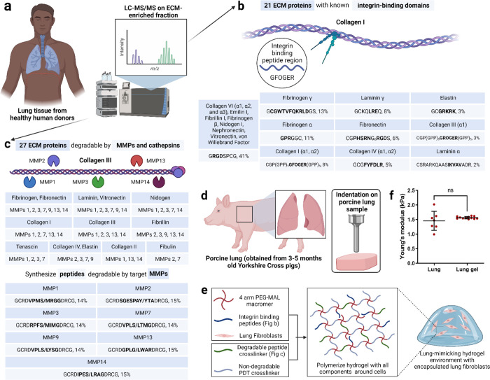
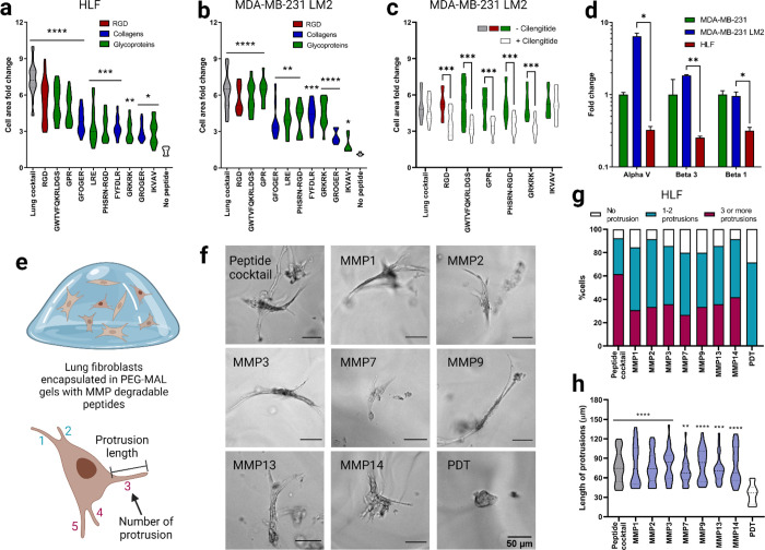
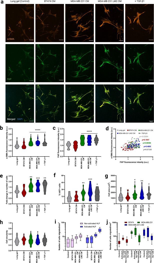
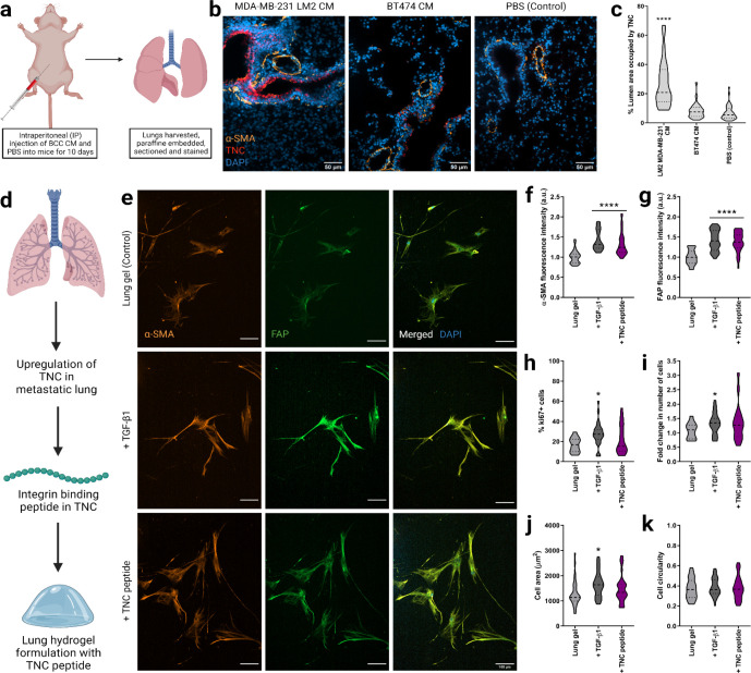
The lung extracellular matrix (ECM) maintains the structural integrity of the tissue and regulates the phenotype and functions of resident fibroblasts. Lung-metastatic breast cancer alters these cell-ECM interactions, promoting fibroblast activation. There is a need for bio-instructive ECM models that contain the ECM composition and biomechanics of the lung to study these cell-matrix interactions in vitro . Here, we developed a synthetic, bioactive hydrogel that mimics the native lung modulus, and includes a representative distribution of the most abundant ECM peptide motifs responsible for integrin binding and matrix metalloproteinase (MMP)-mediated degradation in the lung, which promotes quiescence of human lung fibroblasts (HLFs). Stimulation with transforming growth factor β1 (TGF-β1), metastatic breast cancer conditioned media (CM), or tenascin-C activated these hydrogel-encapsulated HLFs in a manner reflective of their native in vivo responses. We propose this lung hydrogel platform as a tunable, synthetic approach to study the independent and combinatorial effects of ECM in regulating fibroblast quiescence and activation.


Conflict of interest statement

Competing financial interests

The authors declare no competing financial interests.

Figures

Figure 1.
Figure 1.. Design of synthetic 3D lung ECM hydrogel.
a) Schematic of characterization of lung ECM using LC-MS/MS on ECM-enriched lung tissue samples from healthy human donors. b) Illustration of integrin-mediated binding to collagen I and the list of identified integrin-binding peptide domains with their relative quantities corresponding to ECM proteins. c) Illustration of MMP-mediated degradation of collagen III and the list of MMPs responsible for ECM protein degradation along with the list of identified MMP-degradable peptide domains with their relative quantities corresponding to the different MMPs. d) Schematic of mechanical characterization of porcine lung using indentation method. e) Schematic of lung ECM hydrogel preparation with encapsulated lung fibroblasts. f) Young’s modulus of lung hydrogel measured using indentation method and its comparison with the lung modulus using the same method.
Figure 2.
Figure 2.. Functional validation of lung integrin-binding and MMP-degradable peptides.
a) Cell area fold change 2 hours after seeding human lung fibroblasts (HLFs) onto glass coverslips functionalized with intergrin-binding peptides relative to a negative control (glass coverslip treated with silane but no peptides). b) Cell area fold change for lung tropic MDA-MB-231 breast cancer cells in comparison to negative control. c) Cell area fold change on a subset of the integrin-binding peptides in the presence of cilengitide, reported in comparison to positive control (no cilengitide). d) Fold change in gene expression, measured via qRT-PCR, for αv, β3 and β1 integrins in lung tropic MDA-MB-231 breast cancer cells and HLFs, each gene is normalized to the parental MDA-MB-231 breast cancer cells. e) Illustration of HLFs in 3D hydrogels and measurement of cell protrusions quantification. f) Representive images of HLFs after 6 days of encapsulation. g) Quantification of HLF populations with different number of protrusions for individual MMP degradable peptides and lung MMP degradable peptide cocktail. h) HLF protrusion length for individual MMP degradable peptides and lung MMP degradable peptide cocktail. All data are mean + s.d. Statistical analyses were performed using Prism (GraphPad). Data in (a), (b), (c), (d), (g) and (h) were analyzed using a one-way analysis of variance followed by a Dunnett’s multiple comparison test with 95% confidence interval. *, **, ***, and **** indicate P < 0.05, P < 0.01, P < 0.001, and P < 0.0001.
Figure 3.
Figure 3.. Fibroblast phenotype and activation in lung hydrogels in breast cancer conditioned media.
a) Representative fluorescent images of HLFs cultured in 3D lung hydrogels with breast cancer conditioned media showing α-SMA (orange), and FAP (green) expressions along with merged HLF images with nuclei staining with DAPI (blue). Scale bar: 100 μm. b) Quantification of α-SMA expression from HLFs cultured in lung gels with breast cancer conditioned media MDA-MB-231 CM, MDA-MB-231 LM2 CM and BT474 CM along with controls. c) Quantification of FAP expression from HLFs cultured in lung gels with the different media conditions. d) Correlation between α-SMA and FAP expressions from HLFs cultured in lung gels with the different media. e) Fold change in cell count for HLFs cultured in lung gels with the different media conditions. f) Percentage of proliferative Ki67+ cells in HLFs cultured in lung gels with the different media conditions. g) Cell area for HLFs cultured in lung gels the different media conditions. h) Cell circularity for HLFs cultured in lung gels with the different media conditions. i) Quantification of number of non-activated and activated cells migrated towards breast cancer conditioned media in trans-well cell migration assay. j) Quantification of number of breast cancer cells migrated towards non-activated and activated HLF conditioned media. All data are mean + s.d. Statistical analyses were performed using Prism (GraphPad). Data in (b), (c), (e), (f), (g), (h), (i) and (j) were analyzed using a one-way analysis of variance followed by a Dunnett’s multiple comparison test with 95% confidence interval. *, **, ***, and **** indicate P < 0.05, P < 0.01, P < 0.001, and P < 0.0001.
Figure 4.
Figure 4.. Fibroblast phenotype and activation in lung hydrogels with TNC peptide.
a) Schematic of harvesting the lung tissues from nude BALB/c female mice injected with breast cancer conditioned media or PBS (control) for 10 days. b) Representative fluorescent images of lung sections with MDA-MB-231 LM2 CM, BT474 CM and PBS (control) showing DAPI staining for nuclei (blue), a-SMA (orange), and TNC (red) expressions along with merged images. Scale bar: 50 μm. c) Quantification of area coverage by TNC within the lung sections from mice injected with breast cancer conditioned media or PBS. d) Schematic of lung hydrogel design with TNC peptide. e) Representative fluorescent images of HLFs cultured in 3D lung hydrogels with TNC peptide showing a-SMA (orange), and FAP (green) expressions along with merged HLF images with nuclei staining with DAPI (blue). Scale bar: 50 μm. f) Quantification of a-SMA expression from HLFs cultured in lung gels with TNC peptide along with controls. g) Quantification of FAP expression from HLFs cultured in lung gels with TNC peptide along with controls. h) Percentage of proliferative ki67+ cells in HLFs cultured in lung gels with TNC peptide along with controls. i) Fold change in cell count for HLFs cultured in lung gels with TNC peptide, along with HLFs cultured in growth media with or without TGF-β1 as controls. j) Cell area for HLFs cultured in lung gels with TNC peptide along with controls. k) Cell circularity for HLFs cultured in lung gels with TNC peptide along with controls.tide cocktail. All data are mean + s.d. Statistical analyses were performed using Prism (GraphPad). Data in (a), (b), (c), (d), (g) and (h) were analyzed using a one-way analysis of variance followed by a Dunnett’s multiple comparison test with 95% confidence interval. *, **, ***, and **** indicate P < 0.05, P < 0.01, P < 0.001, and P < 0.0001.

References

    1. Dunsmore S. E. & Rannels D. E. Extracellular matrix biology in the lung. Am J Physiol 270, L3–27 (1996). - PubMed
    1. Grinnell F. Fibroblast biology in three-dimensional collagen matrices. Trends Cell Biol 13, 264–269 (2003). - PubMed
    1. Du P. et al. Human lung fibroblast-derived matrix facilitates vascular morphogenesis in 3D environment and enhances skin wound healing. Acta Biomater 54, 333–344 (2017). - PubMed
    1. Darby I. A., Laverdet B., Bonté F. & Desmoulière A. Fibroblasts and myofibroblasts in wound healing. Clinical, Cosmetic and Investigational Dermatology vol. 7 301–311 Preprint at 10.2147/CCID.S50046 (2014). - DOI - PMC - PubMed
    1. Kanta J. Collagen matrix as a tool in studying fibroblastic cell behavior. Cell Adh Migr 9, 308–316 (2015). - PMC - PubMed

Publication types