. 2023 Feb 14:14:1111419.
doi: 10.3389/fimmu.2023.1111419. eCollection 2023.

Donor selection for adoptive immunotherapy with NK cells in AML patients: Comparison between analysis of lytic NK cell clones and phenotypical identification of alloreactive NK cell repertoire

Affiliations

Donor selection for adoptive immunotherapy with NK cells in AML patients: Comparison between analysis of lytic NK cell clones and phenotypical identification of alloreactive NK cell repertoire

Raffaella Meazza et al. Front Immunol. .

Abstract

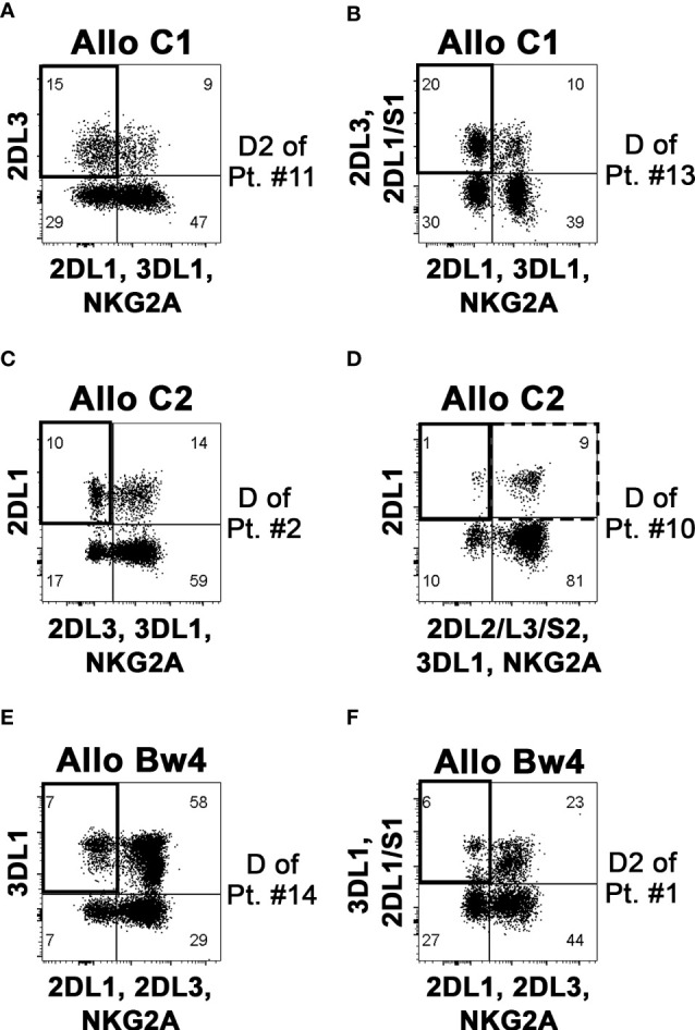
Natural killer (NK) cell-based adoptive immunotherapy in leukemia patients is an emerging field of interest based on clinical evidence of efficacy and safety. Elderly acute myeloid leukemia (AML) patients have been successfully treated with NK cells from HLA-haploidentical donors, especially when high amounts of alloreactive NK cells were infused. The aim of this study was comparing two approaches to define the size of alloreactive NK cells in haploidentical donors for AML patients recruited in two clinical trials with the acronym "NK-AML" (NCT03955848), and "MRD-NK". The standard methodology was based on the frequency of NK cell clones capable of lysing the related patient-derived cells. The alternative approach consisted of the phenotypic identification of freshly derived NK cells expressing, as inhibitory receptors, only the inhibitory KIR(s) specific for the mismatched KIR-Ligand(s) (HLA-C1, HLA-C2, HLA-Bw4). However, in KIR2DS2+ donors and HLA-C1+ patients, the unavailability of reagents staining only the inhibitory counterpart (KIR2DL2/L3) may lead to an underestimated identification of the alloreactive NK cell subset. Conversely, in the case of HLA-C1 mismatch, the alloreactive NK cell subset could be overestimated due to the ability of KIR2DL2/L3 to recognize with low-affinity also HLA-C2. Especially in this context, the additional exclusion of LIR1-expressing cells might be relevant to refine the size of the alloreactive NK cell subset. We could also associate degranulation assays, using as effector cells IL-2 activated donor peripheral blood mononuclear cells (PBMC) or NK cells upon co-culture with the related patient target cells. The donor alloreactive NK cell subset always displayed the highest functional activity, confirming its identification accuracy by flow cytometry. Despite the phenotypic limitations and considering the proposed corrective actions, a good correlation was shown by the comparison of the two investigated approaches. In addition, the characterization of receptor expression on a fraction of NK cell clones revealed expected but also few unexpected patterns. Thus, in most instances, the quantification of phenotypically defined alloreactive NK cells from PBMC can provide data similar to the analysis of lytic clones, with several advantages, such as a shorter time to achieve the results and, perhaps, higher reproducibility/feasibility in many laboratories.

Keywords: NK alloreactivity; acute myeloid leukemia (AML); adoptive immunotherapy; donor selection; human leucocyte antigen (HLA); killer immunoglobulin-like receptors (KIR); natural killer cells (NK cells).


Conflict of interest statement

The authors declare that the research was conducted in the absence of any commercial or financial relationship that could be construed as potential conflict of interest.

Figures

Figure 1
Figure 1
Evaluation of KIR3DL1 NK surface expression to confirm possible NK alloreactivity in case of HLA-Bw4 mismatch. By flow-cytometry, NK cells (gating on CD3 CD56+ cells of PBMC, left panels) from two potential Allo Bw4 donors were analyzed using DX9 (anti-KIR3DL1) and Z27 (anti-KIR3DL1/S1) mAbs to detect the surface expression of KIR3DL1 (i.e., the relevant iKIR specific for HLA-Bw4) and/or KIR3DS1 (right panels). The “KIR” acronym has been omitted in the receptors identified within the quadrants. (A) NK cells from donor of patient #5 did not express KIR3DL1 (DX9), while KIR3DS1 (Z27+/DX9) was present. This donor couldn’t be considered as NK alloreactive. (B) a portion of NK cells from the donor of patient #6 expressed KIR3DL1 (Z27+/DX9+), confirming the NK alloreactivity.
Figure 2
Figure 2
Representative analyses of KIR gene profiles and KIR phenotype. In the KIR gene profiles (top), the KIR presence or absence is indicated with grey or white boxes, respectively. KIR2DS4 alleles coding for surface receptors are reported as F, while those coding for truncated receptors as D. KIR2DS3 and KIR2DS5 are indicated by S3 and S5, respectively. Cytofluorimetric analysis of PBMC (bottom) shows the surface expression of various iKIR and aKIRs on NK cells (gating on CD3 CD56+ cells), using different mAb combinations (see mAb specificity in Supplementary Table 1 ). The “KIR” acronym has been omitted for both genes and molecules. Two representative donors were evaluated. (A) D1 of patient #12 was characterized by KIR A/A genotype and the expression of all analyzed iKIR, and only KIR2DS4 as aKIR. (B) D2 of patient #8 was characterized by KIR B/x genotype, and the expression of all detectable iKIR and aKIR (excepted KIR2DS4). Gating on GL183+ NK cells, 1F12/ECM-41 mAb combination also allowed the discrimination of KIR2DS2+, KIR2DL2+, and KIR2DL3+ NK cell subsets.
Figure 3
Figure 3
Identification and quantification of the alloreactive NK cell subset on donor PBMC. By flow-cytometry, NK cells (gating on CD3 CD56+ cells of PBMC) were analyzed to define the size of the alloreactive subset (indicated as a square in upper left quadrants). Representative donors characterized to be Allo C1 (A, B), Allo C2 (C, D), and Allo Bw4 (E, F), considering the KIR-L present in the donor and missing in the recipient and the presence of donor iKIR specific for the mismatched KIR-L, are shown. Three donors were without (A, C, E), and three donors with the relevant aKIR (B, D, F) as indicated in Table 2 . The presence of KIR2DS2 can determine an underestimation of the alloreactive NK cell subset, as shown for the Allo C1 donor of patient #10 (D); the square with a dashed line (upper right quadrant) includes the possible presence of alloreactive KIR2DL1+ KIR2DS2+ cells that cannot be properly quoted.
Figure 4
Figure 4
The phenotypically identified donor alloreactive NK cell subsets display the highest degranulation capability against the related patient target cells. (A) Degranulation activity of all NK (CD3 CD56+ cells) and different NK cell subsets from three donors representative for each type of alloreactivity (D of patient #16, D1 of patient#12, D of patient#14) evaluated upon incubation in the absence or in the presence of PHA-blasts derived from the related patients (Target). Allo Subset consists of NK cells expressing only the iKIR specific for the KIR-L that is missing in target cells, No Allo Subset represents NK cells expressing iKIRs specific for KIR-L on target cells, and KIR NKG2A+ represents NK cells expressing only NKG2A and no KIRs. E:T ratio 2:1. Numbers indicate the percentage of surface CD107a+ cells, and the percentages of Δ CD107 are indicated in brackets. The samples of each donor were concatenated (data aggregated into one file) and analyzed using Flowjo. (B) Cumulative data of specific NK cell degranulation activity (Δ CD107a) from different donors after stimulation with patient-derived PHA-blasts. Different NK cell subsets were considered. E:T ratio 2:1. n=9 (Allo C1 = 2, Allo C2 = 2, Allo Bw4 = 5). Not significant (ns), *p< 0.05, ***p< 0.001, and ****p< 0.0001 (Kruskal-Wallis followed by Dunn’s multiple comparison test). Mean + SEM are reported. (C) Degranulation activity of different NK cell subsets from two representative Allo C2 donors was tested upon incubation in the absence or in the presence of the MOLM-14 target cell line. (D) Specific degranulation activity (Δ CD107a) of NK cells from different donors after stimulation with K562 cell line. n=10 (Allo C1 = 6, Allo Bw4 = 4). E:T ratio 2:1.
Figure 5
Figure 5
NK alloreactivity data from cytofluorimetric analysis of PBMC and lytic NK cell clones. Correlation analysis between the values obtained by the two different approaches, considering either the whole cohort (A, B), Allo Bw4, and Allo C2 only (C, D), or Allo C1 only (E, F). The size of the alloreactive NK cell subset phenotypically defined without (A, C, E) or with the further exclusion of LIR1+ cells (B, D, F). In panels (A, E) the % NK alloreactive subset of D of patient #3 (Allo C1, 48% without the exclusion of LIR1+ cells) is not shown because its value is out of the range of the axis, but it was considered in the correlation analysis.
Figure 6
Figure 6
Cytofluorimetric analysis of lytic versus non lytic NK cell clones. NK cell clones derived from four donors, selected to be lytic (alloreactive, A) and non-lytic (non-alloreactive, NA) against the related patient PHA-blasts were analyzed, by flow cytometry, for the expression of relevant KIRs, NKG2A, and in one case LIR1. Considering the type of alloreactivity, the presence of the permissive iKIR(s) and relevant aKIR are written in bold and indicated as green, the blocking iKIR(s) as red, NKG2A as grey, and LIR1 as orange boxes, while their absence as white boxes; a question mark indicates uncertainty due to the lack of mono-specificity of certain mAbs (i.e., GL-183, CH-L and 1F12). Representative stainings (A) and cumulative data (B) of clones derived from D of patient #10 (Allo C2). (C) summary of clones from D1 (up) and D2 (bottom) of patient #11 (Allo C1). (D) summary of clones from D of patient #16 (Allo C1).

References

    1. Vivier E, Raulet DH, Moretta A, Caligiuri MA, Zitvogel L, Lanier LL, et al. . Innate or adaptive immunity? the example of natural killer cells. Science (2011) 331(6013):44–9. doi: 10.1126/science.1198687 - DOI - PMC - PubMed
    1. Quatrini L, Della Chiesa M, Sivori S, Mingari MC, Pende D, Moretta L. Human NK cells, their receptors and function. Eur J Immunol (2021) 51(7):1566–79. doi: 10.1002/eji.202049028 - DOI - PMC - PubMed
    1. Moretta A, Bottino C, Vitale M, Pende D, Biassoni R, Mingari MC, et al. . Receptors for HLA class-I molecules in human natural killer cells. Annu Rev Immunol (1996) 14:619–48. doi: 10.1146/annurev.immunol.14.1.619 - DOI - PubMed
    1. Elliott JM, Yokoyama WM. Unifying concepts of MHC-dependent natural killer cell education. Trends Immunol (2011) 32(8):364–72. doi: 10.1016/j.it.2011.06.001 - DOI - PMC - PubMed
    1. Ljunggren HG, Karre K. In search of the 'missing self': MHC molecules and NK cell recognition. Immunol Today (1990) 11(7):237–44. doi: 10.1016/0167-5699(90)90097-S - DOI - PubMed

Publication types