Institutional Evaluation of Fetal Neurology Consults and Postnatal Outcomes: A 10-Year Retrospective Cohort Review
- PMID: 36865645
- PMCID: PMC9973289
- DOI: 10.1212/CPJ.0000000000200100
Institutional Evaluation of Fetal Neurology Consults and Postnatal Outcomes: A 10-Year Retrospective Cohort Review
Abstract
Background and objectives: An increasing number of centers are offering fetal neurology consultation services; however, there is limited information available in overall institutional experiences. Data are lacking on the fetal characteristics, pregnancy course, and the influence of fetal consultation on perinatal outcomes. The aim of this study is to provide insight on the institutional fetal neurology consult process and areas of strengths and weaknesses.
Methods: We performed a retrospective electronic chart review of fetal consults from April 2, 2009, to August 8, 2019, at Nationwide Children's Hospital. The objectives were to summarize clinical characteristics, agreement of prenatal and postnatal diagnoses based on best available imaging, and postnatal outcomes.
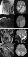
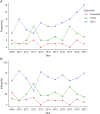
Results: Of the 174 maternal-fetal neurology consults placed, 130 qualified for inclusion based on data available for review. Of the 131 anticipated fetuses, 5 experienced fetal demise, 7 underwent elective termination, and 10 died in the postnatal period. The majority were admitted to the neonatal intensive care unit; 34 (31%) required supportive intervention for feeding, breathing, or hydrocephalus, and 10 (8%) experienced seizures during their neonatal intensive care unit (NICU) stay. Imaging results from 113 babies who had prenatal and postnatal imaging of the brain were analyzed based on the primary diagnosis. The most common malformations were as follows (prenatal % vs postnatal %): midline anomalies (37% vs 29%), posterior fossa abnormalities (26% vs 18%), and ventriculomegaly (14% vs 8%). Additional disorders of neuronal migration were not seen on fetal imaging but were present in 9% of the postnatal studies. Analysis of agreement between prenatal and postnatal diagnostic imaging for the 95 babies who had MRIs at both time points found moderate concordance (Cohen kappa: 0.62, 95% CI 0.5-0.73; percent agreement: 69%, 95% CI 60%-78%). Consult recommendations for neonatal blood tests affected postnatal care in 64 of 73 cases in which the infant survived and data were available.
Discussion: Establishing a multidisciplinary fetal clinic can provide timely counseling and create rapport with families to have continuity of care for birth planning and postnatal management. Prognosis based on radiographic prenatal diagnosis requires caution as some neonatal outcomes may vary considerably.
© 2023 American Academy of Neurology.
Figures


Similar articles
-
The Role of Fetal MRI for Suspected Anomalies of the Posterior Fossa.Pediatr Neurol. 2021 Apr;117:10-18. doi: 10.1016/j.pediatrneurol.2021.01.002. Epub 2021 Jan 10. Pediatr Neurol. 2021. PMID: 33607354
-
Outcomes of congenital diaphragmatic hernia: a population-based study in Western Australia.Pediatrics. 2005 Sep;116(3):e356-63. doi: 10.1542/peds.2004-2845. Pediatrics. 2005. PMID: 16140678
-
Role of magnetic resonance imaging in fetuses with mild or moderate ventriculomegaly in the era of fetal neurosonography: systematic review and meta-analysis.Ultrasound Obstet Gynecol. 2019 Aug;54(2):164-171. doi: 10.1002/uog.20197. Epub 2019 Jul 11. Ultrasound Obstet Gynecol. 2019. PMID: 30549340
-
Neurological and clinical outcomes in infants and children with a fetal diagnosis of asymmetric ventriculomegaly, interhemispheric cyst, and dysgenesis of the corpus callosum.J Neurosurg Pediatr. 2021 Nov 19;29(3):283-287. doi: 10.3171/2021.9.PEDS21252. Print 2022 Mar 1. J Neurosurg Pediatr. 2021. PMID: 34798596
-
Evaluation of prenatally diagnosed structural congenital anomalies.J Obstet Gynaecol Can. 2009 Sep;31(9):875-881. doi: 10.1016/S1701-2163(16)34307-9. J Obstet Gynaecol Can. 2009. PMID: 19941713 Review. English, French.
Cited by
-
The science of uncertainty guides fetal-neonatal neurology principles and practice: diagnostic-prognostic opportunities and challenges.Front Neurol. 2024 Jan 30;15:1335933. doi: 10.3389/fneur.2024.1335933. eCollection 2024. Front Neurol. 2024. PMID: 38352135 Free PMC article. Review.
-
Communicating neurological prognosis in the prenatal period: a narrative review and practice guidelines.Pediatr Res. 2025 Jan 14. doi: 10.1038/s41390-025-03805-8. Online ahead of print. Pediatr Res. 2025. PMID: 39809859 Review.
References
LinkOut - more resources
Full Text Sources
Miscellaneous
