. 2023 Jan 23;7(2):100058.
doi: 10.1016/j.rpth.2023.100058. eCollection 2023 Feb.

Tethered platelet capture provides a mechanism for restricting circulating platelet activation to the wound site

Affiliations

Tethered platelet capture provides a mechanism for restricting circulating platelet activation to the wound site

Irina D Pokrovskaya et al. Res Pract Thromb Haemost. .

Abstract

Background: Puncture wounding is a longstanding challenge to human health for which understanding is limited, in part, by a lack of detailed morphological data on how the circulating platelet capture to the vessel matrix leads to sustained, self-limiting platelet accumulation.

Objectives: The objective of this study was to produce a paradigm for self-limiting thrombus growth in a mouse jugular vein model.

Methods: Data mining of advanced electron microscopy images was performed from authors' laboratories.

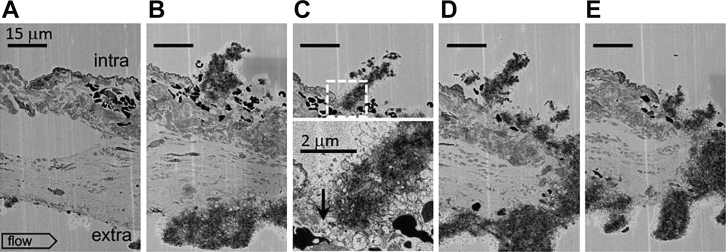
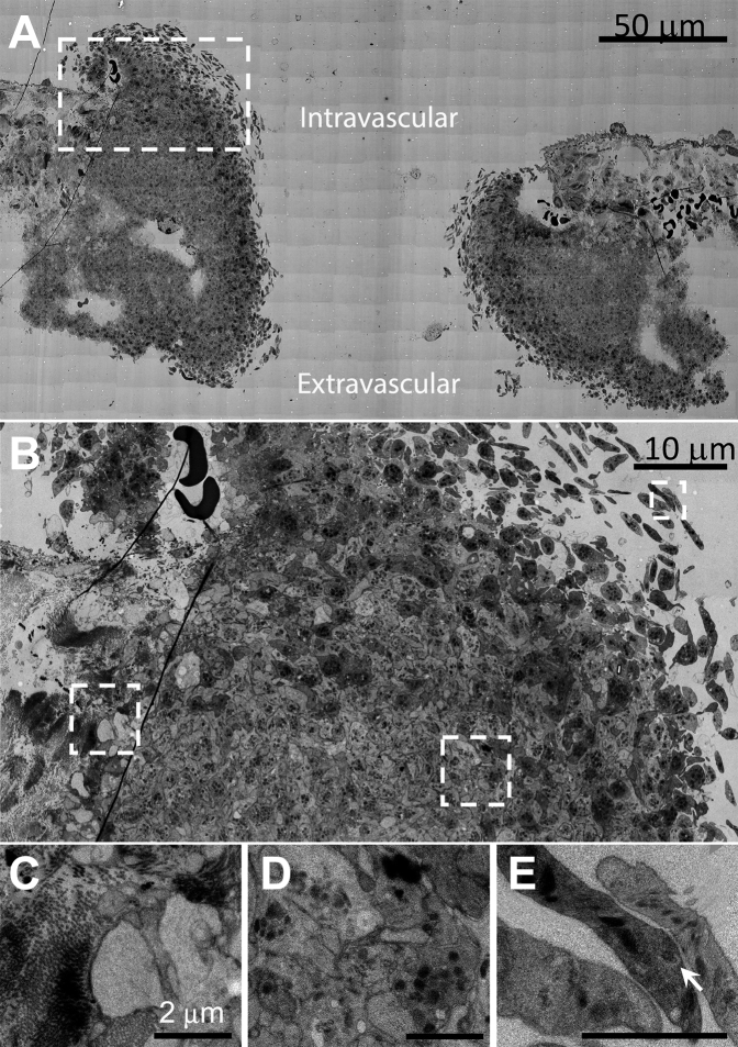
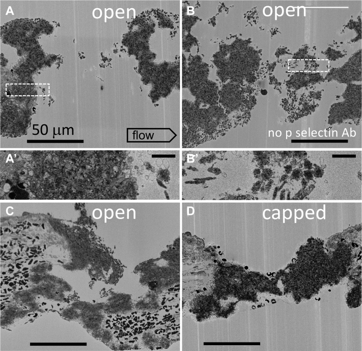
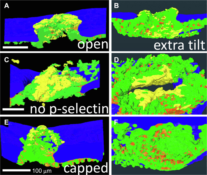
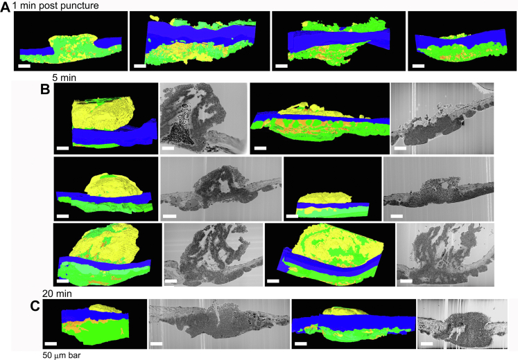
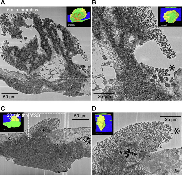
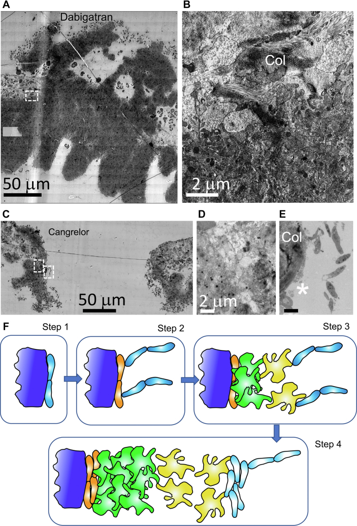
Results: Wide-area transmission electron mcrographs revealed initial platelet capture to the exposed adventitia resulted in localized patches of degranulated, procoagulant-like platelets. Platelet activation to a procoagulant state was sensitive to dabigatran, a direct-acting PAR receptor inhibitor, but not to cangrelor, a P2Y12 receptor inhibitor. Subsequent thrombus growth was sensitive to both cangrelor and dabigatran and sustained by the capture of discoid platelet strings first to collagen-anchored platelets and later to loosely adherent peripheral platelets. Spatial examination indicated that staged platelet activation resulted in a discoid platelet tethering zone that was pushed progressively outward as platelets converted from one activation state to another. As thrombus growth slowed, discoid platelet recruitment became rare and loosely adherent intravascular platelets failed to convert to tightly adherent platelets.

Conclusions: In summary, the data support a model that we term Capture and Activate, in which the initial high platelet activation is directly linked to the exposed adventitia, all subsequent tethering of discoid platelets is to loosely adherent platelets that convert to tightly adherent platelets, and self-limiting, intravascular platelet activation over time is the result of decreased signaling intensity.

Keywords: image analysis; platelets; puncture wound hemostasis; serial block face electron microscopy; thrombus formation; wide-area transmission electron microscopy.


Figures

Figure 1
Figure 1
1-minute thrombus formation in response to a small scratch in the jugular vein endothelial cell layer exposing the collagen-rich adventitia to the lumen of the blood vessel. A-E. sequential ortho block face views, spaced 20 μm in Z, 20 nm XY raw pixel size, across a ∼60 μm wide scratch in the endothelial layer of the mouse jugular vein. As shown in B, C, D, and E, a columnar accumulation of platelets fills the damaged area. At its intravascular tip, the column is marked by discoid platelets (C). Furthermore, as shown in a zoom of the dotted square in C, the platelet column is at its base (arrow, C’) and sides rich in procoagulant-like platelets nearly devoid of cytoplasm. The scratch is presumably the results of a needle slip and is distal from any puncture hole. Intravascular (top), extravascular (bottom), flow: left to right. A total of four 1-minute point samples were performed for SBF-SEM. In addition, 20 nm XY images were taken every 20 mm across the full thrombus width.
Figure 2
Figure 2
A 1-minute mouse jugular vein puncture wound thrombus showing both intravascular and extravascular areas of peripheral, discoid platelet recruitment to a forming thrombus. A. Montaged WA-TEM electron micrographs taken at 3.185 nm raw XY pixel size across the full puncture wound width, mid-puncture hole, and low image zoom. B. Image taken from left side of (A) (left side, upper, dashed box) showing at a higher zoom a full width image of one side of the thrombus from anchoring to collagen (left) to the recruitment of discoid platelets within the puncture hole. C. A high-zoom image taken from right side of B) (dashed box) showing the appearance of platelets at the platelet/exposed collagen-rich adventitia interface. These platelets appear nearly devoid of cytoplasm and procoagulant-like platelets. Note that collagen presents as bundles of fibers. These bundles are sometimes perpendicular (end on) to the platelets and sometimes parallel to the platelets. The collagen bundling results in the adventitia being a highly heterogeneous in its presentation to any locally adherent, ie, anchored platelets. D. High zoom, mid-thrombus example. Note the presence of α-granules. E. A high-zoom, hole peripheral image of newly captured discoid platelets. Arrow points to an example dense granule. A total of three 5-minute point samples were performed for WA-TEM. In addition, 3.185 nm XY images were at 3 separate depths across the full thrombus width.
Figure 3
Figure 3
Single-slice, SBF-SEM, 1-minute thrombus images (20 nm XY raw pixel size) from four separate examples (A, B, C, and D) within a jugular vein, puncture hole showing peripheral, discoid-shaped platelet recruitment on both intravascular and extravascular surfaces of open, but not capped puncture wounds. A’, B’ show the platelet appearance of dashed box areas at a 2× higher zoom. In addition, 20 nm pixel size image slices are taken every 20 μm across the thrombus. Intravascular (top) and extravascular (bottom of frame). A total of four 1-minute point samples were performed for SBF-SEM. The examples shown are at full cross shown at approximately mid-thrombus depth.
Figure 4
Figure 4
Near full thrombus renderings of split open (A, C, E) and extravascular surface (B, D, F) indicating platelet adherence state (loosely adherent to discoid, yellow vs tightly adherent, green) and pockets of highly activated, degranulated platelets (orange). Renderings are from SBF-SEM images (100 nm XY raw pixel size) taken across full thrombus widths every 200 nm. Once the thrombus caps, loosely adherent/discoid-shaped platelet accumulation is restricted to intravascular accessible thrombus surfaces. A total of four 1-minute point samples were performed for SBF-SEM. In addition, 100 nm XY images were taken every 200 nm across the full thrombus width.
Figure 5
Figure 5
Side-viewed renderings (A. 1-minute, one-split open example) and thrombus cross section slice as a raw micrograph and side-viewed renderings (B. 5 minutes and C. 20 minutes) of all SBF-SEM imaged samples. SBF-SEM images were collected across full thrombus width every 200 nm. Raw SBF-SEM micrographs for 1-minute thrombi are shown in Figure 3. Loosely adherent extravascular platelets in these side-view renderings are only seen in the 5- and 20-minute examples when such areas extend into the puncture hole. Color coding: loosely adherent to discoid, yellow vs tightly adherent, green and pockets of highly activated, degranulated platelets, orange. Activation-based segmentation and renderings are from SBF-SEM images (100 nm XY raw pixel size) taken across full thrombus widths every 200 nm. Once the thrombus caps, loosely adherent/discoid-shaped platelet accumulation is restricted to intravascular accessible thrombus surfaces. For more detail, please see [1]. Total number of SBF-SEM samples shown for each time postpuncture wounding.
Figure 6
Figure 6
Quantitation of thrombus formation parameters: volume, extravascular vs intravascular platelet distribution, platelet type, and relative volume % of degranulated platelets, tightly adherent platelets, and loosely adherent platelets within the total thrombus volume sectioned. Intravascular thrombus volume appeared to peak at 5 minutes after puncture, whereas the extravascular volume continued to increase. The portion of a given platelet activation type that was extravascular increased over time. The data were derived from the segmentation, rendering and quantification of the examples shown in Figure 5. P value comparisons with a value of <.05 are indicated with an ∗ and those with a value of < .001 are indicated with a ∗∗. P indicators placed on the graph indicate significant time series events, and those placed vertically, between data points, indicate significant differences in location or between platelet activation type at a given time point. Dot plots include the total sample sets performed.
Figure 7
Figure 7
Peripheral, discoid platelet accumulation in 5- (A,B) and 20-minute (C,D) thrombi is restricted to small, flow protected patches. A, C) near full thrombi cross sections. B, D) 2× higher zoom of downstream protected areas. Dashed boxes mark areas of peripheral, discoid-shaped platelet accumulation. Insets show the overall thrombus arrangement as rendered from 2000 to 2500 images spaced 200 nm apart. Yellow, loosely adherent platelets; green, tightly adherent platelets; orange, stripes of highly degranulated, procoagulant-like platelets. Images from SBF-SEM micrographs, 20 nm raw pixel size. Five-minute postpuncture, N = 6; 20-minute postpuncture, N = 2.
Figure 8
Figure 8
The DOAC, dabigatran, and the acute acting, anti-platelet drug, cangrelor, differentially affect puncture wound thrombus formation and platelet activation at the collagen-rich adventitia/thrombus interface. WA-TEM, 3.185 nm XY raw pixel size. A. Overall appearance of a 5-minute postpuncture thrombus from a dabigatran pre-treated mouse. Montaged image, 5-minute postpuncture, approximately mid-thrombus, postbleeding cessation. The thrombus consisted mainly of tightly adherent platelets with limited intravascular accumulation of loosely adherent platelets. Vessel walls are to the left and right of the thrombus. Top, intravascular and bottom, extravascular. B. Zoom of collagen/platelet interface of dabigatran treated jugular thrombus (dashed box in A). The platelets at the interface possess are degranulated with a cytoplasm/cytosol rich in proteins and hence one that has electron density. Control, 5-minute, WT platelets show both degranulation and extensive cytosol loss at the adventitial interface (Figure 7B, see also [1] ie, dabigatran-inhibited cytosol loss. C. 5-minute cangrelor-treated thrombus showing abundant peripheral, discoid-shaped platelets and progressive increase in apparent activation state within the thrombus. Left and right boxes in (C) mark the position of adventitial interface (left box) and a platelet string (right box) shown at higher zoom in (E). D. Frame showing that cangrelor has little to no inhibitory effect on platelet degranulation/cytosol loss at the collagen/platelet interface. E. Zoom of platelet string at position marked by right box in (C). The string is tethered to a thin collagen anchored platelet layer (white asterisks, see also Supplementary Figure 4). In both cangrelor and dabigatran platelets, rounded, darkly staining mitochondria were the prominent cytosolic organelle. Col, collagen. F. Tethered Capture and Activate Model for platelet recruitment in a venous puncture wound thrombus. Step (1)—early discoid platelet recruitment/capture to generate a monolayer of discoid platelets (light blue) directly bound to adventitia (dark blue). An important role for collagen and GPVI as the major collagen receptor is postulated. Collagen bound VWF may also be contributory; step (2)—initial platelet anchoring directly to collagen leads to extensive platelet degranulation and cytosol loss and subsequent step 2 capture of discoid platelet strings is to the thin layer of degranulated platelets (orange). Step (3)—discoid platelets rapidly convert to loosely adherent (yellow) and tightly adherent (green) platelets. As shown in step 3, subsequent capture of discoid platelet strings to loosely attached platelets. Circulating platelet capture in steps 3 and 4 lead to the formation of platelet pedestals and columns within the growing thrombus. Step (4)—repeated rounds of discoid platelet recruitment with conversion to loosely adherent platelets and tightly adherent platelets accompanied by some increase in the number of degranulated platelets at the adventitial interface leads to full thrombus formation. Note that in this model, both thrombin and P2Y12 signaling happen early and are rate contributing. The data suggest that there is no qualitative change in molecular signaling but rather a decrease in the signaling intensity with time. Whether discoid platelet, ie, circulating platelet capture to different surfaces occurs through identical molecular mechanisms is an open question to be addressed in future experimentation. Consistent with previous literature, we propose that VWF is the likely long tethering protein involved in the formation of platelet strings. Dabigatran – WA-TEM concentration series of 15 mg/kg, 50 mg/kg, and 150 mg/kg dabigatran was performed with 2 to 3 samples at each concentration. N = 3 at 150 mg/kg dabigatran. Cangrelor – Concentration per Materials and Methods. N = 2.

Similar articles

Cited by

References

    1. Rhee S.W., Pokrovskaya I.D., Ball K.K., Ling K., Vedanaparti Y., Cohen J., et al. Venous puncture wound hemostasis results in a vaulted thrombus structured by locally nucleated platelet aggregates. Commun Biol. 2021;4:1090. - PMC - PubMed
    1. Chattaraj S.C. Cangrelor AstraZeneca. Curr Opin Investig Drugs. 2001;2:250–255. - PubMed
    1. Eisert W., Hauel N., Stagier J., Wienen W., Clemens A., van Ryn J. Dabigatran: an oral novel potent reversible nonpeptide inhibitor of thrombin. Arterioscler Thromb Vasc Biol. 2010;30:1885–1889. - PubMed
    1. Tomaiuolo M., Matzko C.N., Poventud-Fuentes I., Weisel J.W., Brass L.F., Stalker T.J. Interrelationships between structure and function during the hemostatic response to injury. Proc Natl Acad Sci USA. 2019;116:2243–2252. - PMC - PubMed
    1. Pokrovskaya I., Joshi S., Tobin M., Desai R., Aronova M.A., Kamykowski J.A., et al. SNARE-dependent membrane fusion initiates α-granule matrix decondensation in mouse platelets. Blood Adv. 2018;2:2947–2958. - PMC - PubMed