

Structural differences among children, adolescents, and adults with attention-deficit/hyperactivity disorder and abnormal Granger causality of the right pallidum and whole-brain
- PMID: 36866118
- PMCID: PMC9971633
- DOI: 10.3389/fnhum.2023.1076873
Structural differences among children, adolescents, and adults with attention-deficit/hyperactivity disorder and abnormal Granger causality of the right pallidum and whole-brain
Abstract
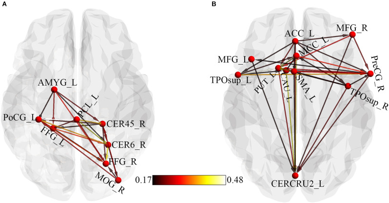
Attention-deficit/hyperactivity disorder (ADHD) is a childhood mental health disorder that often persists to adulthood and is characterized by inattentive, hyperactive, or impulsive behaviors. This study investigated structural and effective connectivity differences through voxel-based morphometry (VBM) and Granger causality analysis (GCA) across child, adolescent, and adult ADHD patients. Structural and functional MRI data consisting of 35 children (8.64 ± 0.81 years), 40 adolescents (14.11 ± 1.83 years), and 39 adults (31.59 ± 10.13 years) was obtained from New York University Child Study Center for the ADHD-200 and UCLA dataset. Structural differences in the bilateral pallidum, bilateral thalamus, bilateral insula, superior temporal cortex, and the right cerebellum were observed among the three ADHD groups. The right pallidum was positively correlated with disease severity. The right pallidum as a seed precedes and granger causes the right middle occipital cortex, bilateral fusiform, left postcentral gyrus, left paracentral lobule, left amygdala, and right cerebellum. Also, the anterior cingulate cortex, prefrontal cortex, left cerebellum, left putamen, left caudate, bilateral superior temporal pole, middle cingulate cortex, right precentral gyrus, and the left supplementary motor area demonstrated causal effects on the seed region. In general, this study showed the structural differences and the effective connectivity of the right pallidum amongst the three ADHD age groups. Our work also highlights the evidence of the frontal-striatal-cerebellar circuits in ADHD and provides new insights into the effective connectivity of the right pallidum and the pathophysiology of ADHD. Our results further demonstrated that GCA could effectively explore the interregional causal relationship between abnormal regions in ADHD.
Keywords: Granger causality analysis; age; attention deficit/hyperactivity disorder; resting-state fMRI; voxel-based morphometry.
Copyright © 2023 Agoalikum, Klugah-Brown, Wu, Hu, Jing and Biswal.
Conflict of interest statement
The authors declare that the research was conducted in the absence of any commercial or financial relationships that could be construed as a potential conflict of interest.
Figures


Similar articles
-
Abnormal resting-state functional connectivity patterns of the putamen in medication-naïve children with attention deficit hyperactivity disorder.Brain Res. 2009 Dec 15;1303:195-206. doi: 10.1016/j.brainres.2009.08.029. Epub 2009 Aug 20. Brain Res. 2009. PMID: 19699190
-
[Altered patterns of functional connectivity of posterior cingulate cortex on resting-state magnetic resonance imaging in children with attention-deficit or hyperactivity disorder].Zhonghua Yi Xue Za Zhi. 2013 Jun 25;93(24):1881-5. Zhonghua Yi Xue Za Zhi. 2013. PMID: 24124739 Chinese.
-
Shared and Distinct Neurobiological Bases of Bipolar Disorder and Attention-Deficit/Hyperactivity Disorder in Children and Adolescents: A Comparative Meta-Analysis of Structural Abnormalities.J Am Acad Child Adolesc Psychiatry. 2024 Jun;63(6):586-604. doi: 10.1016/j.jaac.2023.09.551. Epub 2023 Dec 8. J Am Acad Child Adolesc Psychiatry. 2024. PMID: 38072245
-
A multimodal neuroimaging meta-analysis of functional and structural brain abnormalities in attention-deficit/hyperactivity disorder.Prog Neuropsychopharmacol Biol Psychiatry. 2025 Jan 10;136:111199. doi: 10.1016/j.pnpbp.2024.111199. Epub 2024 Nov 29. Prog Neuropsychopharmacol Biol Psychiatry. 2025. PMID: 39615871
-
[Structural and functional neuroanatomy of attention-deficit hyperactivity disorder (ADHD)].Encephale. 2009 Apr;35(2):107-14. doi: 10.1016/j.encep.2008.01.005. Epub 2008 Jul 7. Encephale. 2009. PMID: 19393378 Review. French.
Cited by
-
Abnormal and Changing Information Interaction in Adults with Attention-Deficit/Hyperactivity Disorder Based on Network Motifs.Brain Sci. 2023 Sep 15;13(9):1331. doi: 10.3390/brainsci13091331. Brain Sci. 2023. PMID: 37759932 Free PMC article.
-
Brain structural differences between fibromyalgia patients and healthy control subjects: a source-based morphometric study.Sci Rep. 2025 May 20;15(1):17446. doi: 10.1038/s41598-025-01070-7. Sci Rep. 2025. PMID: 40394071 Free PMC article.
-
White and Gray Matter Abnormalities in Young Adult Females with Dependent Personality Disorder: A Diffusion-Tensor Imaging and Voxel-Based Morphometry Study.Brain Topogr. 2024 Jan;37(1):102-115. doi: 10.1007/s10548-023-01013-3. Epub 2023 Oct 13. Brain Topogr. 2024. PMID: 37831323
-
Exploring Heterogeneity in Vestibular Migraine Using Individualized Differential Structural Covariance Network Analysis.CNS Neurosci Ther. 2025 Sep;31(9):e70599. doi: 10.1111/cns.70599. CNS Neurosci Ther. 2025. PMID: 40916972 Free PMC article.
-
Investigating the neural correlates of the left thalamus in women with fibromyalgia: A Granger causality and voxel-based morphometry approach.SAGE Open Med. 2025 Jul 3;13:20503121251352360. doi: 10.1177/20503121251352360. eCollection 2025. SAGE Open Med. 2025. PMID: 40620333 Free PMC article.
References
-
- Agoalikum E., Klugah-Brown B., Yang H., Wang P., Varshney S., Niu B., et al. . (2021). Differences in disrupted dynamic functional network connectivity among children, adolescents and adults with attention deficit/hyperactivity disorder: a resting-state fMRI study. Front. Hum. Neurosci. 15:697696. 10.3389/fnhum.2021.697696 - DOI - PMC - PubMed
-
- American Psychiatry Association. (2000). Diagnostic and Statistical Manual of Mental Disorders. 4th Text Revision Ed. Washington, DC: American Psychiatry Association.
-
- Barbaresi W. J., Katusic S. K., Colligan R. C., Pankratz V. S., Weaver A. L., Weber K. J., et al. . (2002). How common is attention-deficit/hyperactivity disorder? Incidence in a population-based birth cohort in rochester, minn. Arch. Pediatr. Adolesc. Med. 156, 217–224. 10.1001/archpedi.156.3.217 - DOI - PubMed
LinkOut - more resources
Full Text Sources