Review
. 2023 Jun;57(6):1655-1675.
doi: 10.1002/jmri.28662. Epub 2023 Mar 3.

Advanced MR Techniques for Preoperative Glioma Characterization: Part 1

Affiliations
Review

Advanced MR Techniques for Preoperative Glioma Characterization: Part 1

Lydiane Hirschler et al. J Magn Reson Imaging. 2023 Jun.

Erratum in

Abstract

Preoperative clinical magnetic resonance imaging (MRI) protocols for gliomas, brain tumors with dismal outcomes due to their infiltrative properties, still rely on conventional structural MRI, which does not deliver information on tumor genotype and is limited in the delineation of diffuse gliomas. The GliMR COST action wants to raise awareness about the state of the art of advanced MRI techniques in gliomas and their possible clinical translation or lack thereof. This review describes current methods, limits, and applications of advanced MRI for the preoperative assessment of glioma, summarizing the level of clinical validation of different techniques. In this first part, we discuss dynamic susceptibility contrast and dynamic contrast-enhanced MRI, arterial spin labeling, diffusion-weighted MRI, vessel imaging, and magnetic resonance fingerprinting. The second part of this review addresses magnetic resonance spectroscopy, chemical exchange saturation transfer, susceptibility-weighted imaging, MRI-PET, MR elastography, and MR-based radiomics applications. Evidence Level: 3 Technical Efficacy: Stage 2.

Keywords: GliMR 2.0; brain; contrasts; glioma; level of clinical validation; preoperative.


Figures

FIGURE 1
FIGURE 1
Elevated perfusion according to dynamic susceptibility contrast (DSC) MRI (a) in a 55‐year‐old male patient with a left frontal high‐grade glioma (HGG) that showed high signal on fluid‐attenuated inversion recovery (FLAIR); (b) imaging and contrast enhancement on T1‐weighted imaging (c, axial non‐contrast, and d, axial contrast‐enhanced images). The borders of the lesion with contrast‐enhancing tumor parts, in particular, showed hyperperfusion on DSC MRI (red circle, a).
FIGURE 2
FIGURE 2
Patient with recurrent glioblastoma. (a) T1w MRI with CEA, (b) corresponding map of fractional tumor burden (FTB) showing regions of zero‐low (blue), intermediate (yellow), and high rCBV (red)within the contrast agent enhancing region.
FIGURE 3
FIGURE 3
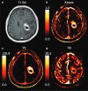
The contrast‐enhanced T1‐weighted image (a) and dynamic contrast‐enhanced‐derived vascular parameter maps: K trans (b), V e (c), and V p (d) of a glioblastoma patient treated with concurrent radiation therapy and temozolomide chemotherapy.
FIGURE 4
FIGURE 4
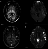
MRI results from a 48‐year‐old patient with a biopsy‐proven grade 4 glioblastoma. (Left‐to‐right) pre‐contrast T1‐weighted; post‐contrast T1‐weighted; T2‐weighted FLAIR images; relative cerebral blood volume (rCBV) map derived from dynamic susceptibility contrast (DSC) sequence; and cerebral blood flow (CBF) map derived from arterial spin labeling (ASL) are shown for a representative image slice. This example illustrates that the ASL CBF map is comparable to DSC rCBV imaging, showing high perfusion values at the periphery of the lesion. Note the partial volume effect on the central portion of the lesion on the CBF map that underestimates the necrotic area.
FIGURE 5
FIGURE 5
Diffusion restriction in a 45‐year‐old female patient with a left temporal high‐grade glioma (HGG) that showed contrast enhancement on T1‐weighted imaging (a; axial non‐contrast and contrast‐enhanced images). On diffusion‐weighted imaging (DWI, b = 1000 s/mm2), the borders of the lesion that primarily overlap with the contrast‐enhancing tumor parts show high signal intensity (b) that spatially corresponded to areas with a signal drop on apparent diffusion coefficient (ADC) maps (c), indicating restricted diffusion most probably due to focal high cellularity of the glioma.
FIGURE 6
FIGURE 6
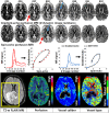
Perfusion MRI and vessel‐architectural imaging (VAI). Merging two perfusion readouts (gradient‐ and spin‐echo MRI) creates unique signal curve “loops” that scale with vessel calibers (slope) and vessel type (loop direction). Note the impaired, venous‐like dominance of the peri‐tumoral area (yellow box) on the vessel type map of a glioblastoma not observed on other images. Adapted from Emblem et al, Nature Medicine 2003.
FIGURE 7
FIGURE 7
Schematic depicting an example of a fingerprint (a) with its corresponding best‐acquired signal and matched dictionary entry (b). Reproduced with permission from Jiang et al Magn Reson Med 2015.
FIGURE 8
FIGURE 8
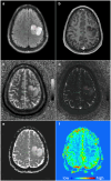
Quantitative maps derived from MRF in an astrocytoma, IDH‐mutant, show increased T1 and T2 values within the tumor mass. The MRF T1 map (c) and T2 map (d) are compared to FLAIR (a), T1w contrast‐enhanced MRI (b), an ADC map (e), and perfusion‐weighted imaging (f). Reproduced from Springer et al 2022 under CC license.

References

    1. Miller KD, Ostrom QT, Kruchko C, et al. Brain and other central nervous system tumor statistics, 2021. CA Cancer J Clin 2021;71:381‐406. - PubMed
    1. Louis DN, Perry A, Wesseling P, et al. The 2021 WHO classification of tumors of the central nervous system: A summary. Neuro Oncol 2021;23:1231‐1251. - PMC - PubMed
    1. Horbinski C, Berger T, Packer RJ, Wen PY. Clinical implications of the 2021 edition of the WHO classification of central nervous system tumours. Nat Rev Neurol 2022;18:515‐529. - PubMed
    1. Louis DN, Perry A, Reifenberger G, et al. The 2016 World Health Organization classification of tumors of the central nervous system: A summary. Acta Neuropathol 2016;131:803‐820. - PubMed
    1. Ellingson BM, Bendszus M, Boxerman J, et al. Consensus recommendations for a standardized brain tumor imaging protocol in clinical trials. Neuro Oncol 2015;17:1188‐1198. - PMC - PubMed

Publication types

MeSH terms