. 2023 Mar 28;42(3):112201.
doi: 10.1016/j.celrep.2023.112201. Epub 2023 Mar 2.

Structural basis of Janus kinase trans-activation

Affiliations

Structural basis of Janus kinase trans-activation

Nathanael A Caveney et al. Cell Rep. .

Abstract

Janus kinases (JAKs) mediate signal transduction downstream of cytokine receptors. Cytokine-dependent dimerization is conveyed across the cell membrane to drive JAK dimerization, trans-phosphorylation, and activation. Activated JAKs in turn phosphorylate receptor intracellular domains (ICDs), resulting in the recruitment, phosphorylation, and activation of signal transducer and activator of transcription (STAT)-family transcription factors. The structural arrangement of a JAK1 dimer complex with IFNλR1 ICD was recently elucidated while bound by stabilizing nanobodies. While this revealed insights into the dimerization-dependent activation of JAKs and the role of oncogenic mutations in this process, the tyrosine kinase (TK) domains were separated by a distance not compatible with the trans-phosphorylation events between the TK domains. Here, we report the cryoelectron microscopy structure of a mouse JAK1 complex in a putative trans-activation state and expand these insights to other physiologically relevant JAK complexes, providing mechanistic insight into the crucial trans-activation step of JAK signaling and allosteric mechanisms of JAK inhibition.

Keywords: CP: Molecular biology; cryo-EM; cytokine; janus kinase; phosphorylation.


Conflict of interest statement

Declaration of interests K.C.G. is the founder of Synthekine. S.R.H. is a co-founder and scientific advisory board member of Ajax Therapeutics, Inc.

Figures
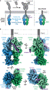
Figure 1.
Figure 1.. Composition and cryo-EM structure of the trans-activation state JAK complex
(A) Cartoon representation of the components of a signaling cytokine receptor complex and the mini-IFNλR1-mJAK1 complex. The two mJAK1 are colored in blue and green, with different shades representing the FERM-SH2, pseudokinase (PK), and tyrosine kinase (TK) domains. Cytokine receptor and mini-IFNλR1 colored in gray and purple. (B) Refined and sharpened cryo-EM density maps of active state mini-IFNλR1-mJAK1 complex, colored as in (A).
Figure 2.
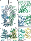
Figure 2.. Structure of the active mini-IFNλR1-mJAK1 complex
(A) Ribbon representation of a model of mini-IFNλR1-mJAK1 complex in an active conformation. mJAK1 colored in blue and green, with different shades representing the FERM-SH2, PK, and TK domains. Regions highlighted in insets (B) and (C) are boxed in gray. (B) Ribbon representation of the PK-TK interaction interface. (C) Ribbon representation of the TK-TK interaction interface, viewed from the bottom of the complex. (D) A structural overlay comparing mini-IFNλR1-mJAK1 active complex to human JAK1 (hJAK1) TK domain crystal structure (PDB: 3EYG). Mini-IFNλR1-mJAK1 colored as in (A)–(C), and hJAK1 colored in yellow and cyan. (E) Ribbon representation of the TK-TK interaction interface of hJAK1 crystal structure, from viewpoint as in (C) and colored as in (D).
Figure 3.
Figure 3.. AlphaFold predictions for JAK homo- and heterodimers
(A) Ribbon representation of a prediction of the PK and TK domains of a human JAK2 homodimer. JAK2 in red and purple, with lighter and darker coloring for PK and TK, respectively. (B) PK interface of the JAK2 homodimer, colored as in (A). Predicted salt bridges depicted with dashed lines. (C) TK interface of the JAK2 homodimer, colored as in (A). (D) Ribbon representation of a prediction of the PK and TK domains of a human JAK1-JAK3 heterodimer. JAK1 in yellow and JAK3 in orange, with lighter and darker coloring for PK and TK, respectively. (E) PK interface of the JAK1-JAK3 heterodimer, colored as in (D). (F) TK interface of the JAK1-JAK3 heterodimer, colored as in (D). Predicted salt bridges depicted with dashed lines. (G) Sequence alignment of key dimerization regions of human JAK1, JAK2, JAK3, and TYK2. Key residues in bold, with basic residues in blue, acidic residues in red, and hydrophobic residues in green. mJAK1 V657F activating mutation marked by an asterisk (*), showing conservation of hydrophobicity at this position among all JAKs.
Figure 4.
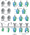
Figure 4.. Structural dynamics of the mini-IFNλR1-mJAK1 complex
(A) 3D variability analysis of the kinase domains of the mini-IFNλR1-mJAK1 complex. Variability components with significant movement and reorientation are shown with their representative initial and final reconstructions. (B) Schematic representations of the movements depicted in (A). Mini-IFNλR1-mJAK1 colored in gray and purple, and mJAK1 colored in blue and green. (C) A schematic representation of the role of the observed flexibility in activation of a cytokine receptor complex. Colored as in (B), with trans-phosphorylation of the TK activation loop represented in red and receptor phosphorylation represented in yellow.
Figure 5.
Figure 5.. Allosteric JAK inhibition via the PK domain
(A) Structural overlay of the mouse JAK1 cryo-EM structure and human TYK2 PK with bound deucravacitinib (PDB: 6NZP). mJAK1 in green and blue, IFNλR1 in purple, and hTYK2 in gray. (B) Structural overlay of the mouse JAK1 cryo-EM structure and human TYK2 PK with bound deucravacitinib. Colored as in (A). Key residues displayed, with mJAK1 trans-PK-TK hydrogen bonding, as predicted by AlphaFold guided modeling, and hTYK2 cis-PK hydrogen bonding from the crystal structure shown with dashed lines. (C) mJAK1 cryo-EM structure in green with AlphaFold modeled PK-TK linker region in gray. C816, which is modified by the inhibitor VVD-118313, is depicted, and the region the compound would bind is colored in red.

References

    1. Philips RL, Wang Y, Cheon H, Kanno Y, Gadina M, Sartorelli V, Horvath CM, Darnell JE, Stark GR, and O’Shea JJ (2022). The JAK-STAT pathway at 30: Much learned, much more to do. Cell 185, 3857–3876. 10.1016/J.CELL.2022.09.023. - DOI - PMC - PubMed
    1. Wilmes S, Hafer M, Vuorio J, Tucker JA, Winkelmann H, Löchte S, Stanly TA, Pulgar Prieto KD, Poojari C, Sharma V, et al. (2020). Mechanism of homodimeric cytokine receptor activation and dysregulation by oncogenic mutations. Science 367, 643–652. 10.1126/science.aaw3242. - DOI - PMC - PubMed
    1. Hubbard SR (2017). Mechanistic insights into regulation of JAK2 tyrosine kinase. Front. Endocrinol 8, 361–367. 10.3389/fendo.2017.00361. - DOI - PMC - PubMed
    1. Bousoik E, and Montazeri Aliabadi H (2018). “Do we know jack” about JAK? A closer look at JAK/STAT signaling pathway. Front. Oncol 8, 287. 10.3389/fonc.2018.00287. - DOI - PMC - PubMed
    1. Lupardus PJ, Skiniotis G, Rice AJ, Thomas C, Fischer S, Walz T, and Garcia KC (2011). Structural snapshots of full-length Jak1, a transmembrane gp130/IL-6/IL-6Rα cytokine receptor complex, and the receptor-Jak1 holocomplex. Structure 19, 45–55. 10.1016/j.str.2010.10.010. - DOI - PMC - PubMed

Publication types