. 2023 Mar 28;42(3):112171.
doi: 10.1016/j.celrep.2023.112171. Epub 2023 Mar 2.

CD169+ macrophage intrinsic IL-10 production regulates immune homeostasis during sepsis

Affiliations

CD169+ macrophage intrinsic IL-10 production regulates immune homeostasis during sepsis

Stephen T Yeung et al. Cell Rep. .

Abstract

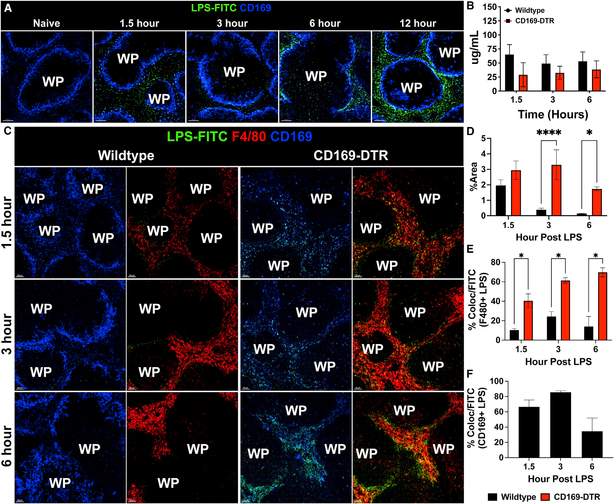
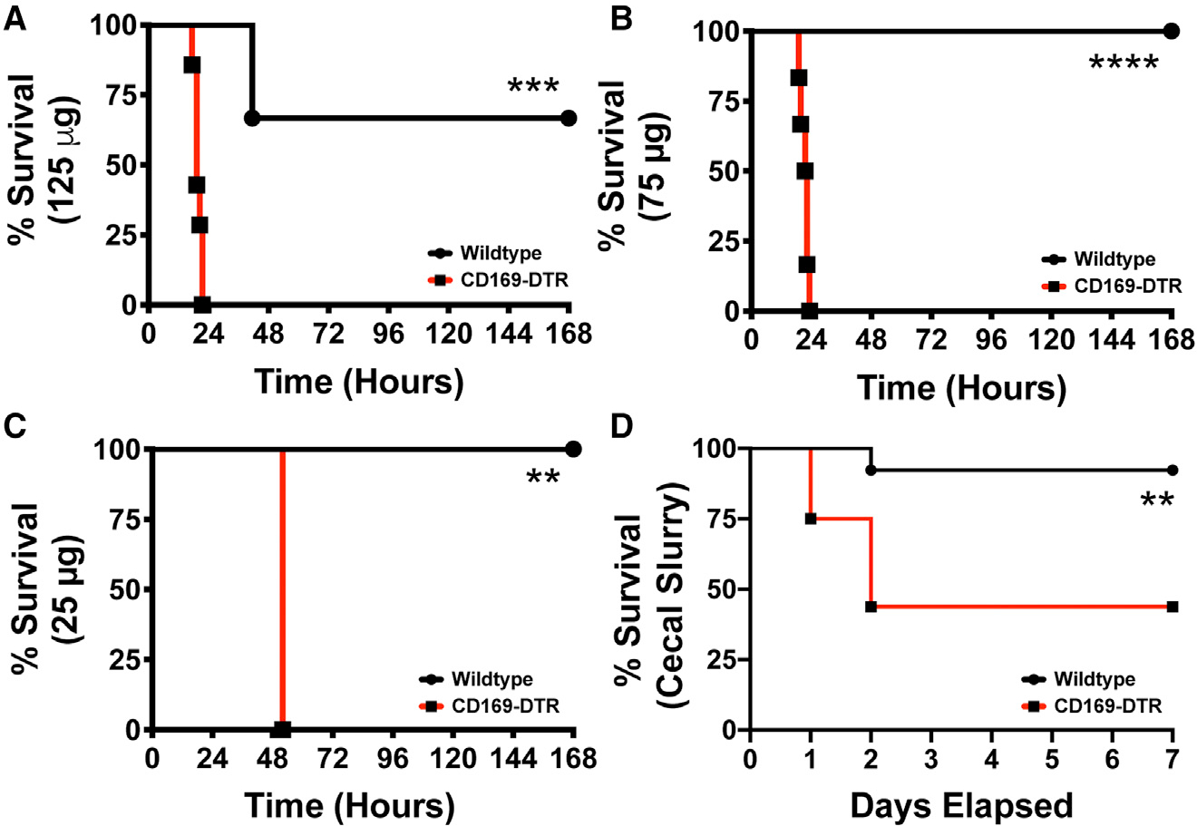
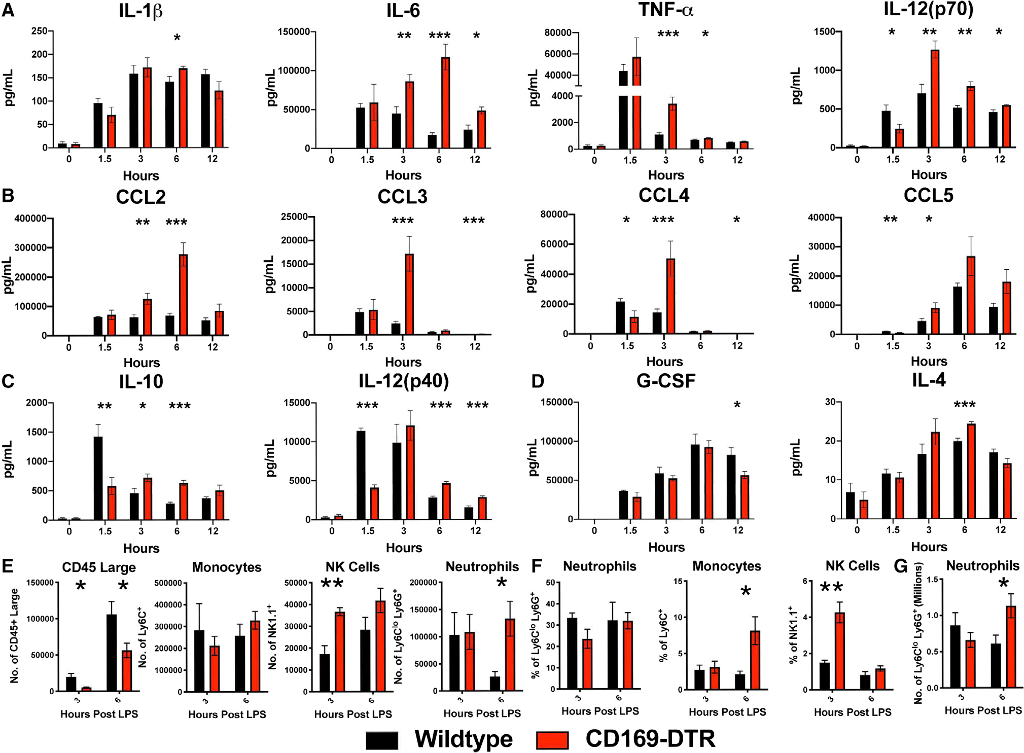
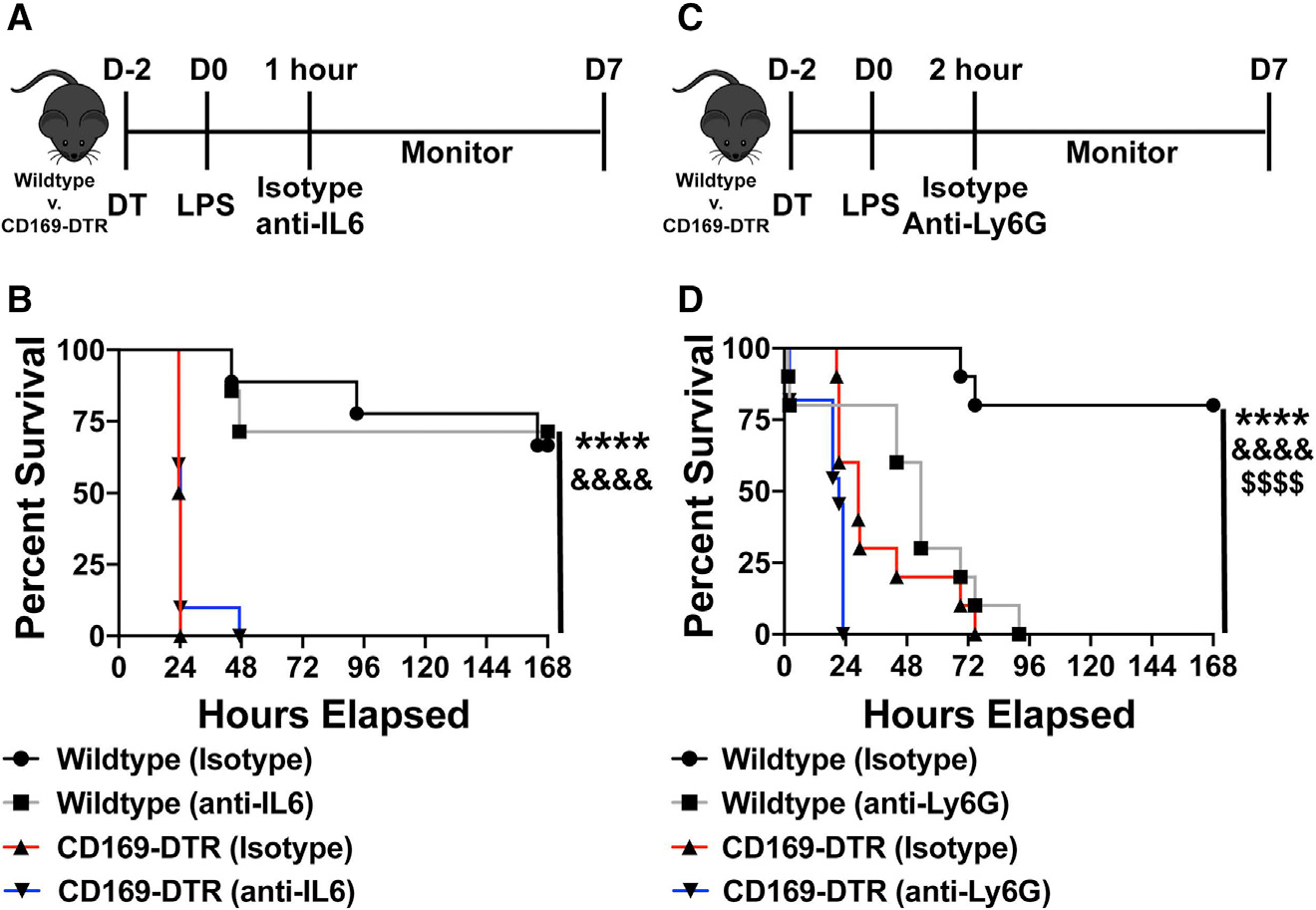
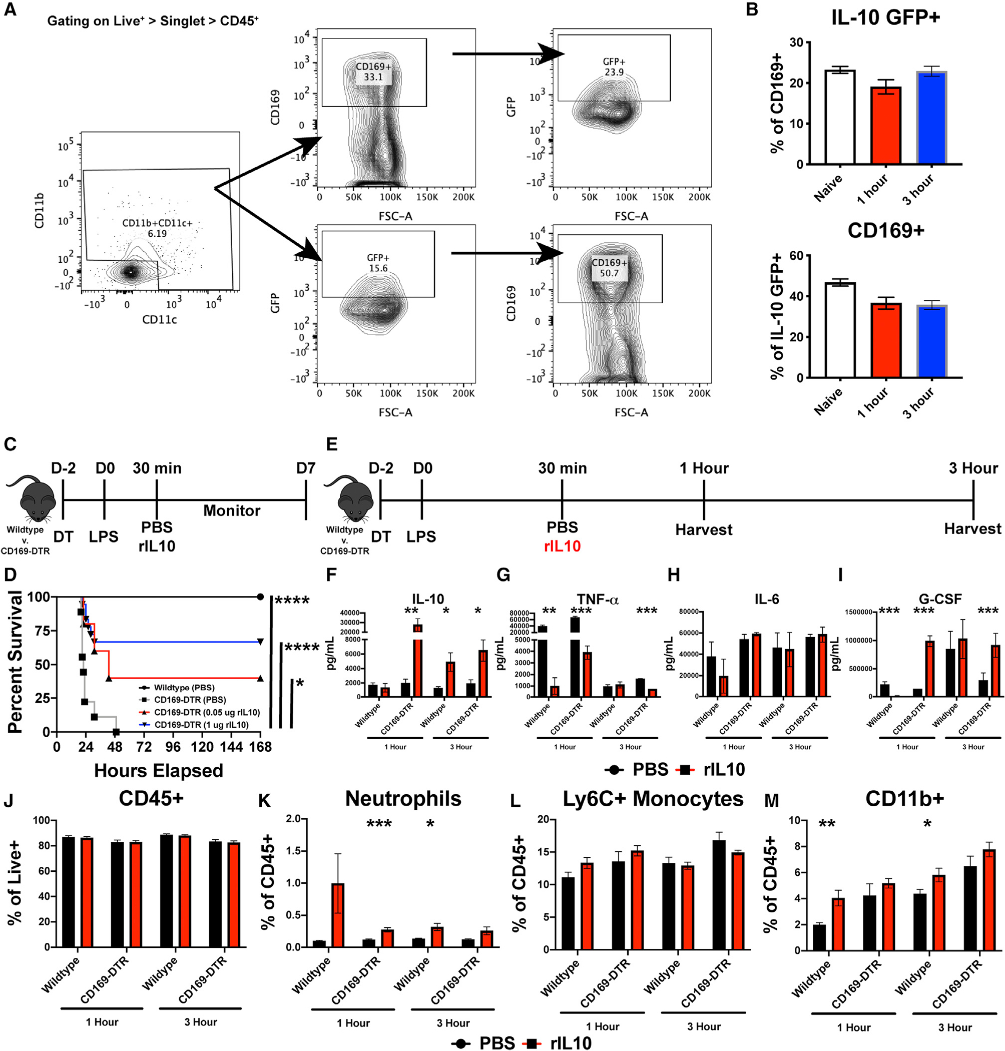
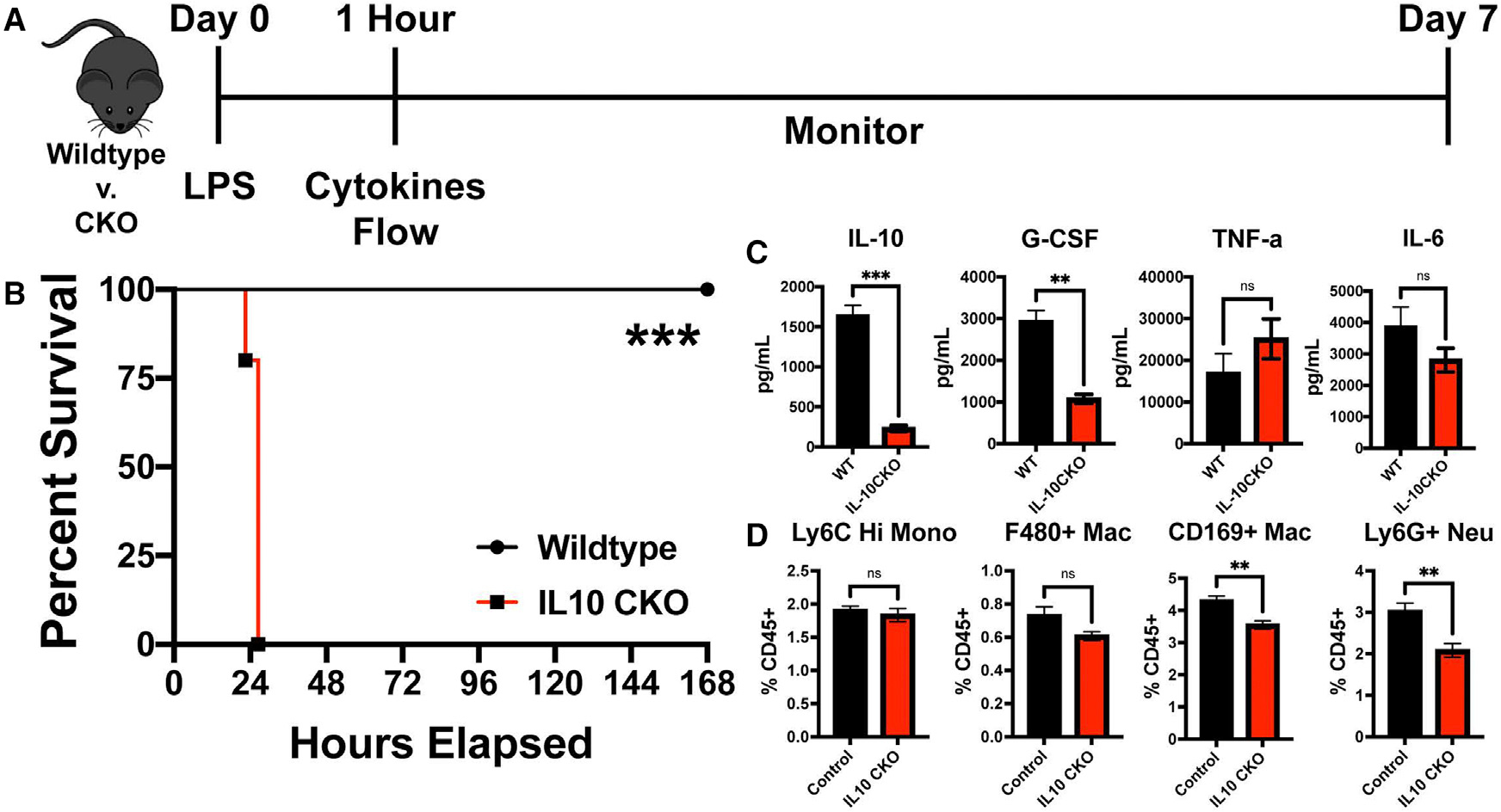
Macrophages facilitate critical functions in regulating pathogen clearance and immune homeostasis in tissues. The remarkable functional diversity exhibited by macrophage subsets is dependent on tissue environment and the nature of the pathological insult. Our current knowledge of the mechanisms that regulate the multifaceted counter-inflammatory responses mediated by macrophages remains incomplete. Here, we report that CD169+ macrophage subsets are necessary for protection under excessive inflammatory conditions. We show that in the absence of these macrophages, even under mild septic conditions, mice fail to survive and exhibit increased production of inflammatory cytokines. Mechanistically, CD169+ macrophages control inflammatory responses via interleukin-10 (IL-10), as CD169+ macrophage-specific deletion of IL-10 was lethal during septic conditions, and recombinant IL-10 treatment reduced lipopolysaccharide (LPS)-induced lethality in mice lacking CD169+ macrophages. Collectively, our findings show a pivotal homeostatic role for CD169+ macrophages and suggest they may serve as an important target for therapy under damaging inflammatory conditions.

Keywords: CD169; CP: Immunology; G-CSF; IL-10; LPS; Siglec-1; macrophages; septic shock.


Conflict of interest statement

Declaration of interests The authors declare no competing interests.

Figures

Figure 1.
Figure 1.. CD169+ macrophages closely associate with LPS and regulate LPS clearance from tissue
(A) Confocal imaging of splenic sections immunostained with CD169 (blue) and FITC-LPS (green) at various time points from naive to 12 h post 200 μg LPS treatment. Scale bar: 80 um. (B) LAL assay to quantify free LPS in serum. (C) Immunofluorescence staining in the spleen from WT (left) and CD169-DTR (right) mice at 1.5 (top), 3 (middle), and 6 (bottom) h post 75 μg LPS treatment. Tissue were stained for CD169 (blue), F4/80 (red), and LPS (green). Scale bar: 50 um (D) Percentage of area of the spleen occupied by LPS at the indicated time after LPS treatment between WT (black) and CD169-DTR (red) mice. (E) Percentage of LPS colocalized with F4/80+ macrophages at the indicated time points after LPS treatment between WT (black) and CD169-DTR (red) mice. (F) Percentage of LPS colocalized with CD169+ macrophages at the indicated time points after LPS treatment in WT mice. The images and graphs are representative of 2–4 independent experiments. WP, white pulp. **p < 0.0, *p < 0.001, p < 0.01. n = 3–5/group ad of 2 independent experiments.
Figure 2.
Figure 2.. CD169+ macrophages are required for the host survival during sepsis
(A–C) WT (black) vs. CD169-DTR (red) mice challenged with Escherichia coli O111:B4 phenol LPS (A) 125, (B) 75, and (C) 25 μg O111:B4 Phenol LPS. (D) Survival curve of WT (black) and CD169-DTR (red) mice following 150 μL cecal slurry injection. **p < 0.01, ***p < 0.001, ****p < 0.0001. n = 3–5/group and of 2 independent experiments.
Figure 3.
Figure 3.. CD169-DTR mice exhibit increased pro-inflammatory cytokines and chemokines, impaired anti-inflammatory cytokines, and infiltration of pro-inflammatory cells in the peritoneal cavity, spleen, and circulation
(A–D) Cytokines and chemokines levels between WT (black) and CD169-DTR (red) mice challenged with 75 μg LPS from E. coli O111:B4 intraperitoneally (i.p.). (A) Pro-inflammatory cytokines (left to right) IL-1β, IL-6, TNF-α, and IL-12(p70), (B) chemokines (left to right) CCL2, CCL3, CCL4, and CCL5, and (C) anti-inflammatory cytokines IL-10 and IL-12(p40) and (D) G-CSF and IL-4. (E–G) Time course cellularity of (E) peritoneal exudate cells (PECs), (F) blood, and (G) spleen of WT (black) and CD169-DTR (red) mice 3 and 6 h post treatment with 75 μg LPS from E. coli O111:B4 i.p. *p < 0.05, **p < 0.01, ***p < 0.001. n = 3–5/group and of 2 independent experiments.
Figure 4.
Figure 4.. Neutrophil depletion or IL-6 neutralization fails to rescue CD169-DTR susceptibility to LPS
(A) Experimental timeline of IL-6 neutralization following LPS challenge. (B) Survival curve between WT and CD169-DTR mice given isotype control or anti-IL-6 antibody. (C) Experimental timeline of neutrophil depletion following LPS challenge. (D) Survival curve between WT and CD169-DTR mice given isotype control or anti-Ly6G antibody. ****p < 0.0001, &&&&p < 0.0001, $$$$p < 0.0001. n = 3–5/group and of 2 independent experiments.
Figure 5.
Figure 5.. CD169+ macrophages actively produce IL-10, and recombinant IL-10 is sufficient to rescue CD169-DTR sensitivity to LPS
(A) Flow cytometry gating strategy from IL-10-GFP mice. (B) Percentage of CD169+ cells that are IL-10 GFP+ and IL-10 GFP+ cells that are CD169+ (top) and percentage of IL-10 GFP+ cells that are CD169+ (bottom). (C) Experimental timeline of recombinant IL-10 (rIL-10) treatment following LPS challenge. (D) Survival curve between WT and CD169-DTR mice following 75 μg LPS and dose-dependent rIL-10 treatment. (E) Experimental timeline of organ harvest at 1 and 3 h post LPS stimulation. (F–I) Cytokine levels following 1 and 3 h post LPS and 1 μg rIL-10, (F) IL-10, (G) TNF-α, (H) IL-6, and (I) G-CSF. (J–M) Flow cytometric analysis following 1 and 3 h post LPS and 1 μg rIL-10, (J) CD45+, (K) neutrophils, (L) Ly6C + monocytes, and (M) CD11b+ myeloid cells. Scale bar: 80 μm. *p < 0.05, **p < 0.01, ***p < 0.001, ****p < 0.0001. n = 3–5/group and of 2 independent experiments.
Figure 6.
Figure 6.. IL-10 production by CD169+ macrophages is required for LPS-induced sepsis protection
(A) Experimental timeline of LPS challenge of control vs. CD169-IL-10 CKO (IL-10 CKO). (B) Survival curve between control and IL-10 CKO mice following 75 μg LPS. (C) (Left to right) IL-10, G-CSF, TNF-α, and IL-6 cytokine levels following 1 h post LPS between control vs. IL-10 CKO. (D) (Left to right) Splenic Ly6C Hi monocytes, F4/80+ macrophages, CD169+ macrophages, and Ly6G+ neutrophil flow cytometric analysis following 1 h post LPS between control vs. IL-10 CKO mice. **p < 0.01, ***p < 0.001, ns, not significant. n = 3–5/group and of 2 independent experiments.

References

    1. Amit I, Winter DR, and Jung S (2016). The role of the local environment and epigenetics in shaping macrophage identity and their effect on tissue homeostasis. Nat. Immunol. 17, 18–25. 10.1038/ni.3325. - DOI - PubMed
    1. Blériot C, Dupuis T, Jouvion G, Eberl G, Disson O, and Lecuit M (2015). Liver-resident macrophage necroptosis orchestrates type 1 microbicidal inflammation and type-2-mediated tissue repair during bacterial infection. Immunity 42, 145–158. - PubMed
    1. Glass CK, and Natoli G (2016). Molecular control of activation and priming in macrophages. Nat. Immunol. 17, 26–33. 10.1038/ni.3306. - DOI - PMC - PubMed
    1. Kierdorf K, Prinz M, Geissmann F, and Gomez Perdiguero E (2015). Development and function of tissue resident macrophages in mice. Semin. Immunol. 27, 369–378. 10.1016/j.smim.2016.03.017. - DOI - PMC - PubMed
    1. Asano K, Takahashi N, Ushiki M, Monya M, Aihara F, Kuboki E, Moriyama S, Iida M, Kitamura H, Qiu CH, et al. (2015). Intestinal CD169(+) macrophages initiate mucosal inflammation by secreting CCL8 that recruits inflammatory monocytes. Nat. Commun. 6, 7802. 10.1038/ncomms8802. - DOI - PMC - PubMed

Publication types