. 2023 Mar 3;19(3):e1011171.
doi: 10.1371/journal.ppat.1011171. eCollection 2023 Mar.

HPV upregulates MARCHF8 ubiquitin ligase and inhibits apoptosis by degrading the death receptors in head and neck cancer

Affiliations

HPV upregulates MARCHF8 ubiquitin ligase and inhibits apoptosis by degrading the death receptors in head and neck cancer

Mohamed I Khalil et al. PLoS Pathog. .

Abstract

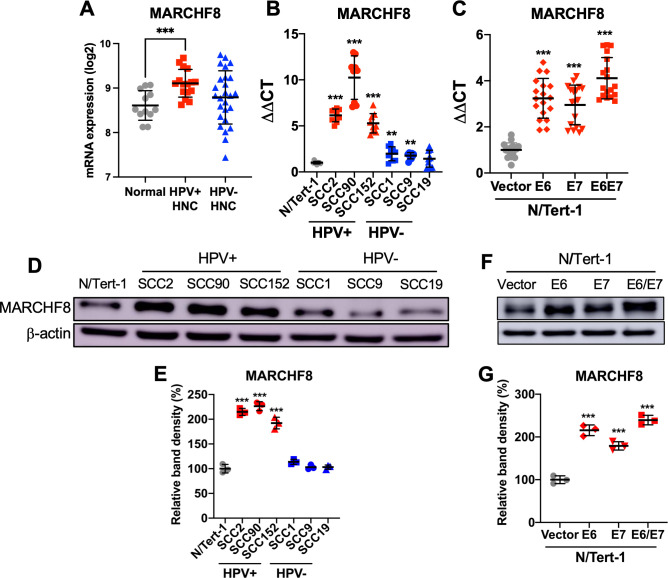
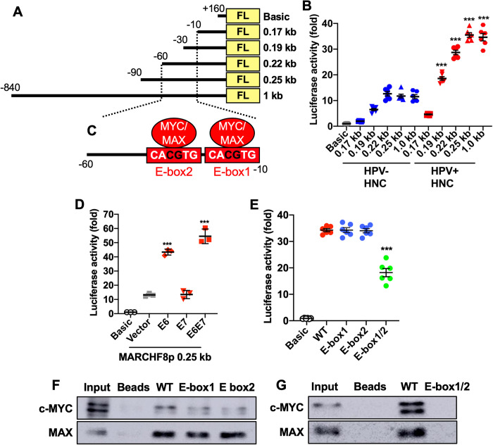
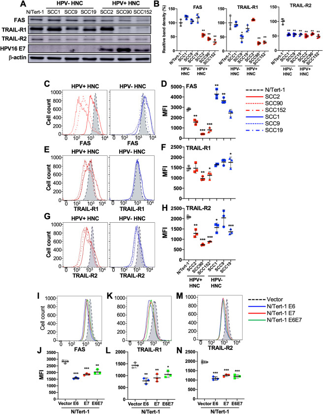
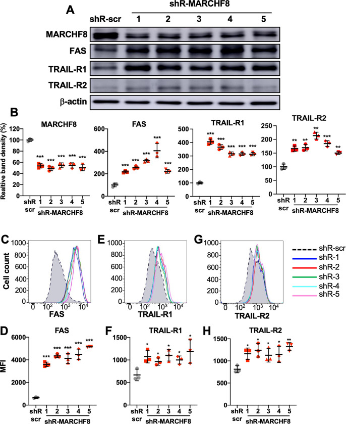
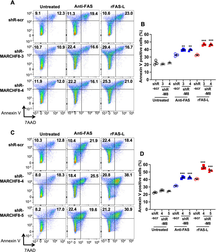
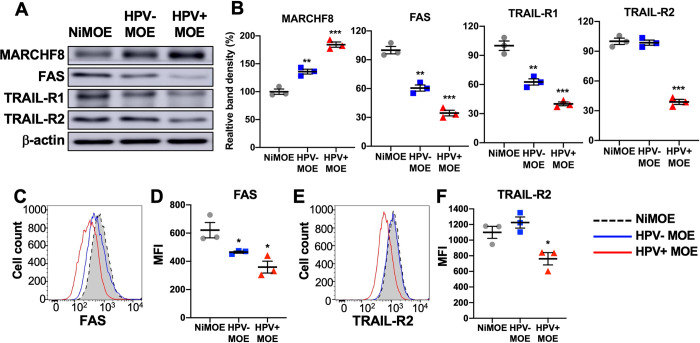
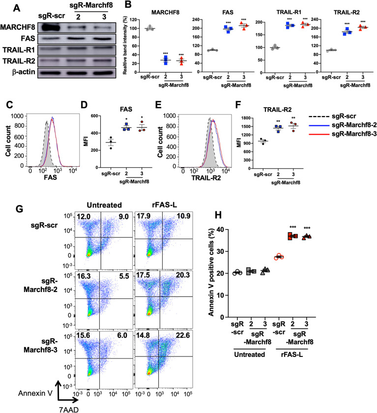
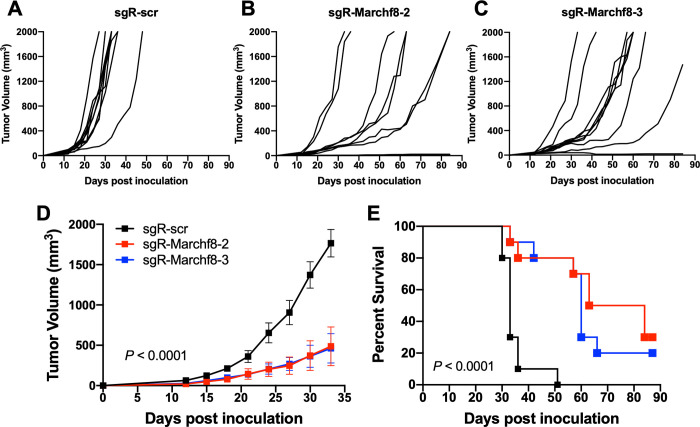
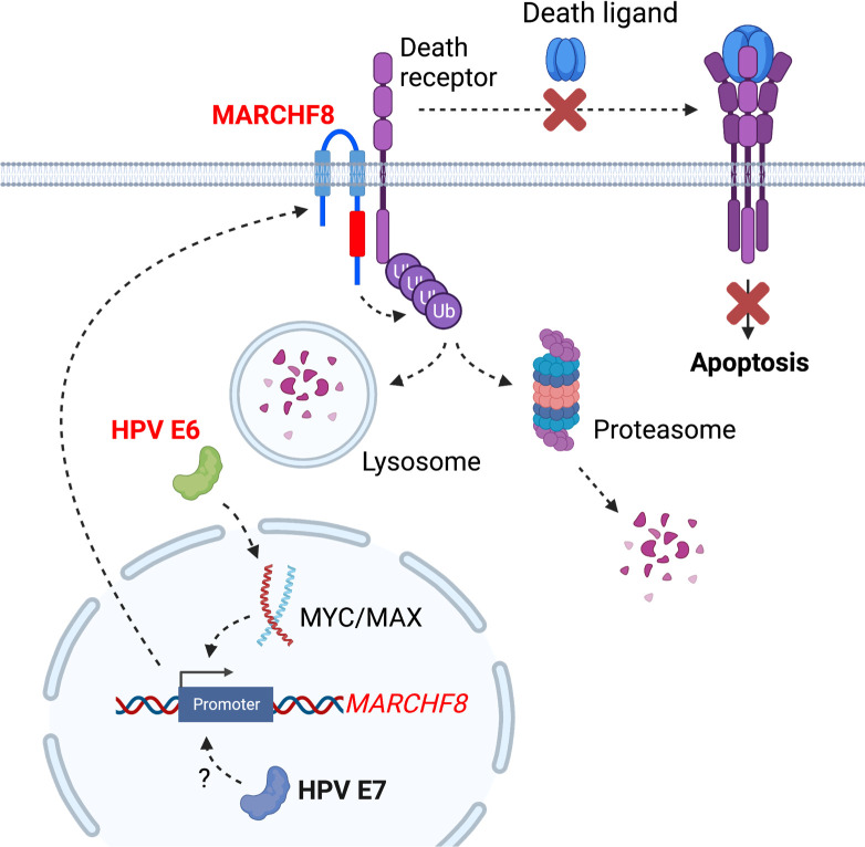
The membrane-associated RING-CH-type finger ubiquitin ligase MARCHF8 is a human homolog of the viral ubiquitin ligases Kaposi's sarcoma herpesvirus K3 and K5 that promote host immune evasion. Previous studies have shown that MARCHF8 ubiquitinates several immune receptors, such as the major histocompatibility complex II and CD86. While human papillomavirus (HPV) does not encode any ubiquitin ligase, the viral oncoproteins E6 and E7 are known to regulate host ubiquitin ligases. Here, we report that MARCHF8 expression is upregulated in HPV-positive head and neck cancer (HNC) patients but not in HPV-negative HNC patients compared to normal individuals. The MARCHF8 promoter is highly activated by HPV oncoprotein E6-induced MYC/MAX transcriptional activation. The knockdown of MARCHF8 expression in human HPV-positive HNC cells restores cell surface expression of the tumor necrosis factor receptor superfamily (TNFRSF) death receptors, FAS, TRAIL-R1, and TRAIL-R2, and enhances apoptosis. MARCHF8 protein directly interacts with and ubiquitinates the TNFRSF death receptors. Further, MARCHF8 knockout in mouse oral cancer cells expressing HPV16 E6 and E7 augments cancer cell apoptosis and suppresses tumor growth in vivo. Our findings suggest that HPV inhibits host cell apoptosis by upregulating MARCHF8 and degrading TNFRSF death receptors in HPV-positive HNC cells.


Conflict of interest statement

The authors have declared that no competing interests exist.

Figures

Fig 1
Fig 1. MARCHF8 expression is upregulated in HPV+ HNC.
MARCHF8 mRNA expression levels in microdissected human tissue samples from HPV+ (n = 16) and HPV- (n = 26) HNC patients, and normal individuals (n = 12) were analyzed using our previous gene expression data (GSE6791) [28] and shown as fluorescence intensity (log2) (A). The MARCHF8 mRNA expression was validated in normal (N/Tert-1), HPV+ HNC (SCC2, SCC90, and SCC152), and HPV- HNC (SCC1, SCC9, and SCC19) cells (B) by RT-qPCR. MARCHF8 mRNA levels were determined in N/Tert-1 cells expressing E6 and/or E7 by RT-qPCR (C). The data shown are ΔΔCT values normalized by the GAPDH mRNA level as an internal control. MARCHF8 protein levels were determined in HPV+ and HPV- HNC cells (D and E) and N/Tert-1 cells expressing HPV16 E6, E7, or E6 and E7 (F and G) by western blotting. β-actin was used as a loading control. The relative band density was quantified using the NIH ImageJ program (E and G). All experiments were repeated at least three times, and the data shown are means ± SD. P values were determined by Student’s t-test. *p < 0.05, **p < 0.01, ***p < 0.001.
Fig 2
Fig 2. The HPV oncoprotein E6 induces the MARCHF8 promoter activity mediated by the MYC/MAX complex.
Schematic representation of the MARCHF8 promoter regions (-840 to +160) in the firefly luciferase (FL) reporter plasmid pGL4.2 (A) and two E-boxes (C). The promoter-reporter constructs were transfected into HPV+ (SCC152) and HPV- (SCC1) cells (B) and cotransfected into SCC1 cells with HPV16 E6, E7, or E6 and E7 expression plasmids (D). SCC152 cells were transfected with the 0.25 kb promoter (-90 to +160) reporter constructs of wildtype (WT) and E-box mutants containing single or double CG deletion in E-box1, E-box-2, and E-box1/2 (B and E). Luciferase activity was measured 48 h post transfection. Representative data from three independent experiments are shown as a fold change relative to the empty pGL4.2 vector (Basic). Shown are representative data of three repeats. P values were determined by Student’s t-test. *p < 0.05, **p < 0.01, ***p < 0.001. A DNA-protein pulldown assay was performed using biotinylated 90 bp oligonucleotides containing wildtype or E-boxes mutants (-85 to +5) incubated with nuclear extracts from SCC152 cells. The DNA-bound proteins were analyzed using anti-c-MYC and MAX antibodies (F and G) by western blotting.
Fig 3
Fig 3. Expression of FAS, TRAIL-R1, and TRAIL-R2 is downregulated in HPV+ HNC cells.
Total protein expression of FAS, TRAIL-R1, and TRAIL-R2 in HPV- (SCC1, SCC9, and SCC19) and HPV+ (SCC2, SCC90, and SCC152) HNC cells were determined by western blotting (A). Relative band density was quantified using NIH ImageJ (B). HPV16 E7 and β-actin were used as viral and internal controls, respectively. Cell surface expression of FAS (C and D), TRAIL-R1 (E and F), and TRAIL-R2 (G and H) proteins on HPV+ (SCC2, SCC90, and SCC152) and HPV- (SCC1, SCC9, and SCC19) HNC cells was analyzed by flow cytometry. Cell surface expression of FAS (I and J), TRAIL-R1 (K and L), and TRAIL-R2 (M and N) proteins on N/Tert-1 cells expressing HPV16 E6, E7, and E6E7 was analyzed by flow cytometry. Mean fluorescence intensities (MFI) of three independent experiments are shown (D, F, H, J, L, and N). All experiments were repeated at least three times, and the data shown are means ± SD. P values were determined by Student’s t-test. *p < 0.05, **p < 0.01, ***p < 0.001.
Fig 4
Fig 4. Knockdown of MARCHF8 expression increases FAS, TRAIL-R1, and TRAIL-R2 protein expression in HPV+ HNC cells.
HPV+ HNC (SCC152) cells were transduced with one of five lentiviral shRNAs against MARCHF8 (shR-MARCHF8 clones 1–5) or scrambled shRNA (shR-scr) as a control. Protein expression of MARCHF8, FAS, TRAIL-R1, and TRAIL-R2 was determined by western blotting (A). Relative band density was quantified using NIH ImageJ (B). β-actin was used as an internal control. The data shown are means ± SD of three independent experiments. Cell surface expression of FAS (C and D), TRAIL-R1 (E and F), and TRAIL-R2 (G and H) proteins were analyzed by flow cytometry. Mean fluorescence intensities (MFI) of three independent experiments are shown (D, F, and H). P values were determined by Student’s t-test. *p < 0.05, **p < 0.01, ***p < 0.001.
Fig 5
Fig 5. MARCHF8 protein interacts with and ubiquitinates FAS, TRAIL-R1, and TRAIL-R2 proteins.
MARCHF8 (A) and FAS (B) were pulled down from the cell lysate of HPV+ HNC (SCC152) cells treated with a proteasome inhibitor MG132 using anti-MARCHF8 (A) and anti-FAS (B) antibodies, respectively. Western blotting detected FAS, TRAIL-R1, TRAIL-R2, and MARCHF8 proteins in the immunoprecipitated proteins. Ubiquitinated proteins were pulled down from the cell lysate of HPV+ HNC (SCC152) cells with scrambled shRNA (shR-scr) or shRNA against MARCHF8 (shR-MARCHF8 clone 3) treated with a proteasome inhibitor MG132 using an anti-ubiquitin antibody (C—H). FAS (C and F), TRAIL-R1 (D and G), and TRAIL-R2 (E and H) proteins were detected in the immunoprecipitated proteins by western blotting. Relative band density was quantified using ImageJ. The band densities of the shR-MARCHF8 are normalized to the shR-scr. The data shown are means ± SD from three repeats (Figs 5C–5E and S7A–S7F). P values were determined by Student’s t-test. *p < 0.05, **p < 0.01, ***p < 0.001.
Fig 6
Fig 6. Knockdown of MARCHF8 enhances apoptosis of HPV+ HNC cells.
Two HPV+ HNC cells, SCC152 (A and B) and SCC2 (C and D), with scrambled shRNA (shR-scr) or two shRNAs against MARCHF8 (shR-MARCHF8), were treated with an anti-human FAS antibody (Anti-FAS, clone EOS9.1, eBioscience) or the recombinant FAS ligand (rFAS-L, BioLegend #585404). The cells were stained with an anti-annexin V antibody and 7-AAD and analyzed by flow cytometry. The percentage of cells with positive staining is indicated (A and C). The data shown are means ± SD of three independent experiments (B and D). P values were determined by Student’s t-test. **p < 0.01, ***p < 0.001.
Fig 7
Fig 7. MARCHF8 is upregulated, and death receptor expression is downregulated in HPV+ mouse oral cancer cells.
Mouse MARCHF8, FAS, TRAIL-R1, and TRAIL-R2 protein levels in mouse normal immortalized (NiMOE), HPV- transformed (HPV- MOE), and HPV+ transformed (HPV+ MOE) oral epithelial cells were determined by western blotting (A). β-actin was used as a loading control. The relative band density was quantified using NIH ImageJ (B). Cell surface expression of FAS (C and D) and TRAIL-R2 (E and F) proteins on NiMOE (dotted black line), HPV- MOE (blue line), and HPV+ MOE (red line) cells were analyzed by flow cytometry. Mean fluorescence intensities (MFI) of three independent experiments are shown (D and F). All experiments were repeated at least three times, and the data shown are means ± SD. P values were determined by Student’s t-test. *p < 0.05, ***p < 0.001.
Fig 8
Fig 8. Knockout of Marchf8 expression increases FAS, TRAIL-R1, and TRAIL-R2 protein levels and enhances apoptosis of HPV+ mouse oral cancer cells.
mEERL cells were transduced with lentiviral Cas9 and one of two sgRNAs against Marchf8 (sgR-Marchf8-2 and sgR-Marchf8-3) or scrambled sgRNA (sgR-scr). Protein levels of MARCHF8, FAS, TRAIL-R1, and TRAIL-R2 were determined by western blotting (A). Relative band density was quantified using NIH ImageJ (B). β-actin was used as a loading control. The data shown are means ± SD of three independent experiments. Cell surface expression of FAS (C and D) and TRAIL-R2 (E and F) proteins were analyzed by flow cytometry. Mean fluorescence intensities (MFI) of three independent experiments are shown (D and F). P values were determined by Student’s t-test. *p < 0.05, **p < 0.01, ***p < 0.001. Untreated and rFAS-L-treated mEERL cells (G) with sgR-Marchf8-2, sgR-Marchf8-3, or sgR-scr were stained with an anti-annexin V antibody and 7-AAD and analyzed by flow cytometry. The percentage of cells with positive staining is indicated (G). The data shown are means ± SD of three independent experiments (H). P values were determined by Student’s t-test. ***p < 0.001.
Fig 9
Fig 9. Knockout of Marchf8 expression suppresses HPV+ HNC tumor growth in vivo.
Marchf8-knockout mEERL cells (mEERL/Marchf8-/-) were generated by lentiviral Cas9, and two sgRNAs targeting Marchf8 (sgR-Marchf8-2 and sgR-Marchf8-3) or scrambled sgRNA (sgR-scr). mEERL/scr (A) or mEERL/Marchf8-/- (B and C) cells were injected into the rear right flank of C57BL/6J mice (n = 10 per group). Tumor volume was measured twice a week (A-D). Survival rates of mice were analyzed using a Kaplan-Meier estimator (E). The time to event was determined for each group, with the event defined as a tumor size larger than 2000 mm3. The data shown are means ± SD. P values of mice injected with mEERL/Marchf8-/- cells compared with mice injected with mEERL/scr cells were determined for tumor growth (D) and survival (E) by two-way ANOVA analysis. Shown are representative of two independent experiments.
Fig 10
Fig 10. The schematic diagram summarizes that HPV E6-induced MARCHF8 expression inhibits host cell apoptosis by degrading the TNFRSF death receptors.
The HPV oncoprotein E6 activates the MARCHF8 promoter activity through the MYC/MAX transcription factor complex (A) and upregulates cell surface expression of the MARCHF8 protein (B). MARCHF8 protein binds to and ubiquitinates the TNFRSF death receptors FAS, TRAIL-R1, and TRAIL-R2 (C). Ubiquitinated TNFRSF death receptors may be degraded by lysosomes (D) or proteasomes in the cytoplasm (E). Created with BioRender.com.

Similar articles

Cited by

References

    1. Gillison ML, Koch WM, Capone RB, Spafford M, Westra WH, Wu L, et al.. Evidence for a causal association between human papillomavirus and a subset of head and neck cancers. J Natl Cancer Inst. 2000;92: 709–20. doi: 10.1093/jnci/92.9.709 - DOI - PubMed
    1. Powell SF, Vu L, Spanos WC, Pyeon D. The Key Differences between Human Papillomavirus-Positive and -Negative Head and Neck Cancers: Biological and Clinical Implications. Cancers (Basel). 2021;13. doi: 10.3390/cancers13205206 - DOI - PMC - PubMed
    1. Yu X, Xu J, Xu D, Bi X, Wang H, Lu Y, et al.. Comprehensive Analysis of the Carcinogenic Process, Tumor Microenvironment, and Drug Response in HPV-Positive Cancers. Front Oncol. 2022;12: 842060. doi: 10.3389/fonc.2022.842060 - DOI - PMC - PubMed
    1. Chow LQM. Head and Neck Cancer. N Engl J Med. 2020;382: 60–72. doi: 10.1056/NEJMra1715715 - DOI - PubMed
    1. Chaturvedi AK, Freedman ND, Abnet CC. The Evolving Epidemiology of Oral Cavity and Oropharyngeal Cancers. Cancer Res. 2022;82: 2821–2823. doi: 10.1158/0008-5472.CAN-22-2124 - DOI - PubMed

Publication types