. 2023 Mar 16;186(6):1263-1278.e20.
doi: 10.1016/j.cell.2023.02.001. Epub 2023 Feb 13.

A pseudovirus system enables deep mutational scanning of the full SARS-CoV-2 spike

Affiliations

A pseudovirus system enables deep mutational scanning of the full SARS-CoV-2 spike

Bernadeta Dadonaite et al. Cell. .

Abstract

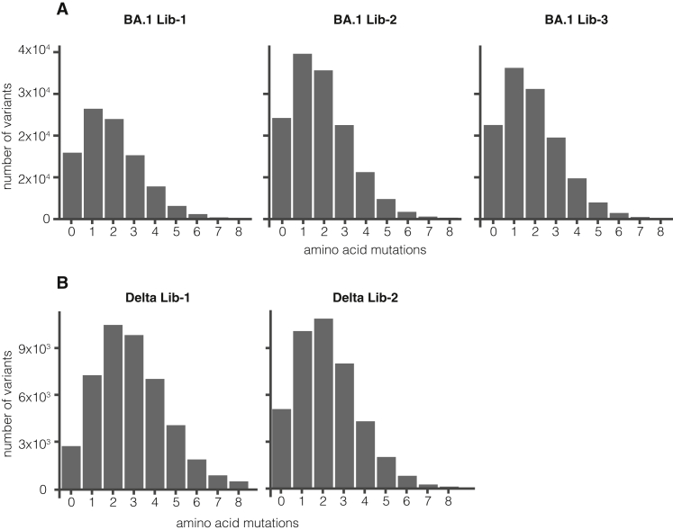
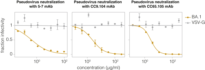
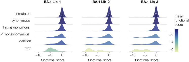
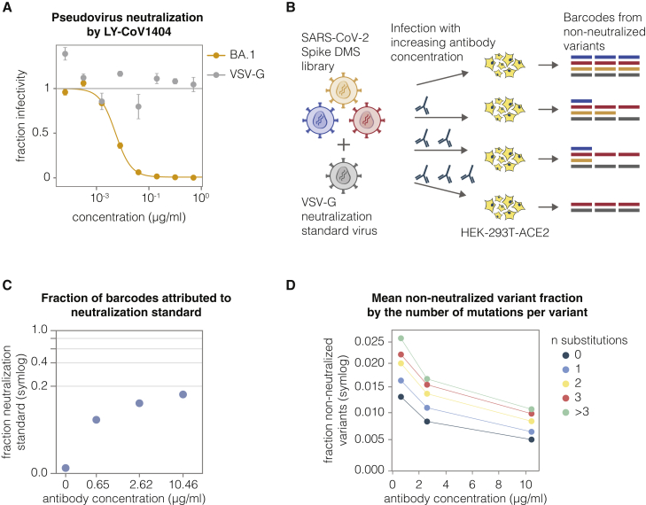
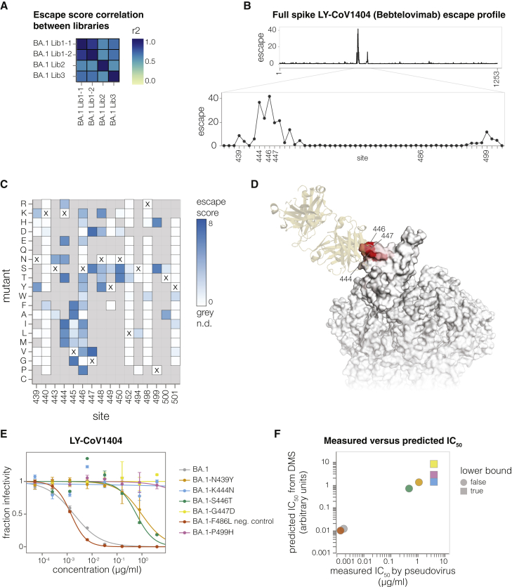
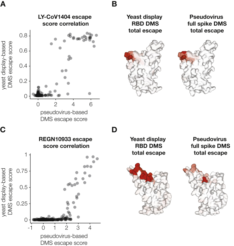
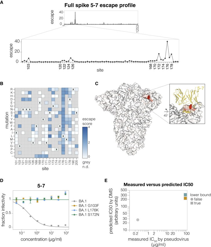
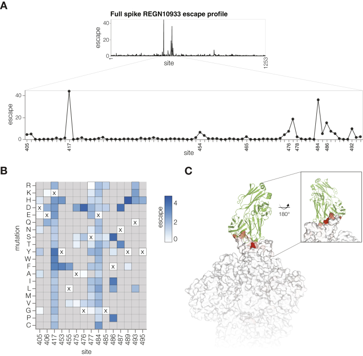
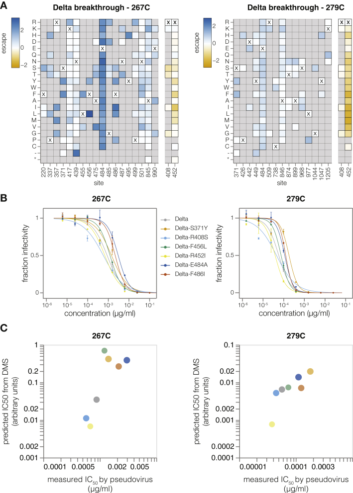
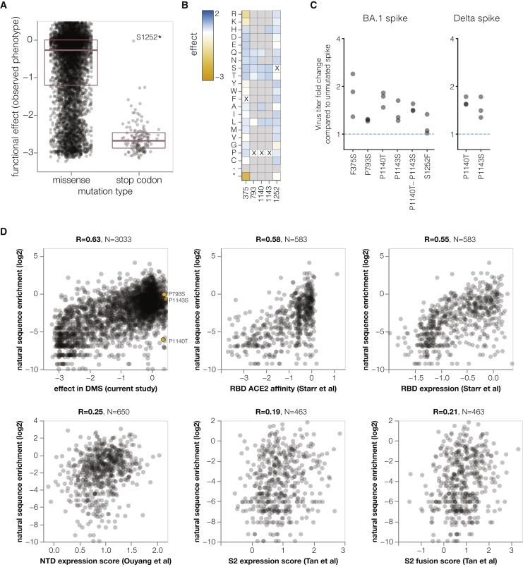
A major challenge in understanding SARS-CoV-2 evolution is interpreting the antigenic and functional effects of emerging mutations in the viral spike protein. Here, we describe a deep mutational scanning platform based on non-replicative pseudotyped lentiviruses that directly quantifies how large numbers of spike mutations impact antibody neutralization and pseudovirus infection. We apply this platform to produce libraries of the Omicron BA.1 and Delta spikes. These libraries each contain ∼7,000 distinct amino acid mutations in the context of up to ∼135,000 unique mutation combinations. We use these libraries to map escape mutations from neutralizing antibodies targeting the receptor-binding domain, N-terminal domain, and S2 subunit of spike. Overall, this work establishes a high-throughput and safe approach to measure how ∼105 combinations of mutations affect antibody neutralization and spike-mediated infection. Notably, the platform described here can be extended to the entry proteins of many other viruses.

Keywords: BA.1; Delta; Omicron; SARS-CoV-2; antibody escape; antibody neutralization; deep mutational scanning; pseudovirus; spike.


Conflict of interest statement

Declaration of interests J.D.B. is on the scientific advisory board of Apriori Bio, Aerium Therapeutics, Invivyd, the Vaccine Company, and Oncorus and has recently consulted on topics related to viral evolution for Moderna and Merck. J.D.B., K.H.D.C., and C.E.R. receive royalty payments as inventors on Fred Hutch licensed patents related to viral deep mutational scanning. J.D.B., K.H.D.C., C.E.R., and B.D. are inventors on a pending patent application relating to the viral deep mutational scanning system described in this paper. R.B. is a consultant for IAVI, Adagio, Adimab, Mabloc, VosBio, Nonigenex, and Radiant. D.D.H. is a co-founder of TaiMed Biologics and RenBio, consultant to WuXi Biologics and Brii Biosciences, and board director for Vicarious Surgical. H.Y.C. consulted with Ellume, Pfizer, The Bill and Melinda Gates Foundation, GlaxoSmithKline, and Merck. H.Y.C. received research funding from Gates Ventures, Sanofi Pasteur, and support and reagents from Ellume and Cepheid outside of the submitted work.

Figures

None
Graphical abstract
Figure S1
Figure S1
Pseudovirus titers from phenotype-genotype linked lentiviruses, related to Figure 1 (A) Traditional lentivirus pseudotyping method. The lentivirus backbone used for pseudotyping does not code for the spike gene. To make spike-pseudotyped lentivirus, lentiviral helper plasmids, backbone, and spike expression plasmid are transfected into producer cells to make spike-pseudotyped lentivirus. This method produces lentiviruses that lack a genotype-phenotype link because the spike expressed on the surface of a viral particle is not coded by the lentiviral genome. (B) Delta spike-pseudotyped lentivirus titers. Viruses were produced under indicated conditions from cells with integrated lentivirus genomes carrying Delta spike. Virus titers for conditions used to generate the actual deep mutational scanning libraries are colored red. Viruses were titrated on ACE2-TMPRSS2-HEK-293T cells. (C) BA.1 or Delta spike-pseudotyped lentivirus titers in the presence or absence of amphotericin B (amphoB). BA.1 virus was titrated on ACE2-HEK-293T cells and Delta virus was titrated on ACE2-TMPRSS2-HEK-293T cells.
Figure 1
Figure 1
Deep mutational scanning platform for spike (A) Lentivirus backbone used for deep mutational scanning. The backbone contains functional lentiviral 5′ and 3′ long terminal repeat (LTR) regions. The spike gene is under an inducible tetresponse element 3rd generation (TRE3G) promoter, and there is a 16-nucleotide barcode (BC) downstream of the stop codon. A CMV promoter drives the expression of the reporter ZsGreen gene that is linked to a puromycin resistance gene (PuR) via a T2A linker. The backbone also contains a woodchuck hepatitis virus posttranscriptional regulatory element (WPRE), Rev response element (RRE), and a central polypurine tract (cPPT). (B) Approach for creating genotype-phenotype linked lentivirus libraries. HEK-293T cells are transfected with spike-carrying lentivirus backbone, VSV-G expression plasmid, and lentiviral helper plasmids to generate VSV-G-pseudotyped lentiviruses. These viruses are used to transduce reverse tetracycline-controlled transactivator (rtTA) expressing HEK-293T cells at low multiplicity of infection (MOI), and successfully transduced cells are selected using puromycin. Selected cells can be transfected with helper plasmids and a VSV-G expression plasmid to produce VSV-G-pseudotyped viruses carrying all genomes present in the deep mutational library or selected cells can be induced with doxycycline (dox) to express spike and transfected with only the helper plasmids to generate spike-pseudotyped lentiviruses that have a genotype-phenotype link. (C) Average number of mutations per barcoded spike in BA.1 libraries. (D) Total number of barcoded variants in each BA.1 library. (E) The coverage of intended mutations across all BA.1 libraries. See also Figures S1, S2, S3, S6, and S7.
Figure S2
Figure S2
Characteristics of Delta spike deep mutational scanning libraries and BA.1 library functional scores, related to Figures 1 and 2 (A) Total number of barcoded variants in each Delta library. (B) Coverage of intended mutations across both Delta libraries. (C) Average number of mutations per barcoded spike in Delta libraries. (D) Distribution of functional scores for variants with different types of mutations in the Delta libraries. (E) Distribution of functional scores for variants in BA.1 libraries stratified by whether they only contained mutations “intended” during the library design or also contain other non-intended mutations.
Figure S3
Figure S3
Distribution of the number of amino acid mutations per variant in full spike deep mutational scanning libraries, related to Figure 1 (A) Number of mutations per variant in BA.1 libraries (B) Number of mutations per variant in Delta libraries.
Figure S4
Figure S4
The VSV-G neutralization standard is not neutralized by antibodies 5–7, CC9.104, and CC65.105, related to Figures 3, 4, 5, and 6 Neutralization assays using NTD-targeting 5–7 mAb and S2-targeting CC9.104 and CC65.105 antibodies against lentivirus pseudotyped with BA.1 spike or VSV-G. Error bars indicate standard error between two technical replicates.
Figure 2
Figure 2
Some mutations tend to impair spike-mediated pseudovirus infection For each barcoded spike variant, we compute a functional score that reflects how well that spike mediates pseudovirus infection relative to the unmutated spike: negative scores indicate impaired infection, and positive scores indicate improved infection. The plots show the distribution of functional scores across all variants in each of the three BA.1 libraries for different categories of variants, with each distribution colored by the mean functional score for that variant type. See also Figure S2.
Figure 3
Figure 3
A VSV-G standard enables the measurement of absolute neutralization by deep sequencing (A) Neutralization assay demonstrating that BA.1-spike-pseudotyped lentivirus is neutralized by antibody LY-CoV1404, but the VSV-G-pseudotyped neutralization standard is not. Error bars indicate standard error between two technical replicates. (B) Use of the VSV-G standard to measure absolute neutralization. Deep mutational scanning libraries are mixed with the VSV-G neutralization standard. The virus mixture is incubated with a no-antibody control or increasing antibody concentrations and infected into ACE2-expressing 293T cells. After ∼12 h, viral genomes are recovered, barcodes are sequenced, and absolute neutralization of each variant is computed by comparing its barcode counts to those from the VSV-G standard. (C) Fraction of barcodes derived from the VSV-G neutralization standard in infections with increasing LY-CoV1404 concentrations. (D) BA.1 deep mutational scanning library non-neutralized fractions averaged across variants with different numbers of amino acid mutations at differentLY-CoV1404 concentrations. Note (C) and (D) use a symlog scale. See also Figures S4, S6, and S7.
Figure 4
Figure 4
Antibody LY-CoV1404 escape mapping (A) Correlation of mutation-escape scores between technical replicates (BA1 Lib-1.1 and BA1 Lib-1.2) and biological replicates (BA1 Lib-1, BA1 Lib-2, and BA1 Lib-3). (B) Total escape scores at each site in the BA.1 spike, and a zoomed-in plot showing the key escape sites. Sites of mutations chosen for validation experiments are labeled on the x-axis. (C) Heatmap of mutation-escape scores at key sites. Residues marked with X are the wild-type amino acids in BA.1. Amino acids not present in our libraries are shown in gray. An interactive heatmap for the entirety of spike is at https://dms-vep.github.io/SARS-CoV-2_Omicron_BA.1_spike_DMS_mAbs/LyCoV-1404_escape_plot.html. (D) Surface representation of spike colored by the sum of escape scores at that site. LY-CoV1404 antibody is in yellow. Only the antibody-bound protomer is colored. PDB: 7MMO and PDB: 6XM4 were aligned to make this structure. (E) Validation pseudovirus neutralization assays of the indicated BA.1 spike mutants against the LY-CoV1404 antibody. Error bars indicate standard error between two technical replicates. (F) Correlation between predicted IC50 values from deep mutational scanning (DMS) data versus the IC50 values measured in the validation assays in (E). The points are colored as in (E). Lower bound indicates that the antibody did not neutralize at the highest concentration tested in the validation neutralization assay. Site numbering in all plots is based on the Wuhan-Hu-1 sequence. See also Figures S4 and S5.
Figure S5
Figure S5
Comparison between antibody-escape mapping using full spike pseudovirus deep mutational scanning versus our previously described yeast-display deep mutational scanning of just the RBD, related to Figure 4 (A) Correlation between measured mutation-level escape scores for LY-CoV1404 antibody in pseudovirus and yeast-display deep mutational scanning experiments. Yeast display data is taken from Starr et al.. (B) Surface representation of SARS-CoV-2 RBD colored by the sum of escape scores for LY-CoV1404 antibody at that site. (C) Correlation between measured mutation-level escape scores for REGN10933 antibody in pseudovirus and yeast-display deep mutational scanning experiments. Yeast display data is taken from Starr et al.,. (D) Surface representation of SARS-CoV-2 RBD colored by the sum of escape scores for REGN10933 at that site. PDB: 6XM4.
Figure 5
Figure 5
Antibody 5–7 escape mapping (A) Total escape scores for each site in the BA.1 spike and a zoomed-in plot showing the key escape sites. (B) Heatmap of mutation-escape scores at key sites. Residues marked with X are the wild-type amino acids in BA.1. Amino acids not present in our libraries are shown in gray. An interactive version of this plot for the entirety of spike is at https://dms-vep.github.io/SARS-CoV-2_Omicron_BA.1_spike_DMS_mAbs/NTD_5-7_escape_plot.html. (C) Surface representation of spike colored by the sum of escape scores at that site. Antibody 5–7 is shown in yellow in the inset. PDB: 7RW2. (D) Validation pseudovirus neutralization assays of the indicated BA.1 spike mutants against antibody 5–7. Error bars indicate standard error between two technical replicates. (E) Correlation between predicted IC50 values from DMS data versus the IC50 values measured in the validation assays in (D). Lower bound indicates that the antibody did not neutralize at the highest concentration tested in the validation neutralization assay. Site numbering in all plots is based on the Wuhan-Hu-1 sequence. See also Figure S4.
Figure 6
Figure 6
Antibody CC9.104 and CC67.105 escape mapping (A and B) Total escape scores for each site in the BA.1 spike for the CC9.104 (A) and CC67.105 (B) antibodies. (C and D) Escape heatmaps for the S2 stem-helix (sites 1146–1163) for CC9.104 (C) and CC67.105 (D) antibodies. Residues marked with X are the wild-type amino acids in the BA.1 sequence. Amino acids that are not present in our libraries are shown in gray. Interactive heatmaps for the entirety of spike are at https://dms-vep.github.io/SARS-CoV-2_Omicron_BA.1_spike_DMS_mAbs/CC67.105_escape_plot.html and https://dms-vep.github.io/SARS-CoV-2_Omicron_BA.1_spike_DMS_mAbs/CC9.104_escape_plot.html. (E) Surface representation of spike colored by the sum of escape scores at that site for CC9.104 (left) and CC67.105 (right) antibodies. Site 1163 is not resolved in the structure. PDB: 6XR8. (F) Alignment of SARS-CoV-2 and MERS-CoV spikes at sites 1146–1163. (G) Validation pseudovirus neutralization assay for CC9.104 (left) and CC67.105 (right) antibodies with BA.1 spike carrying the indicated mutations. Error bars indicate standard error between two technical replicates. (H) Correlation between predicted IC50 values from DMS data versus the IC50 values measured in the validation assays in (G). Lower bound indicates that the antibody did not neutralize at the highest concentration tested in the validation neutralization assay. Site numbering in all plots is based on the Wuhan-Hu-1 sequence. See also Figure S4.
Figure S6
Figure S6
Antibody REGN10933 escape mapping using Delta deep mutational scanning libraries, related to Figures 1 and 3 (A) Total escape scores for each site within Delta spike and a zoomed-in plot showing key escape sites. (B) Heatmap of mutation-escape scores at key sites. Residues marked with X are the wild-type amino acids in the Delta sequence. Amino acids not present in our libraries are shown in gray. An interactive version of this heatmap for the entirety of spike is at https://dms-vep.github.io/SARS-CoV-2_Delta_spike_DMS_REGN10933/REGN10933_escape_plot.html. (C) Surface representation of spike colored by the sum of escape scores at that site. REGN10933 antibody is shown in green. PDB:6XDG and PDB: 6XM4 were aligned to make this figure. Site numbering in all plots is based on the Wuhan-Hu-1 sequence.
Figure S7
Figure S7
Delta breakthrough serum escape mapping using Delta deep mutational scanning libraries, related to Figures 1 and 3 (A) Heatmap of mutation-escape scores at key sites for Delta breakthrough sera 267C (left) and 279C (right). Sites shaded blue are escape mutations and sited shaded orange are sensitizing mutations. Residues marked with X are the wild-type amino acids in Delta sequence. Amino acids not present in our libraries are shown in gray. Interactive plots for these heatmaps can be found at https://dms-vep.github.io/SARS-CoV-2_Delta_spike_DMS_REGN10933/267C_escape_plot.html and https://dms-vep.github.io/SARS-CoV-2_Delta_spike_DMS_REGN10933/279C_escape_plot.html. (B) Validation pseudovirus neutralization assays of the indicated Delta spike mutants against the Delta breakthrough sera. Error bars indicate standard error between two technical replicates. (C) Correlation between predicted IC50 values from DMS data versus the IC50 values measured in the validation assays in (B). The points are colored as in (B). Site numbering in all plots is based on the Wuhan-Hu-1 sequence. Note that site 452 likely contains many sensitizing mutations because it is mutated in Delta relative to the original vaccine received by the individuals from which the sera is derived.
Figure 7
Figure 7
Functional effects of mutations on spike-mediated Pseudovirus infection (A) Distribution of functional effects of mutations in BA.1 deep mutational scanning libraries. Negative values indicate mutations are deleterious for viral entry. The stop codon mutation with a neutral functional effect of ∼0 is at the last codon of the spike used in our experiments. (B) Heatmap showing functional effects at sites of mutations with beneficial functional effects that were chosen for validation assays in (C). An interactive version of this heatmap for the entire spike is at https://dms-vep.github.io/SARS-CoV-2_Omicron_BA.1_spike_DMS_mAbs/muteffects_observed_heatmap.html. (C) Fold change in virus entry titer for spike mutants relative to unmutated spike. There are three points for each mutant, reflecting triplicate measurements. (D) Correlation between enrichment of mutations during actual evolution of human SARS-CoV-2 and functional effects from our lentivirus-based deep mutational scanning or previous RBD expression or ACE2 affinity for yeast-based deep mutational scanning Starr et al., and S2 Tan et al. or Ouyang et al. expression for mammalian display-based deep mutational scanning. Interactive plots that enable mouseovers and show correlations among experiments are at https://dms-vep.github.io/SARS-CoV-2_Omicron_BA.1_spike_DMS_mAbs/all_natural_enrichment_vs_dms.html. Interactive plots with correlations of enrichments during natural evolution calculated only from BA.1 sequences are at https://dms-vep.github.io/SARS-CoV-2_Omicron_BA.1_spike_DMS_mAbs/21K_natural_enrichment_vs_dms.html and have a lower correlation with the deep mutational scanning probably because a smaller dataset leads to less accurate estimates of enrichment during natural evolution.

Update of

References

    1. Cao Y., Jian F., Wang J., Yu Y., Song W., Yisimayi A., Wang J., An R., Chen X., Zhang N., et al. Imprinted SARS-CoV-2 humoral immunity induces convergent Omicron RBD evolution. Nature. 2022 doi: 10.1038/s41586-022-05644-7. - DOI - PMC - PubMed
    1. Liu L., Iketani S., Guo Y., Chan J.F.-W., Wang M., Liu L., Luo Y., Chu H., Huang Y., Nair M.S., et al. Striking antibody evasion manifested by the Omicron variant of SARS-CoV-2. Nature. 2022;602:676–681. doi: 10.1038/s41586-021-04388-0. - DOI - PubMed
    1. Wang Q., Guo Y., Iketani S., Nair M.S., Li Z., Mohri H., Wang M., Yu J., Bowen A.D., Chang J.Y., et al. Antibody evasion by SARS-CoV-2 Omicron subvariants BA.2.12.1, BA.4 and BA.5. Nature. 2022;608:603–608. doi: 10.1038/s41586-022-05053-w. - DOI - PMC - PubMed
    1. Wang Q., Iketani S., Li Z., Guo Y., Yeh A.Y., Liu M., Yu J., Sheng Z., Huang Y., Liu L., Ho D.D. Antigenic characterization of the SARS-CoV-2 Omicron subvariant BA.2.75. Cell Host Microbe. 2022;30:1512–1517.e4. doi: 10.1016/j.chom.2022.09.002. - DOI - PMC - PubMed
    1. Starr T.N., Greaney A.J., Addetia A., Hannon W.W., Choudhary M.C., Dingens A.S., Li J.Z., Bloom J.D. Prospective mapping of viral mutations that escape antibodies used to treat COVID-19. Science. 2021;371:850–854. doi: 10.1126/science.abf9302. - DOI - PMC - PubMed

Publication types