. 2023 Apr;64(4):100351.
doi: 10.1016/j.jlr.2023.100351. Epub 2023 Mar 2.

Ichthyosis linked to sphingosine 1-phosphate lyase insufficiency is due to aberrant sphingolipid and calcium regulation

Affiliations

Ichthyosis linked to sphingosine 1-phosphate lyase insufficiency is due to aberrant sphingolipid and calcium regulation

Christopher J Smith et al. J Lipid Res. 2023 Apr.

Abstract

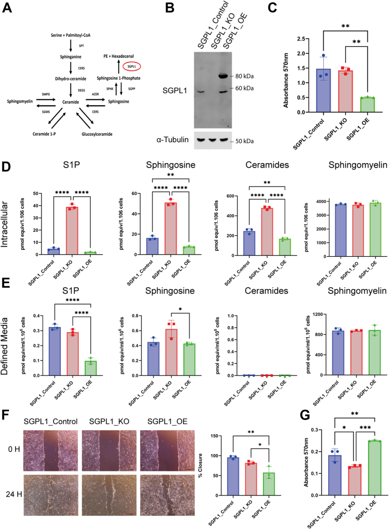
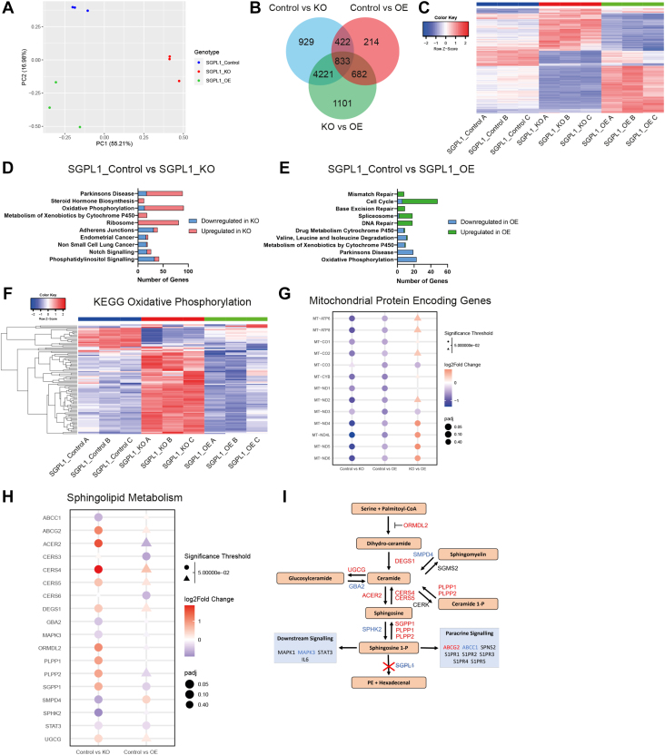
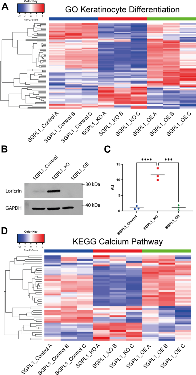
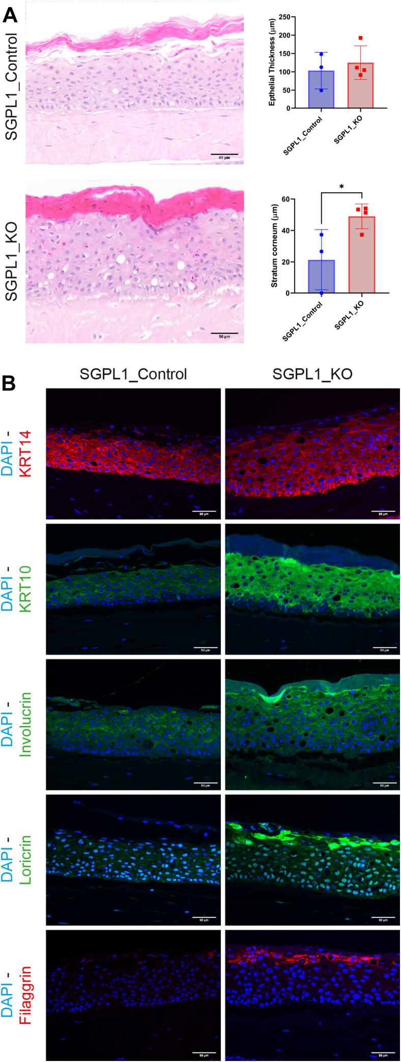
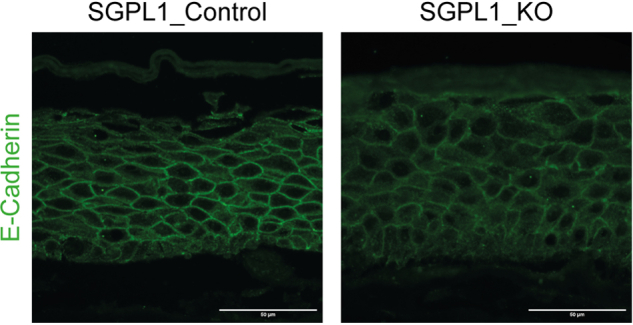
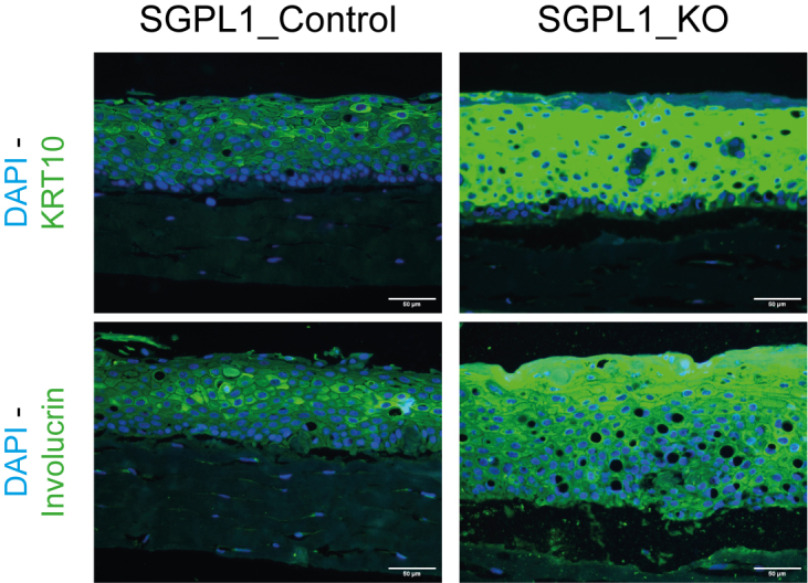
Sphingosine 1-phosphate lyase (SGPL1) insufficiency (SPLIS) is a syndrome which presents with adrenal insufficiency, steroid-resistant nephrotic syndrome, hypothyroidism, neurological disease, and ichthyosis. Where a skin phenotype is reported, 94% had abnormalities such as ichthyosis, acanthosis, and hyperpigmentation. To elucidate the disease mechanism and the role SGPL1 plays in the skin barrier we established clustered regularly interspaced short palindromic repeats-Cas9 SGPL1 KO and a lentiviral-induced SGPL1 overexpression (OE) in telomerase reverse-transcriptase immortalised human keratinocytes (N/TERT-1) and thereafter organotypic skin equivalents. Loss of SGPL1 caused an accumulation of S1P, sphingosine, and ceramides, while its overexpression caused a reduction of these species. RNAseq analysis showed perturbations in sphingolipid pathway genes, particularly in SGPL1_KO, and our gene set enrichment analysis revealed polar opposite differential gene expression between SGPL1_KO and _OE in keratinocyte differentiation and Ca2+ signaling genesets. SGPL1_KO upregulated differentiation markers, while SGPL1_OE upregulated basal and proliferative markers. The advanced differentiation of SGPL1_KO was confirmed by 3D organotypic models that also presented with a thickened and retained stratum corneum and a breakdown of E-cadherin junctions. We conclude that SPLIS associated ichthyosis is a multifaceted disease caused possibly by sphingolipid imbalance and excessive S1P signaling, leading to increased differentiation and an imbalance of the lipid lamellae throughout the epidermis.

Keywords: calcium signaling; ceramides; differentiation; epidermis; gene set enrichment analysis; keratinocytes; lipid lamellae imbalance; organotypics; pyridoxal 5′-phosphate-dependent aldehyde lyase; sphingolipid accumulation.


Conflict of interest statement

Conflict of interest The authors declare that there is no conflict of interest that could be perceived as prejudicing the impartiality of the research reported.

Figures

Fig. 1
Fig. 1
Loss and overexpression of SGPL1 results in a perturbance of sphingolipid species levels and significant changes in growth and migration. A: Schematic of the sphingolipid pathway where SGPL1 performs the final degradative process. B: SGPL1_Control shows a detectable band at 60 KDa, SGPL1_KO show no detectable band and SGPL1_OE show endogenous SGPL1 at 60 KDa and a band at 90 KDa representing SGPL1 linked to GFP. C: Proliferation rate measured of cells grown in serum-free media by MTT assay after 96 h. D, E: Levels of intracellular and extracellular sphingolipid intermediates in SGPL1_Control, SGPL1_KO and SGPL1_OE grown in FBS-containing media. F: Migration assay and quantification of wound gap closure (%) of cell grown in FBS media. (All statistical analyses were conducted using one-way ANOVAs with Tukey’s multiple comparison test, n = 3. Scale bar = 500 μm). G: Proliferation rates measured by MTT assay of cells grown in FBS after 96 h. SGPL1, Sphingosine 1-phosphate lyase.
Fig. 2
Fig. 2
RNAseq analysis shows loss and overexpression of SGPL1 causes a dichotomy of gene expression. A: Principal component analysis (PCA plot) displaying the nine samples used for RNAseq analysis with samples clustered by genotype. B: Venn diagram of differentially expressed genes displaying the number of genes present in each comparison SGPL1_Control versus SGPL1_KO, SGPL1_Control versus SGPL1_OE, and SGPL1_KO versus SGPL1_OE. C: Heatmap showing the z-scores of all genes common between all three above comparisons. D, E: The top five enriched pathways in each direction for SGPL1_KO and SGPL1_OE compared to SGPL1_Control. The number of DEGs in each gene set is demonstrated on the x-axis. F: Heatmap showing the z scores of all genes in the KEGG oxidative phosphorylation. G: Dotplot showing the P values and log2Fold change of mitochondrial encoded genes in all comparisons. H: Dotplot showing the P values and log2Fold change of SGPL1_Control versus SGPL1_KO and SGPL1_Control versus SGPL1_OE of genes encoding enzymes involved in the sphingolipid metabolism pathway. I: Sphingolipid metabolism pathway with enzymes colour coded according to log2fold change of SGPL1_Control versus SGPL1_KO. Genes upregulated in SGPL1_KO are red and genes downregulated are blue. SGPL1, Sphingosine 1-phosphate lyase.
Fig. 3
Fig. 3
Loss and overexpression of SGPL1 causes a dichotomy in growth rate and overexpression results in a lower differentiated state. A: Heatmap showing the z scores of all genes in the “GO Keratinocyte Differentiation” gene set. B: Western blot of terminal differentiation marker loricrin of cell lysates extracted from cells grown in monolayer in FBS media. C: Densitometry analysis of loricrin Western blot. (One-way ANOVA with Tukey’s multiple comparison test, n = 3). D: Heatmap showing the z scores of all genes in the “KEGG Calcium Pathway” gene set. SGPL1, Sphingosine 1-phosphate lyase.
Fig. 4
Fig. 4
SGPL1_KO models present with a thickened stratum corneum and increased differentiation. A: Representative H&E-stained images of SGPL1_Control and SGPL1_KO 3D organotypic models. Total epidermal and stratum corneum thickness were measured in μm using ImageJ. Scale bar = 50 μm. B: Immunofluorescent staining of organotypic models for differentiation markers KRT14, KRT10, involucrin, loricrin, and filaggrin. Images were captured at the same exposure and gain for comparison. Scale bar = 50 μm. (Statistical analysis was conducted using unpaired t test, n = three-fourths, ∗P < 0.05.). SGPL1, Sphingosine 1-phosphate lyase.
Fig. 5
Fig. 5
SGPL1_KO organotypic models display abnormal intercellular junctions. Immunofluorescent staining of SGPL1_Control and SGPL1_KO 3D organotypic models for junctional marker E-cadherin. Scale bar = 50 μm. SGPL1, Sphingosine 1-phosphate lyase.
Supplemental Figure S2

Similar articles

Cited by

References

    1. Prasad R., Hadjidemetriou I., Maharaj A., Meimaridou E., Buonocore F., Saleem M., et al. Sphingosine-1-phosphate lyase mutations cause primary adrenal insufficiency and steroid-resistant nephrotic syndrome. J. Clin. Invest. 2017;127:942–953. - PMC - PubMed
    1. Lovric S., Goncalves S., Gee H.Y., Oskouian B., Srinivas H., Choi W.I., et al. Mutations in sphingosine-1-phosphate lyase cause nephrosis with ichthyosis and adrenal insufficiency. J. Clin. Invest. 2017;127:912–928. - PMC - PubMed
    1. Janecke A.R., Xu R., Steichen-Gersdorf E., Waldegger S., Entenmann A., Giner T., et al. Deficiency of the sphingosine-1-phosphate lyase SGPL1 is associated with congenital nephrotic syndrome and congenital adrenal calcifications. Hum. Mutat. 2017;38:365–372. - PMC - PubMed
    1. Choi Y.J., Saba J.D. Sphingosine phosphate lyase insufficiency syndrome (SPLIS): a novel inborn error of sphingolipid metabolism. Adv. Biol. Regul. 2019;71:128–140. - PMC - PubMed
    1. Linhares N.D., Arantes R.R., Araujo S.A., Pena S.D.J. Nephrotic syndrome and adrenal insufficiency caused by a variant in SGPL1. Clin. Kidney J. 2018;11:462–467. - PMC - PubMed

Publication types