. 2023 Jul;79(1):93-108.
doi: 10.1016/j.jhep.2023.02.027. Epub 2023 Mar 1.

Liquid biopsy-based protein biomarkers for risk prediction, early diagnosis, and prognostication of cholangiocarcinoma

Affiliations

Liquid biopsy-based protein biomarkers for risk prediction, early diagnosis, and prognostication of cholangiocarcinoma

Ainhoa Lapitz et al. J Hepatol. 2023 Jul.

Abstract

Background & aims: Cholangiocarcinoma (CCA), heterogeneous biliary tumours with dismal prognosis, lacks accurate early diagnostic methods especially important for individuals at high-risk (i.e. those with primary sclerosing cholangitis [PSC]). Here, we searched for protein biomarkers in serum extracellular vesicles (EVs).

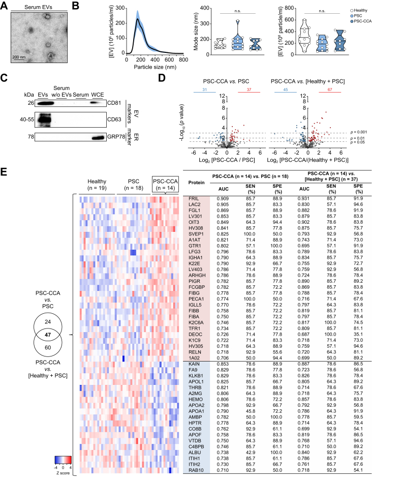
Methods: EVs from patients with isolated PSC (n = 45), concomitant PSC-CCA (n = 44), PSC who developed CCA during follow-up (PSC to CCA; n = 25), CCAs from non-PSC aetiology (n = 56), and hepatocellular carcinoma (n = 34) and healthy individuals (n = 56) were characterised by mass spectrometry. Diagnostic biomarkers for PSC-CCA, non-PSC CCA, or CCAs regardless of aetiology (Pan-CCAs) were defined and validated by ELISA. Their expression was evaluated in CCA tumours at a single-cell level. Prognostic EV biomarkers for CCA were investigated.

Results: High-throughput proteomics of EVs identified diagnostic biomarkers for PSC-CCA, non-PSC CCA, or Pan-CCA, and for the differential diagnosis of intrahepatic CCA and hepatocellular carcinoma, which were cross-validated by ELISA using total serum. Machine learning-based algorithms disclosed CRP/FIBRINOGEN/FRIL for the diagnosis of PSC-CCA (local disease [LD]) vs. isolated PSC (AUC = 0.947; odds ratio [OR] =36.9) and, combined with carbohydrate antigen 19-9, overpowers carbohydrate antigen 19-9 alone. CRP/PIGR/VWF allowed the diagnosis of LD non-PSC CCAs vs. healthy individuals (AUC = 0.992; OR = 387.5). It is noteworthy that CRP/FRIL accurately diagnosed LD Pan-CCA (AUC = 0.941; OR = 89.4). Levels of CRP/FIBRINOGEN/FRIL/PIGR showed predictive capacity for CCA development in PSC before clinical evidence of malignancy. Multi-organ transcriptomic analysis revealed that serum EV biomarkers were mostly expressed in hepatobiliary tissues, and single-cell RNA sequencing and immunofluorescence analysis of CCA tumours showed their presence mainly in malignant cholangiocytes. Multivariable analysis unveiled EV prognostic biomarkers, with COMP/GNAI2/CFAI and ACTN1/MYCT1/PF4V associated negatively and positively with patients' survival, respectively.

Conclusions: Serum EVs contain protein biomarkers for the prediction, early diagnosis, and prognostication of CCA that are detectable using total serum, representing a tumour cell-derived liquid biopsy tool for personalised medicine.

Impact and implications: The accuracy of current imaging tests and circulating tumour biomarkers for cholangiocarcinoma (CCA) diagnosis is far from satisfactory. Most CCAs are considered sporadic, although up to 20% of patients with primary sclerosing cholangitis (PSC) develop CCA during their lifetime, constituting a major cause of PSC-related death. This international study has proposed protein-based and aetiology-related logistic models with predictive, diagnostic, or prognostic capacities by combining two to four circulating protein biomarkers, moving a step forward into personalised medicine. These novel liquid biopsy tools may allow the (i) easy and non-invasive diagnosis of sporadic CCAs, (ii) identification of patients with PSC with higher risk for CCA development, (iii) establishment of cost-effective surveillance programmes for the early detection of CCA in high-risk populations (e.g. PSC), and (iv) prognostic stratification of patients with CCA, which, altogether, may increase the number of cases eligible for potentially curative options or to receive more successful treatments, decreasing CCA-related mortality.

Keywords: Cholangiocarcinoma; Extracellular vesicles; Liquid biopsy; Mass spectrometry; Primary sclerosing cholangitis; Protein biomarkers; Single-cell RNA-sequencing.


Figures

None
Graphical abstract
Fig. 1
Fig. 1
Isolated serum EV fractions are enriched in exosomes and microvesicles, and contain proteins with diagnostic capacity for PSC-CCA. Characterisation of serum EVs by (A) TEM, (B) NTA, and (C) immunoblotting. (D) Volcano plot of identified proteins in PSC-CCA vs. PSC and in PSC-CCA vs a ‘non-malignant control group’ including healthy individuals and patients with PSC. Significantly enriched proteins are coloured in red and proteins with lower abundance in blue (parametric Student's t test). (E) Venn diagram with the number of proteins with significant AUC values in both comparatives. Heatmap and diagnostic values of EV proteins altered in patients with concomitant PSC-CCA compared with PSC and to ‘the non-malignant control group’. Enriched proteins are coloured in red, and proteins with lower abundance in blue. n.s. (Kruskal–Wallis test). CCA, cholangiocarcinoma; ER, endoplasmic reticulum; EV, extracellular vesicle; NTA, nanoparticle tracking analysis; PSC-CCA, concomitant PSC and CCA; PSC, primary sclerosing cholangitis; SEN, sensitivity; SPE, specificity; TEM, transmission electron microscopy; WCE, whole cell extract.
Fig. 2
Fig. 2
Serum EV protein biomarkers for the diagnosis of CCA according to tumour aetiology. Biomarkers for the specific diagnosis of (A) CCA in patients with PSC (specific PSC-CCA biomarkers), (B) CCA in patients without PSC (non-PSC CCA biomarkers), and (C) CCA regardless of aetiology (Pan-CCA biomarkers; the best 53 biomarkers are displayed). Enriched proteins are coloured in red, and proteins with lower abundance in blue. ‘–’, n.s. AUC. CCA, cholangiocarcinoma; EV, extracellular vesicle; LD, local disease; PSC-CCA, concomitant PSC-CCA; PSC, primary sclerosing cholangitis; SEN, sensitivity; SPE, specificity.
Fig. 3
Fig. 3
Candidate EV protein biomarkers are detected using total serum and aid the diagnosis of CCA. (A) Representative immunoblots of selected biomarkers in serum EVs, serum without EVs, and total serum subfractions (10 μg protein loaded per column) of patients with CCA and healthy individuals. (B) Representative immunoblots of selected biomarkers in total serum (50 μg protein loaded per column) of patients with iCCA, pCCA, or dCCA, and healthy individuals. (C) Levels of CRP, FRIL, FGL1, VWF, PIGR, FIBRINOGEN, and OIT3 measured by ELISA in serum samples from patients with PSC, PSC-CCA, and non-PSC CCA and healthy individuals and diagnostic values for Pan-CCA vs. the non-malignant group (healthy + PSC), PSC-CCA vs. PSC, and CCA vs. healthy individuals. n.s.; ∗p <0.05; ∗∗p <0.01; ∗∗∗∗p <0.0001 (Kruskal–Wallis test). AI, accuracy index; CCA, cholangiocarcinoma; dCCA, distal CCA; EV, extracellular vesicle; iCCA, intrahepatic CCA; NPV, negative predictive value; OR, odds ratio; Pan-CCA, CCA regardless of aetiology; pCCA, perihiliar CCA; PPV, positive predictive value; PSC-CCA, concomitant PSC and CCA; PSC, primary sclerosing cholangitis; ROC, receiver operating characteristic; SEN, sensitivity; SPE, specificity.
Fig. 4
Fig. 4
LMs combining ELISA-validated serum protein biomarkers enable the accurate diagnosis of CCA in patients with or without PSC. Binary logistic regression models in the training (70%) cohort, as well as in the testing 30% and LD cohorts for CCA diagnosis (A) regardless of disease aetiology, (B) in patients with PSC, and (C) in patients without PSC. AI, accuracy index; CCA, cholangiocarcinoma; LD, local disease; LM, logistic model; OR, odds ratio; Pan-CCA, CCA regardless of aetiology; PSC, primary sclerosing cholangitis; ROC, receiver operating characteristic; SEN, sensitivity; SPE, specificity.
Fig. 5
Fig. 5
Serum EV protein biomarkers for the differential diagnosis of iCCA vs. HCC. (A) Levels and diagnostic values of FGL1, CRP, PIGR, FIBRINOGEN, VWF, FRIL, and OIT3 measured by ELISA in serum samples from patients with iCCA and HCC. Heatmaps, Venn diagrams, and diagnostic values of specific EV proteins for the diagnosis of (B) iCCA and (C) HCC. Enriched proteins are coloured in red, and proteins with lower abundance in blue. ∗p <0.05; ∗∗p <0.01; ∗∗∗p <0.001; ∗∗∗∗p <0.0001 (Mann–Whitney test). AI, accuracy index; EV, extracellular vesicle; HCC, hepatocellular carcinoma; iCCA, intrahepatic cholangiocarcinoma; NPV, negative predictive value; PPV, positive predictive value; PSC, primary sclerosing cholangitis; ROC, receiver operating characteristic; SEN, sensitivity; SPE, specificity.
Fig. 6
Fig. 6
Human multi-organ transcriptome and scRNA-seq reveal the potential origin of serum EV protein biomarkers. (A) Normalised expression of candidate serum biomarkers in 61 human tissues/organs from the Consensus dataset of the Human Protein Atlas. (B) tSNE plot and normalised expression of candidate biomarkers in each liver cell type from normal liver scRNA-seq (GSE115469). (C) tSNE plot and cell type proportion from 12 iCCA tumours (GSE151530). Biomarker expressing-positive cells and relative biomarker expression within the cell types of iCCA tumours. (D) Representative IF images of PIGR, FIBG, CRP, and FGL1 and colocalisation with CK19+-positive cells. Scale bars = 50 μm ∗∗∗∗p <0.0001 (Kruskal–Wallis test). CCA, cholangiocarcinoma; CK19, cytokeratin 19; EV, extracellular vesicle; HCC, hepatocellular carcinoma; iCCA, intrahepatic CCA; IF, immunofluorescence; NC, normalised counts; NK, natural killer; PSC-CCA, concomitant PSC and CCA; PSC, primary sclerosing cholangitis; RNA-seq, RNA sequencing; ROC, receiver operating characteristic; sCRNA-seq, single-cell RNA sequencing; SL, surrounding liver; tSNE, t-distributed stochastic neighbour embedding.
Fig. 7
Fig. 7
Serum proteins allow the prediction of CCA development in patients with PSC. (A) Heatmap and diagnostic values of specific EV proteins for the differential identification of patients with PSC who progressed to CCA over time (PSC to CCA) and non-malignant PSC. Enriched proteins in red, and proteins with lower abundance in blue. (B) Levels and diagnostic values of FIBRINOGEN, CRP, PIGR, and FRIL in total serum from patients with PSC to CCA, PSC-CCA, and non-malignant PSC. (C) Binary logistic regression models for the prediction of CCA development in patients with PSC. ‘–’, n.s. AUC; ∗p <0.05; ∗∗∗p <0.001; ∗∗∗∗p <0.0001 (Kruskal–Wallis test). m, months from sampling to CCA diagnosis; AI, accuracy index; CCA, cholangiocarcinoma; EV, extracellular vesicle; iCCA, intrahepatic CCA; LM, logistic model; NPV, negative predictive value; OR, odds ratio; PPV, positive predictive value; PSC-CCA, concomitant PSC and CCA; PSC, primary sclerosing cholangitis; ROC, receiver operating characteristic; SEN, sensitivity; SPE, specificity; CA19-9, carbohydrate antigen 19-9.
Fig. 8
Fig. 8
Association of serum EV protein levels with patients’ outcome. (A) Schematic representation of the strategy used to define prognostic biomarkers. (B) Multivariable analysis of serum EV proteins with prognostic value independent of the clinical/demographic variables sex, age, PSC, cirrhosis, CCA subtype, disease status, CA19-9, and surgical intervention. Kaplan–Meier curves of OS for each prognostic biomarker in 10 years of follow-up. (C) Kaplan–Meier curve, Cox regression analysis, and log-rank test of patients with CCA according to the ‘bad prognostic’ (COMP/GNAI2/CFAI) and ‘good prognostic’ (ACTN1/MYCT1/PF4V) panels. AI, accuracy index; CA19-9, carbohydrate antigen 19-9; CCA, cholangiocarcinoma; dCCA, distal CCA; EV, extracellular vesicle; HR, hazard ratio; iCCA, intrahepatic CCA; LAD, locally advanced disease; LD, local disease; LM, logistic model; MD, metastatic disease; mOS, median overall survival; NPV, negative predictive value; OR, odds ratio; OS, overall survival; pCCA, perihiliar CCA; PPV, positive predictive value; PSC-CCA, concomitant PSC and CCA; PSC, primary sclerosing cholangitis; ROC, receiver operating characteristic; SEN, sensitivity; SPE, specificity.

References

    1. Banales J.M., Marin J.J.G., Lamarca A., Rodrigues P.M., Khan S.A., Roberts L.R., et al. Cholangiocarcinoma 2020: the next horizon in mechanisms and management. Nat Rev Gastroenterol Hepatol. 2020;17:557–588. - PMC - PubMed
    1. Rodrigues P.M., Olaizola P., Paiva N.A., Olaizola I., Agirre-Lizaso A., Landa A., et al. Pathogenesis of cholangiocarcinoma. Annu Rev Pathol. 2021;16:433–463. - PubMed
    1. Izquierdo-Sanchez L., Lamarca A., La Casta A., Buettner S., Utpatel K., Klumpen H.J., et al. Cholangiocarcinoma landscape in Europe: diagnostic, prognostic and therapeutic insights from the ENSCCA Registry. J Hepatol. 2022;76:1109–1121. - PubMed
    1. Macias R.I.R., Cardinale V., Kendall T.J., Avila M.A., Guido M., Coulouarn C., et al. Clinical relevance of biomarkers in cholangiocarcinoma: critical revision and future directions. Gut. 2022;71:1669–1683. - PubMed
    1. Valle J.W., Borbath I., Khan S.A., Huguet F., Gruenberger T., Arnold D., et al. Biliary cancer: ESMO Clinical Practice Guidelines for diagnosis, treatment and follow-up. Ann Oncol. 2016;27:v28–v37. - PubMed

Publication types

MeSH terms