. 2023 Mar 3;13(1):78.
doi: 10.1038/s41398-023-02382-w.

Mu-opioid receptor-expressing neurons in the paraventricular thalamus modulate chronic morphine-induced wake alterations

Affiliations

Mu-opioid receptor-expressing neurons in the paraventricular thalamus modulate chronic morphine-induced wake alterations

Darrell Eacret et al. Transl Psychiatry. .

Abstract

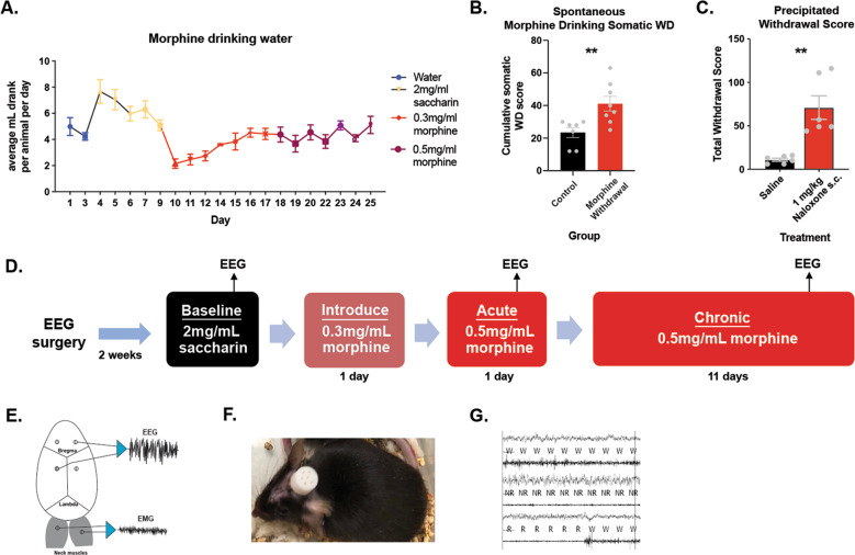
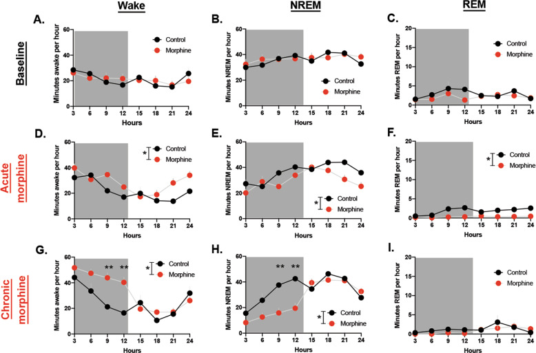
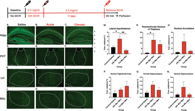
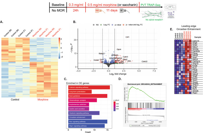
Disrupted sleep is a symptom of many psychiatric disorders, including substance use disorders. Most drugs of abuse, including opioids, disrupt sleep. However, the extent and consequence of opioid-induced sleep disturbance, especially during chronic drug exposure, is understudied. We have previously shown that sleep disturbance alters voluntary morphine intake. Here, we examine the effects of acute and chronic morphine exposure on sleep. Using an oral self-administration paradigm, we show that morphine disrupts sleep, most significantly during the dark cycle in chronic morphine, with a concomitant sustained increase in neural activity in the Paraventricular Nucleus of the Thalamus (PVT). Morphine binds primarily to Mu Opioid Receptors (MORs), which are highly expressed in the PVT. Translating Ribosome Affinity Purification (TRAP)-Sequencing of PVT neurons that express MORs showed significant enrichment of the circadian entrainment pathway. To determine whether MOR + cells in the PVT mediate morphine-induced sleep/wake properties, we inhibited these neurons during the dark cycle while mice were self-administering morphine. This inhibition decreased morphine-induced wakefulness but not general wakefulness, indicating that MORs in the PVT contribute to opioid-specific wake alterations. Overall, our results suggest an important role for PVT neurons that express MORs in mediating morphine-induced sleep disturbance.


Conflict of interest statement

The authors declare no competing interests.

Figures

Fig. 1
Fig. 1. Experimental Design.
A Amount consumed in drinking water in average mL per animal per day. B Spontaneous somatic withdrawal signs between morphine-treated mice and saccharin-treated mice. C Somatic withdrawal signs in mice that drank morphine precipitated by 1 mg/kg Naloxone or Saline. D Timeline of experiments. E Example schematic showing placement of electrodes 1 mm from bregma in each quadrant and on the nuchal muscles with example EEG and EMG traces. F Image of mouse with electrode cap. G Example traces of Wake, NREM, and REM in 4 s epochs.
Fig. 2
Fig. 2. Morphine increases wake during the dark cycle.
A Minutes awake per hour at baseline. B Minutes in NREM per hour at baseline. C Minutes REM per hour at baseline. D Minutes awake per hour at acute morphine timepoint. E Minutes NREM per hour at acute morphine timepoint. F Minutes REM per hour at acute morphine timepoint. G Minutes awake per hour at chronic morphine timepoint. H Minutes NREM per hour at chronic morphine timepoint. I Minutes REM per hour at chronic morphine timepoint.
Fig. 3
Fig. 3. Screen of immediate early gene expression across brain regions.
A Representative image of hippocampus from saline group. B Hippocampus from acute morphine group. C Chronic morphine hippocampus. D Representative image of PVT (paraventricular nucleus of the thalamus) from saline group. E PVT from acute morphine group. F Chronic morphine PVT. G Representative image of LH (lateral hypothalamus) from saline group. H LH from acute morphine group. I Chronic morphine LH. J Representative image of NAc (nucleus accumbens) from saline group. K NAc from acute morphine group. L Chronic morphine LH. M Quantification of cFOS expression in LH across morphine timepoints. N cFOS expression in PVT across morphine timepoints. O cFOS expression in NAc quantified across morphine timepoints. P Quantification of cFOS expression in VTA across morphine timepoints. Q cFOS expression levels in Dorsal Hippocampus across morphine timepoints. R cFOS expression in ventral hippocampus at control, acute, and chronic morphine. Images were taken at 10× quantification and scale bars represent 500 um.
Fig. 4
Fig. 4. Translating ribosome affinity purification on MOR-expressing neurons targeted at the paraventricular nucleus of the thalamus.
A Heatmap of significantly differentially expressed genes in PVT MOR-expressing neurons of controls compared to mice maintained on chronic morphine. B Volcano plot showing −log10P value vs log2 fold change (morphine vs ctl group) for all genes analyzed. Genes corresponding to significant adjusted P values are those above the dashed horizontal line. Dashed vertical lines correspond to a threshold of 1 for the absolute log2 fold change. C Significant KEGG pathways from ORA analyses of DE genes. D Enrichment plot of the Circadian Entrainment pathway. E Heatmap of the genes in the leading edge of the Circadian Entrainment pathway.
Fig. 5
Fig. 5. DREADD inhibition of neurons in the PVT that express mu-opioid receptors.
A Representative image of mu-opioid receptors in the PVT with MOR Cre x GFP reporter mouse. B Virus strategy to target PVT MOR-expressing neurons. A Cre-dependent inhibitory DREADDs construct was injected into the PVT of mice that express Cre recombinase on the nucleus of mu-opioid receptor-expressing neurons. C Mu-opioid receptor-expressing neurons identified by GFP reporter in the PVT at 10× and super-resolution. D hM4Di expression in the PVT identified by mCherry reporter at 10× and super-resolution. E Overlay of MOR-expressing neurons in green and DREADDs construct in red at 10× and super-resolution. F quantification of transfection of MOR + neurons in the PVT that also express hM4Di. G Representative image of cFOS expression in the PVT of mice injected with the DREADDs virus. H Representative image of cFOS expression in the PVT of mice injected with the DREADDs virus after i.p. CNO injection. I Quantification of cFOS expression in the PVT to show DREADD inhibition from CNO. J Activity plot per 10 m bins of mice that received CNO or saline injection with a morphine stimulus. Scale bars represent 100 μm.
Fig. 6
Fig. 6. DREADDs inhibition of PVT MOR-expressing neurons during chronic morphine.
A Representative image showing viral strategy- Cre-dependent DREADDs virus or control mCherry virus was injected into the PVT of MOR-Cre mice. B Representative image of MOR expression in the PVT in mCherry animals. C mCherry expression in the PVT. D Overlay of mCherry and MOR expression. E Representative image of MOR expression in the PVT in DREADDs injected mice. F Representative hM4Di expression in the PVT. G Overlay of PVT MOR and hM4Di expression. H Minutes awake per 12 h during the light cycle. I Minutes awake per 12 h during the dark cycle. CNO was injected 1 h into the dark cycle on the last day (13) of morphine treatment. J Minutes awake per hour in the light cycle immediately before CNO administration. K Minutes awake per hour in the dark cycle, including CNO administration 1 h into the dark period at 8 pm. L Minutes awake per 12 h in the dark cycle preceding CNO administration and the dark cycle including CNO administration in mice that did not receive morphine. M Minutes awake per 12 h in the dark cycle preceding CNO administration and the dark cycle including CNO administration in mice that were treated with morphine. Scale bars represent 100 μm.

References

    1. Eacret D, Veasey SC, Blendy JA. Bidirectional relationship between opioids and disrupted sleep: putative mechanisms. Mol Pharmacol. 2020. 10.1124/mol.119.119107. - PMC - PubMed
    1. Valentino RJ, Volkow ND. Drugs, sleep, and the addicted brain. Neuropsychopharmacology. 2019. 10.1038/s41386-019-0465-x. - PMC - PubMed
    1. Dijkstra BAG, De Jong CAJ, Krabbe PFM, Van Der Staak CPF. Prediction of abstinence in opioid-dependent patients. J Addict Med. 2008. 10.1097/ADM.0b013e31818a6596. - PubMed
    1. Lydon-Staley DM, Cleveland HH, Huhn AS, Cleveland MJ, Harris J, Stankoski D, et al. Daily sleep quality affects drug craving, partially through indirect associations with positive affect, in patients in treatment for nonmedical use of prescription drugs. Addict Behav. 2017;65:275–82. doi: 10.1016/j.addbeh.2016.08.026. - DOI - PMC - PubMed
    1. Kosobud AEK, Gillman AG, Leffel JK, Pecoraro NC, Rebec GV, Timberlake W. Drugs of abuse can entrain circadian rhythms. ScientificWorldJournal. 2007. 10.1100/tsw.2007.234. - PMC - PubMed

Publication types