

First pan-specific vNAR against human TGF-β as a potential therapeutic application: in silico modeling assessment
- PMID: 36869086
- PMCID: PMC9982792
- DOI: 10.1038/s41598-023-30623-x
First pan-specific vNAR against human TGF-β as a potential therapeutic application: in silico modeling assessment
Abstract
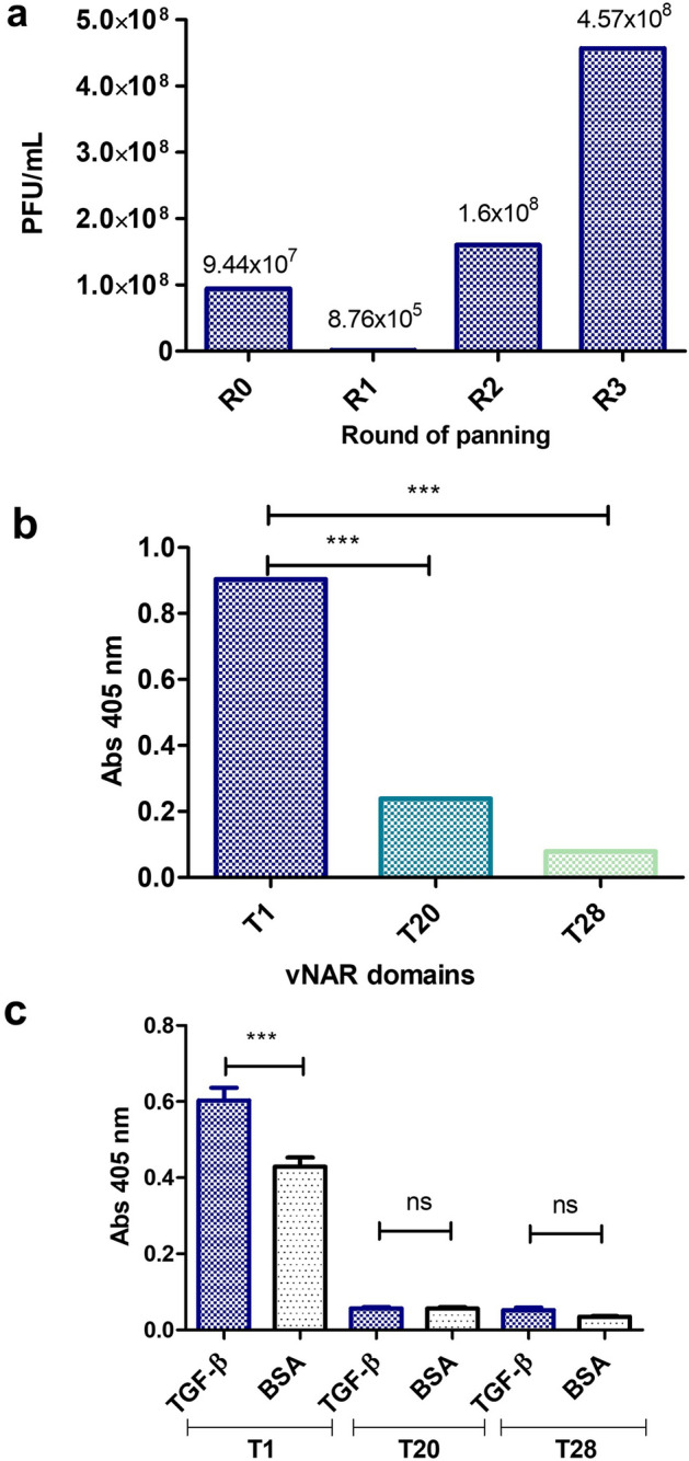
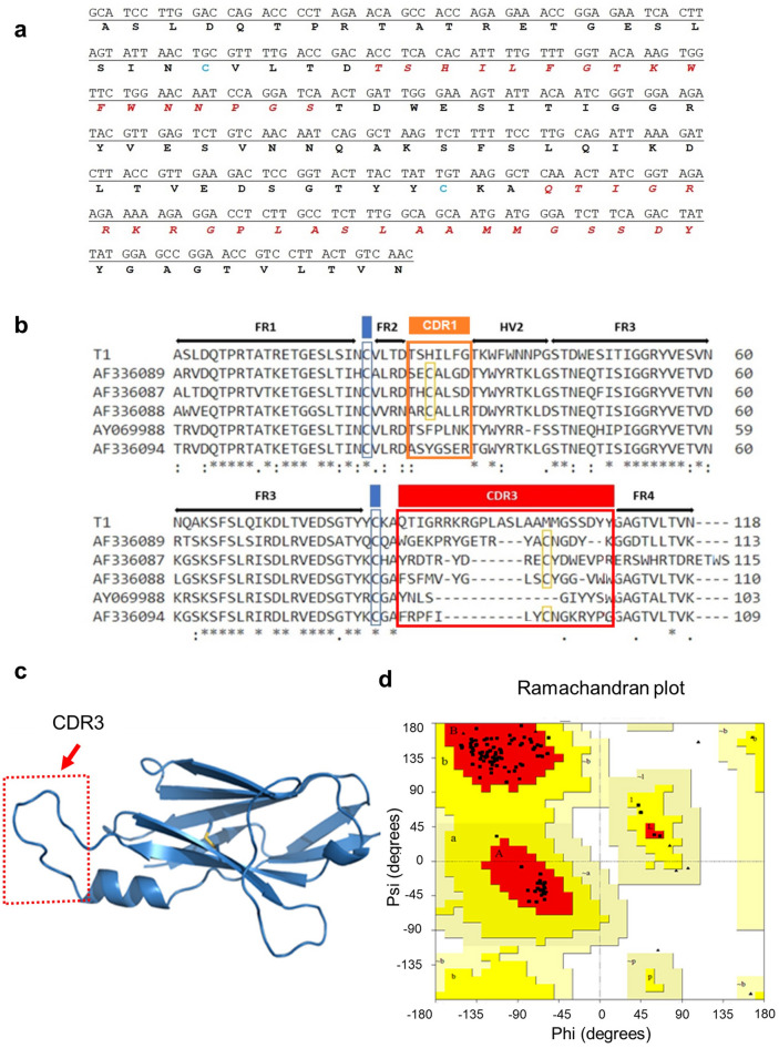
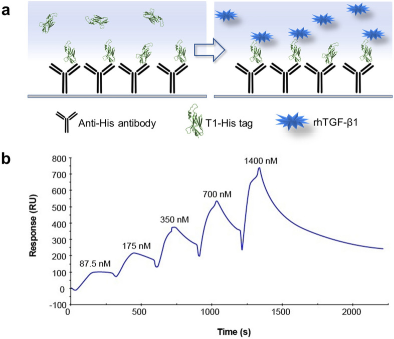
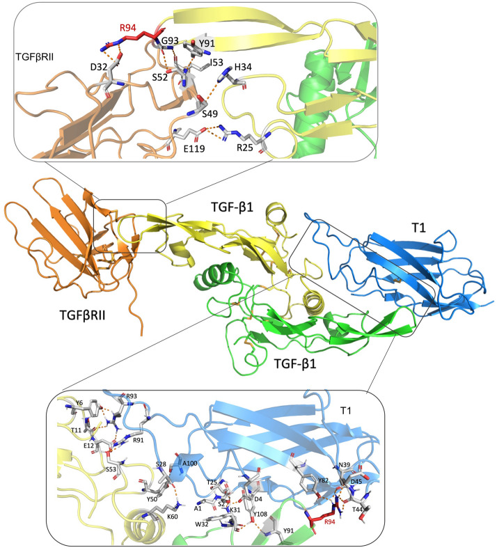
Immunotherapies based on antibody fragments have been developed and applied to human diseases, describing novel antibody formats. The vNAR domains have a potential therapeutic use related to their unique properties. This work used a non-immunized Heterodontus francisci shark library to obtain a vNAR with recognition of TGF-β isoforms. The isolated vNAR T1 selected by phage display demonstrated binding of the vNAR T1 to TGF-β isoforms (-β1, -β2, -β3) by direct ELISA assay. These results are supported by using for the first time the Single-Cycle kinetics (SCK) method for Surface plasmon resonance (SPR) analysis for a vNAR. Also, the vNAR T1 shows an equilibrium dissociation constant (KD) of 9.61 × 10⁻⁸ M against rhTGF-β1. Furthermore, the molecular docking analysis revealed that the vNAR T1 interacts with amino acid residues of TGF-β1, which are essential for interaction with type I and II TGF-β receptors. The vNAR T1 is the first pan-specific shark domain reported against the three hTGF-β isoforms and a potential alternative to overcome the challenges related to the modulation of TGF-β levels implicated in several human diseases such as fibrosis, cancer, and COVID-19.
© 2023. The Author(s).
Conflict of interest statement
The authors declare no competing interests.
Figures


Similar articles
-
Exploring shark VNAR antibody against infectious diseases using phage display technology.Fish Shellfish Immunol. 2023 Sep;140:108986. doi: 10.1016/j.fsi.2023.108986. Epub 2023 Aug 2. Fish Shellfish Immunol. 2023. PMID: 37541634 Review.
-
Intraocular Penetration of a vNAR: In Vivo and In Vitro VEGF165 Neutralization.Mar Drugs. 2018 Mar 31;16(4):113. doi: 10.3390/md16040113. Mar Drugs. 2018. PMID: 29614715 Free PMC article.
-
Transforming growth factor-beta (TGF-beta) binding to the extracellular domain of the type II TGF-beta receptor: receptor capture on a biosensor surface using a new coiled-coil capture system demonstrates that avidity contributes significantly to high affinity binding.J Mol Biol. 2003 May 16;328(5):1173-83. doi: 10.1016/s0022-2836(03)00360-7. J Mol Biol. 2003. PMID: 12729750
-
Construction and next-generation sequencing analysis of a large phage-displayed VNAR single-domain antibody library from six naïve nurse sharks.Antib Ther. 2019 Jan;2(1):1-11. doi: 10.1093/abt/tby011. Epub 2018 Nov 7. Antib Ther. 2019. PMID: 30627698 Free PMC article.
-
Ancient species offers contemporary therapeutics: an update on shark VNAR single domain antibody sequences, phage libraries and potential clinical applications.Antib Ther. 2020 Jan;3(1):1-9. doi: 10.1093/abt/tbaa001. Epub 2020 Jan 21. Antib Ther. 2020. PMID: 32118195 Free PMC article. Review.
Cited by
-
vNARs as Neutralizing Intracellular Therapeutic Agents: Glioblastoma as a Target.Antibodies (Basel). 2024 Mar 18;13(1):25. doi: 10.3390/antib13010025. Antibodies (Basel). 2024. PMID: 38534215 Free PMC article. Review.
-
VNAR development through antigen immunization of Japanese topeshark (Hemitriakis japanica).Front Bioeng Biotechnol. 2023 Sep 12;11:1265582. doi: 10.3389/fbioe.2023.1265582. eCollection 2023. Front Bioeng Biotechnol. 2023. PMID: 37771574 Free PMC article.
-
Unleashing the power of shark variable single domains (VNARs): broadly neutralizing tools for combating SARS-CoV-2.Front Immunol. 2023 Sep 11;14:1257042. doi: 10.3389/fimmu.2023.1257042. eCollection 2023. Front Immunol. 2023. PMID: 37753081 Free PMC article. Review.
-
Shark IgNAR: The Next Broad Application Antibody in Clinical Diagnoses and Tumor Therapies?Mar Drugs. 2023 Sep 16;21(9):496. doi: 10.3390/md21090496. Mar Drugs. 2023. PMID: 37755109 Free PMC article. Review.
-
Isolation and Characterization of the First Antigen-Specific EGFRvIII vNAR from Freshwater Stingray (Potamotrygon spp.) as a Drug Carrier in Glioblastoma Cancer Cells.Int J Mol Sci. 2025 Jan 21;26(3):876. doi: 10.3390/ijms26030876. Int J Mol Sci. 2025. PMID: 39940647 Free PMC article.
References
Publication types
MeSH terms
Substances
LinkOut - more resources
Full Text Sources
Medical
