. 2023 Mar 3;23(1):6.
doi: 10.1186/s12896-023-00776-8.

A fully human connective tissue growth factor blocking monoclonal antibody ameliorates experimental rheumatoid arthritis through inhibiting angiogenesis

Affiliations

A fully human connective tissue growth factor blocking monoclonal antibody ameliorates experimental rheumatoid arthritis through inhibiting angiogenesis

Yang Qin et al. BMC Biotechnol. .

Abstract

Background: Connective tissue growth factor (CTGF) plays a pivotal role in the pathogenesis of rheumatoid arthritis (RA) by facilitating angiogenesis and is a promising therapeutic target for RA treatment. Herein, we generated a fully human CTGF blocking monoclonal antibody (mAb) through phage display technology.

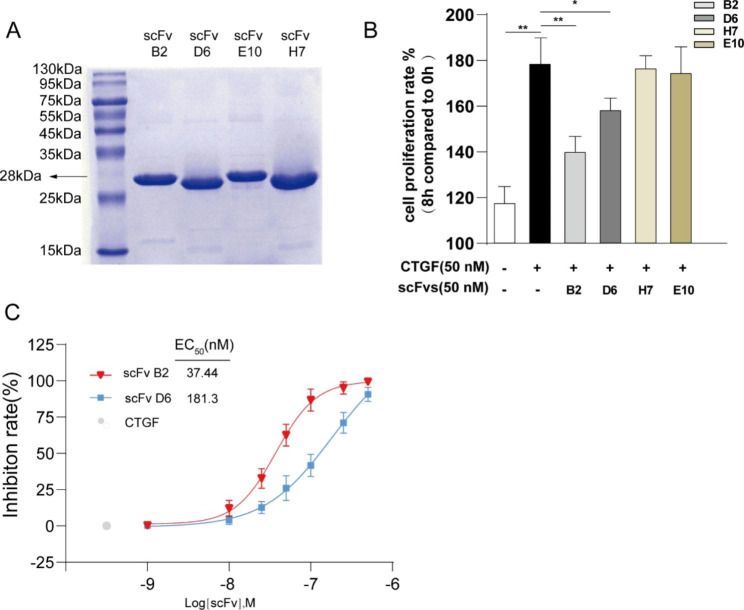
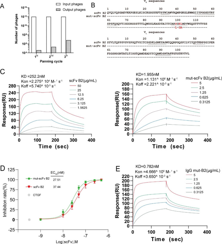
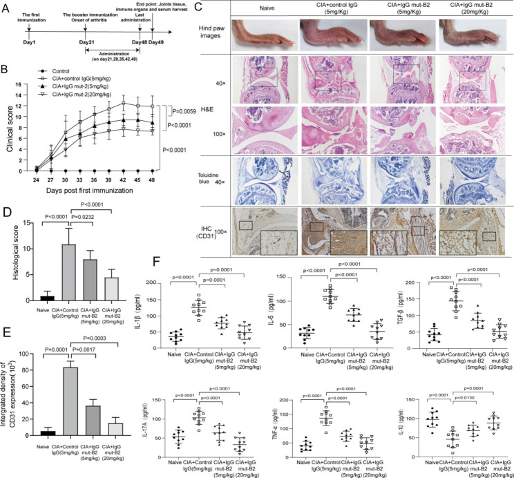
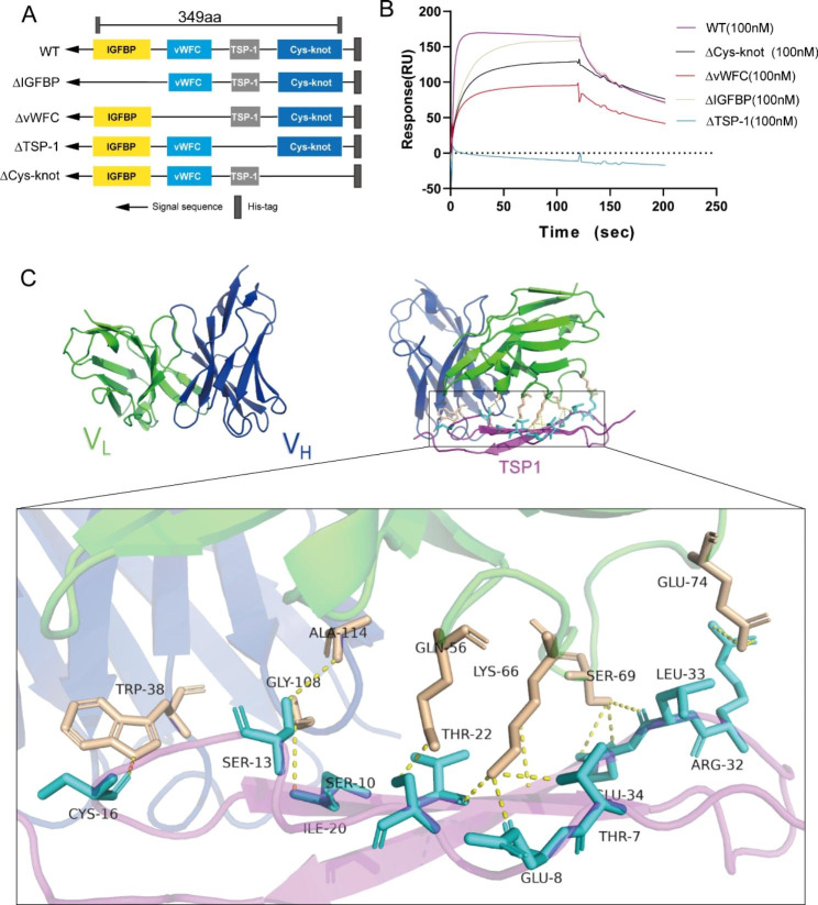
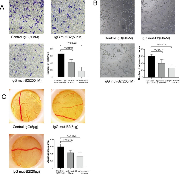
Results: A single-chain fragment variable (scFv) with a high affinity to human CTGF was isolated through screening a fully human phage display library. We carried out affinity maturation to elevate its affinity for CTGF and reconstructed it into a full-length IgG1 format for further optimization. Surface plasmon resonance (SPR) data showed that full-length antibody IgG mut-B2 bound to CTGF with a dissociation constant (KD) as low as 0.782 nM. In the collagen-induced arthritis (CIA) mice, IgG mut-B2 alleviated arthritis and decreased the level of pro-inflammatory cytokines in a dose-dependent manner. Furthermore, we confirmed that the TSP-1 domain of CTGF is essential for the interaction. Additionally, the results of Transwell assays, tube formation experiments, and chorioallantoic membrane (CAM) assays showed that IgG mut-B2 could effectively inhibit angiogenesis.

Conclusion: The fully human mAb that antagonizes CTGF could effectively alleviate arthritis in CIA mice, and its mechanism is tightly associated with the TSP-1 domain of CTGF.

Keywords: Affinity maturation; Angiogenesis; Arthritis; CTGF; Human antibody; Phage display.


Conflict of interest statement

The authors declare no conflict of interest.

Figures

Fig. 1
Fig. 1
Screening and identification of connective tissue growth factor (CTGF) blocking single chain fragment variables (scFvs). (A) SDS-PAGE analysis of purified anti-CTGF scFvs with Coomassie blue staining. (B) Cell proliferation rate of HUVECs was evaluated by CCK-8 assay after being treated with recombinant human CTGF (50 nM) and purified anti-CTGF scFvs (50 nM) for 8 h. HUVECs untreated (White bar) and HUVECs only treated with CTGF (Black bar) were used as controls. Formula used to calculate the cell proliferation rate is shown in Methods. (C) Inhibition rate of CTGF-induced cell proliferation. Cell viability of HUVECs was evaluated by CCK-8 assay after being treated with recombinant human CTGF (50 nM) and scFv B2 or scFv D6 in increasing concentrations (1-500 nM). HUVECs only treated with CTGF (Grey dot) were used as control. Formula used to calculate the inhibition rate is shown in Methods
Fig. 2
Fig. 2
The affinity of anti-CTGF scFv B2 increased after mutagenesis and reconstruction to the IgG1 format. (A) The number of eluted phages. After three rounds of screening for CTGF in phage-displayed scFv library with scFv B2-VH-CDR3 mutagenesis, the number of eluted phages increased 163-fold over that of the first round. (B) Amino acids of the heavy and light chain variable region of scFv B2 and mut-scFv B2. The red letters indicate residues that vary between scFv B2 and mut-scFv B2. Underline: CDR1, CDR2, CDR3 regions of heavy and light chain. (C) Affinity of scFv B2 and mut-scFv B2 determined by surface plasmon resonance (SPR). (D) Inhibition rate of CTGF-induced cell proliferation. Cell viability of HUVECs was evaluated by CCK-8 assay after treated with recombinant human CTGF (50 nM) and scFv B2 or mut-scFv B2 (1-500 nM) for 8 h. HUVECs only treated with CTGF (white dot) were used as control. Formula used to calculate the inhibition rate is shown in Methods. (E) Affinity of IgG mut-B2 determined by SPR.
Fig. 3
Fig. 3
Anti-CTGF IgG mut-B2 ameliorated progression of arthritis in a collagen-induced arthritis (CIA) model. (A) Timeline of anti-CTGF IgG mut-B2 and control IgG intervention in CIA mice. (n = 10 per group). (B) Clinical scores of CIA mice treated with control IgG or IgG mut-B2. Two independent observers who were unaware of the mice’s treatment examined their arthritis severity every 3 days. The scores were determined as follows: 0: no erythema or swelling, 1: erythema and mild swelling limited to the tarsals or ankle joint, 2: erythema and mild swelling extending from the ankle to the tarsals, 3: erythema and moderate swelling extending from the ankle to the metatarsal joints, 4: erythema and severe swelling encompassing the ankle, paw, and digits, or ankylosis of the limb. Statistical significance was determined by analysis of variance (ANOVA) of repeated measurements. (n = 10 per group). (C) Articular tissues of CIA mice were stained by H&E and toluidine blue. Expression of CD31 in paraffin sections of synovium samples of CIA mice was determined by IHC analysis. (D) Histological scores of CIA mice based on analysis of synovial hyperplasia, cartilage and bone erosion, and inflammatory cell infiltration shown in (C). (E) Intergrated density of CD31 expression in synovial tissues of CIA mice. (F) Concentrations of IL-1β, IL-6, TNF-α, IL-17 A, IL-10 in serum of CIA mice were detected by ELISA. (n = 10 per group). All reactions were conducted in triplicate and data were presented with mean ± SD.
Fig. 4
Fig. 4
IgG mut-B2 interacted with CTGF through TSP-1 domain. (A) Schematic of full-length CTGF (WT) and its deletion mutants. (B) Binding affinity of IgG mut-B2 to CTGF and its deletion mutants determined by SPR assay. (C) Molecular modeling of variable regions of IgG mut-B2 and TSP-1 domain of CTGF. Left panel: Three-dimensional model of variable regions of IgG mut-B2 structure predicted by homology modeling. The heavy chains’ variable regions are shown in blue and the light chains’ variable regions in green. Right panel: The intermolecular interaction analyses of the IgG mut-B2 with TSP-1 domain. TSP-1 domain of CTGF is represented by magenta. The sticks model is used to demonstrate the key residues involved in the interactions, and the key residues of TSP-1 domain are marked in cyan, key residues of antibody are marked in light orange. Yellow dotted lines represent hydrogen bonds
Fig. 5
Fig. 5
Effect of IgG mut-B2 on angiogenesis. (A) Cell migration assay used to assess angiogenesis in vitro. Lower right panel: numbers of HUVEC per field. (Original magnifcation, ×200) (B) Tube formation assay used to assess angiogenesis in vitro. Lower right panel: numbers of intersection nodes. (Original magnifcation, ×100) (C) Chorioallantoic membrane (CAM) assay used to assess angiogenesis in vivo. Lower right panel: ratio of vascular area to CAM area

References

    1. Smolen JS, Pangan AL, Emery P, Rigby W, Tanaka Y, Vargas JI, Zhang Y, Damjanov N, Friedman A, Othman AA, et al. Upadacitinib as monotherapy in patients with active rheumatoid arthritis and inadequate response to methotrexate (SELECT-MONOTHERAPY): a randomised, placebo-controlled, double-blind phase 3 study. Lancet. 2019;393(10188):2303–11. doi: 10.1016/S0140-6736(19)30419-2. - DOI - PubMed
    1. McInnes IB, Schett G. The pathogenesis of rheumatoid arthritis. N Engl J Med. 2011;365(23):2205–19. doi: 10.1056/NEJMra1004965. - DOI - PubMed
    1. Tsai CY, Shiau AL, Chen SY, Chen YH, Cheng PC, Chang MY, Chen DH, Chou CH, Wang CR, Wu CL. Amelioration of collagen-induced arthritis in rats by nanogold. Arthritis Rheum-Us. 2007;56(2):544–54. doi: 10.1002/art.22401. - DOI - PubMed
    1. Huh YH, Lee G, Lee KB, Koh JT, Chun JS, Ryu JH. HIF-2alpha-induced chemokines stimulate motility of fibroblast-like synoviocytes and chondrocytes into the cartilage-pannus interface in experimental rheumatoid arthritis mouse models. Arthritis Res Ther. 2015;17:302. doi: 10.1186/s13075-015-0816-x. - DOI - PMC - PubMed
    1. Fearon U, Canavan M, Biniecka M, Veale DJ. Hypoxia, mitochondrial dysfunction and synovial invasiveness in rheumatoid arthritis. Nat Rev Rheumatol. 2016;12(7):385–97. doi: 10.1038/nrrheum.2016.69. - DOI - PubMed

Publication types