Review
. 2023 Mar 3;21(1):77.
doi: 10.1186/s12951-023-01827-0.

Recent advances of CREKA peptide-based nanoplatforms in biomedical applications

Affiliations
Review

Recent advances of CREKA peptide-based nanoplatforms in biomedical applications

Nannan Zhang et al. J Nanobiotechnology. .

Abstract

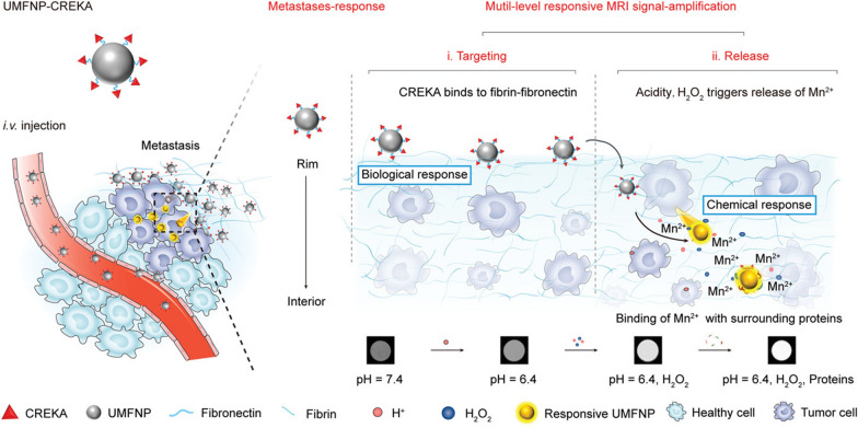
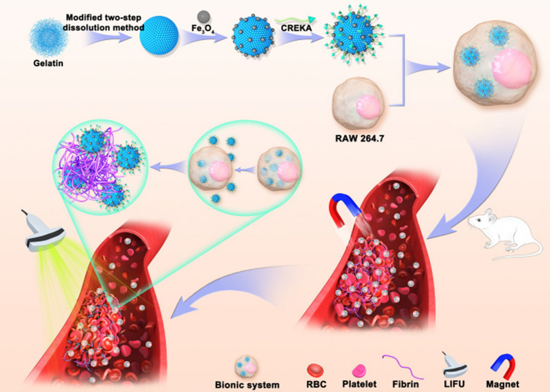
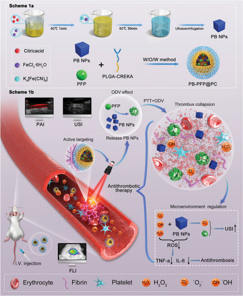
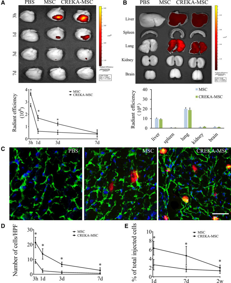
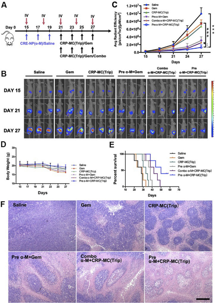
Nanomedicine technology is a rapidly developing field of research and application that uses nanoparticles as a platform to facilitate the diagnosis and treatment of diseases. Nanoparticles loaded with drugs and imaging contrast agents have already been used in clinically, but they are essentially passive delivery carriers. To make nanoparticles smarter, an important function is the ability to actively locate target tissues. It enables nanoparticles to accumulate in target tissues at higher concentrations, thereby improving therapeutic efficacy and reducing side effects. Among the different ligands, the CREKA peptide (Cys-Arg-Glu-Lys-Ala) is a desirable targeting ligand and has a good targeting ability for overexpressed fibrin in different models, such as cancers, myocardial ischemia-reperfusion, and atherosclerosis. In this review, the characteristic of the CREKA peptide and the latest reports regarding the application of CREKA-based nanoplatforms in different biological tissues are described. In addition, the existing problems and future application perspectives of CREKA-based nanoplatforms are also addressed.

Keywords: Active targeting; Biomedical applications; CREKA peptide; Fibrin-fibronectin complexes; Nanoplatform.


Conflict of interest statement

The authors have declared that no competing interest exists.

Figures

Fig. 1
Fig. 1
Scheme illustration of the prepared UMFNP-CREKA nanoprobe for imaging ultrasmall metastases by T1-weighted MRI with multiple levels of response [58]
Fig. 2
Fig. 2
T1-weighted MR imaging of lung metastases in vivo. A MR images and H&E stained images after 20 min of injection of UMFNP-CREKA. B MRI-based proportion of lung metastases area. C Quantified bioluminescence imaging intensity. D Proportion of lung metastases area based on H&E staining [58]
Fig. 3
Fig. 3
CREKA peptides specifically bind fibronectin-associated complexes in metastatic tumors. a Fluorescence imaging of major organ metastatic tissue in mice with spontaneous metastatic 4T1-GFP-Luc2 breast tumors. b Fluorescence intensity ratio between metastatic tumor and normal tissue. c Fibronectin staining of cryosections of metastatic tumors in different organs as shown in a [37]
Fig. 4
Fig. 4
Biodistribution and targeting performance of TG NPs. a In vivo fluorescence images in jugular venous thrombosis model rats treated with TG NPs or non-targeted NPs. b In vivo thrombosis fluorescence intensity at the corresponding time point. c In vivo fluorescence image of major organ after 6 h intravenous injection. d Ex vivo fluorescence intensity of thrombosis 6 h after intravenous administration of TG NPs or non-targeted NPs. e Fluorescence images of thrombus sections after treatment with TG NPs or non-targeted NPs [64]
Fig. 5
Fig. 5
Scheme of the construction of TNPs and their application in targeted imaging of thrombus [65]
Fig. 6
Fig. 6
a Scheme illustration of the preparation of PB-PFP@PC nanodroplets and b its application in specific antithrombotic therapy [73]
Fig. 7
Fig. 7
Distribution of CREKA-MSCs in the area of cardiac infarction. A Ex vivo optical imaging and semi-quantitative results of the heart injected with DiD-labeled CREKA-MSCs at different time points. B Ex vivo optical imaging and semi-quantitative analysis of other major organs in model mice treated with DiD-labeled CREKA-MSCs 3 h. C Fluorescence micrographs of the distribution of CREKA-MSCs in the infarct area. D Quantitative analysis of transplanted cells. E Quantitative PCR of male-specific Sex-determining Region Y gene (SRY) at 1 day, 7 days and 2w after cell infusion [74]
Fig. 8
Fig. 8
CRE-NP (α-M) pretreatment improved the antitumor effect of CRP-MC (Trip) in pancreatic cancer. A Schematic diagram of different treatment modes in situ tumor model. B IVIS photographs of mice after different treatments. C Tumor growth curves of mice in different treatment groups. D Mice weight change curve. E Survival ratio of mice in different treatment groups. F H&E staining of tumor tissues in different treatment groups [82]
Fig. 9
Fig. 9
A Changes in tumor volume in mice after different treatments. B Changes in body weight after different treatments. C Tumor body weight at the end of treatment. D Tumor images of mice in different groups at the end of treatment. E H&E stained sections of tumor tissue. F Photographs of the gross morphology of mouse lungs after different treatments. G H&E stained sections of mouse lung metastases. Arrows indicated metastases [84]
Fig. 10
Fig. 10
Micro-CT imaging of the femur. a Three-dimensional reconstruction and CT images of bone defects in different treatment groups; b Quantification of Micro-CT [93]
Fig. 11
Fig. 11
Scheme illustration of the construction of CREKA-Lip/CEL and its targeting delivery of celastrol to renal interstitial myofibroblasts [40]

References

    1. Ruoslahti E. Peptides as targeting elements and tissue penetration devices for nanoparticles. Adv Mater. 2012;24(28):3747–3756. doi: 10.1002/adma.201200454. - DOI - PMC - PubMed
    1. Hu H, Feng W, Qian X, Yu L, Chen Y, Li Y. Emerging nanomedicine-enabled/enhanced nanodynamic therapies beyond traditional photodynamics. Adv Mater. 2021;33(12):e2005062. doi: 10.1002/adma.202005062. - DOI - PubMed
    1. Han Y, Wen P, Li J, Kataoka K. Targeted nanomedicine in cisplatin-based cancer therapeutics. J Control Rel. 2022;345:709–720. doi: 10.1016/j.jconrel.2022.03.049. - DOI - PubMed
    1. Li J, Kataoka K. Chemo-physical strategies to advance the in Vivo functionality of targeted nanomedicine: the next generation. J Am Chem Soc. 2021;143(2):538–559. doi: 10.1021/jacs.0c09029. - DOI - PubMed
    1. Ehlerding EB, Grodzinski P, Cai W, Liu CH. Big potential from small agents: nanoparticles for imaging-based companion diagnostics. ACS Nano. 2018;12(3):2106–2121. doi: 10.1021/acsnano.7b07252. - DOI - PMC - PubMed