Review
. 2024 Jan;109(1):17-26.
doi: 10.1113/EP090765. Epub 2023 Mar 3.

Proprioceptors in extraocular muscles

Affiliations
Review

Proprioceptors in extraocular muscles

Roland Blumer et al. Exp Physiol. 2024 Jan.

Abstract

Proprioception is the sense that lets us perceive the location, movement and action of the body parts. The proprioceptive apparatus includes specialized sense organs (proprioceptors) which are embedded in the skeletal muscles. The eyeballs are moved by six pairs of eye muscles and binocular vision depends on fine-tuned coordination of the optical axes of both eyes. Although experimental studies indicate that the brain has access to eye position information, both classical proprioceptors (muscle spindles and Golgi tendon organ) are absent in the extraocular muscles of most mammalian species. This paradox of monitoring extraocular muscle activity in the absence of typical proprioceptors seemed to be resolved when a particular nerve specialization (the palisade ending) was detected in the extraocular muscles of mammals. In fact, for decades there was consensus that palisade endings were sensory structures that provide eye position information. The sensory function was called into question when recent studies revealed the molecular phenotype and the origin of palisade endings. Today we are faced with the fact that palisade endings exhibit sensory as well as motor features. This review aims to evaluate the literature on extraocular muscle proprioceptors and palisade endings and to reconsider current knowledge of their structure and function.

Keywords: Golgi tendon organs; eye muscle; muscle spindles; palisade endings; proprioception.


Conflict of interest statement

None declared.

Figures

FIGURE 1
FIGURE 1
Classical proprioceptor organs in skeletal muscles of mammals. (a) Schematic drawing of a muscle spindle. The muscle spindle contains nuclear bag and nuclear chain intrafusal fibres. Both of them are innervated by Ia afferent axons (blue) that form annulospiral endings in the equatorial region. Some intrafusal muscle fibres are also innervated by II afferent axons (purple). Additionally, efferent γ‐motoneurons (yellow) establish motor contacts on the intrafusal muscle fibres in the muscle spindle's polar regions. Outside the muscle spindle, an α‐motoneuron (green) establishes synaptic contacts with the extrafusal muscle fibres. (b) Schematic drawing of a Golgi tendon organ. This organ is located at the muscle–tendon junction and is filled with collagen bundles. A single large Ib afferent axon (red) enters the Golgi tendon organ and after splitting into several branches, nerve terminals establish contact with the collagen fibres of the Golgi tendon organ.
FIGURE 2
FIGURE 2
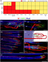
Classical proprioceptors and palisade endings in the EOMs of mammals. (a) Heatmap showing the presence or absence of classical proprioceptors and palisade endings in eye muscles in different mammals. Golgi tendon organs are present in even‐toed ungulates. In monkeys (asterisk), they are present in some, but not all, EOMs. The rest of the species lack Golgi tendon organs. Muscle spindles are only found in humans, monkeys and even‐toed ungulates. Palisade endings are present in all species, except mice and guinea pigs. In rabbits and rats, the palisade endings differ from the canonical palisade endings of higher mammals. (b, c) Visualization of classical proprioceptors in pig EOMs by immunofluorescence. Nerve fibres (red) are labelled with an antibody against neurofilament (NF), and nerve terminal (green) with an antibody against synaptophysin (SYP). Muscle fibres (blue) are counterstained with phalloidin (Phall). (b) The equatorial region of a muscle spindle. The axons (red) spiral around the intrafusal muscle fibres (blue) and establish synaptophysin‐positive contacts (green) on the intrafusal muscle fibres. (c) A Golgi tendon organ at the muscle–tendon junction. The tendon is not labelled and continues to the right of the muscle fibres (blue). The Golgi tendon organ is innervated by a single axon (red) which divides into several branches inside the Golgi tendon organ. Axonal branches establish synaptophysin‐positive nerve terminals (green). MF, muscle fibre; MTJ, muscle–tendon junction; T, tendon. (d–g) Palisade endings in cat EOMs are shown following immunofluorescence staining using the same staining combination as for the muscle spindle and the Golgi tendon organ and in schematic drawing (e). In the fluorescence staining (d, f, g) the tendon is not visible and continues to the right of the muscle fibres (blue). (d) Low magnification image showing palisade endings at the muscle–tendon junction. Palisade endings are formed by nerve fibres (red) which come from the muscle and extend into the tendon. There, they turn back to approach the muscle–tendon junction. By further splitting, axons establish synaptophysin‐positive terminal varicosities (green) around single muscle fibre tips. (e) Schematic drawing of a palisade ending. Terminal varicosities (green) of the palisade ending are found at the level of the tendon (T) and the muscle fibre (MF) tip. (f) The palisade ending from the inset in (d) is shown at high magnification. (g) The nerve fibre (red) that forms a palisade ending establishes multiple synaptophysin‐positive en grappe motor terminals (green) along the muscle fibre (blue). (f, g) From the publication ‘Palisade endings have an exocytotic machinery but lack acetylcholine receptors and distinct acetylcholine esterase activity’ (Blumer et al., 2020).

Similar articles

Cited by

References

    1. Abuel Atta, A. A. , DeSantis, M. , & Wong, A. (1997). Encapsulated sensory receptors within intraorbital skeletal muscles of a camel. Anatomical Record, 247(2), 189–198. - PubMed
    1. Alvarado‐Mallart, R. M. , & Pincon‐Raymond, M. (1979). The palisade endings of cat extraocular muscles: A light and electron microscope study. Tissue & Cell, 11(3), 567–584. - PubMed
    1. Balslev, D. , & Miall, R. C. (2008). Eye position representation in human anterior parietal cortex. Journal of Neuroscience, 28(36), 8968–8972. - PMC - PubMed
    1. Balslev, D. , Mitchell, A. G. , Faria, P. J. M. , Priba, L. , & Macfarlane, J. A. (2022). Proprioceptive contribution to oculomotor control in humans. Human Brain Mapping, 43(16), 5081–5090. - PMC - PubMed
    1. Bewick, G. S. , & Banks, R. W. (2015). Mechanotransduction in the muscle spindle. Pflugers Archiv: European Journal of Physiology, 467(1), 175–190. - PMC - PubMed

Publication types