. 2023 Apr;601(8):1407-1424.
doi: 10.1113/JP284026. Epub 2023 Mar 16.

Antagonism of TRPV4 channels partially reduces mechanotransduction in rat skeletal muscle afferents

Affiliations

Antagonism of TRPV4 channels partially reduces mechanotransduction in rat skeletal muscle afferents

Ayumi Fukazawa et al. J Physiol. 2023 Apr.

Abstract

Mechanical distortion of working skeletal muscle induces sympathoexcitation via thin fibre afferents, a reflex response known as the skeletal muscle mechanoreflex. However, to date, the receptor ion channels responsible for mechanotransduction in skeletal muscle remain largely undetermined. Transient receptor potential vanilloid 4 (TRPV4) is known to sense mechanical stimuli such as shear stress or osmotic pressure in various organs. It is hypothesized that TRPV4 in thin-fibre primary afferents innervating skeletal muscle is involved in mechanotransduction. Fluorescence immunostaining revealed that 20.1 ± 10.1% of TRPV4 positive neurons were small dorsal root ganglion (DRG) neurons that were DiI-labelled, and among them 9.5 ± 6.1% of TRPV4 co-localized with the C-fibre marker peripherin. In vitro whole-cell patch clamp recordings from cultured rat DRG neurons demonstrated that mechanically activated current amplitude was significantly attenuated after the application of the TRPV4 antagonist HC067047 compared to control (P = 0.004). Such reductions were also observed in single-fibre recordings from a muscle-nerve ex vivo preparation where HC067047 significantly decreased afferent discharge to mechanical stimulation (P = 0.007). Likewise, in an in vivo decerebrate rat preparation, the renal sympathetic nerve activity (RSNA) and mean arterial pressure (MAP) responses to passive stretch of hindlimb muscle were significantly reduced by intra-arterial injection of HC067047 (ΔRSNA: P = 0.019, ΔMAP: P = 0.002). The findings suggest that TRPV4 plays an important role in mechanotransduction contributing to the cardiovascular responses evoked by the skeletal muscle mechanoreflex during exercise. KEY POINTS: Although a mechanical stimulus to skeletal muscle reflexively activates the sympathetic nervous system, the receptors responsible for mechanotransduction in skeletal muscle thin fibre afferents have not been fully identified. Evidence suggests that TRPV4 is a mechanosensitive channel that plays an important role in mechanotransduction within various organs. Immunocytochemical staining demonstrates that TRPV4 is expressed in group IV skeletal muscle afferents. In addition, we show that the TRPV4 antagonist HC067047 decreases the responsiveness of thin fibre afferents to mechanical stimulation at the muscle tissue level as well as at the level of dorsal root ganglion neurons. Moreover, we demonstrate that intra-arterial HC067047 injection attenuates the sympathetic and pressor responses to passive muscle stretch in decerebrate rats. These data suggest that antagonism of TRPV4 attenuates mechanotransduction in skeletal muscle afferents. The present study demonstrates a probable physiological role for TRPV4 in the regulation of mechanical sensation in somatosensory thin fibre muscle afferents.

Keywords: group IV muscle afferents; mechanotransduction; muscle mechanoreflex; primary sensory neuron; transient receptor potential vanilloid 4.


Conflict of interest statement

COMPETING INTERESTS

The authors declare that they have no competing interests.

Figures
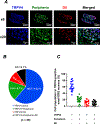
Figure 1.
Figure 1.. Co-localization of transient receptor potential vanilloid 4 (TRPV4) and peripherin in L4-L6 dorsal root ganglion (DRG) neurons subserving skeletal muscle sensory neurons in rats
A, representative images showing the expression of TRPV4, and peripherin-positive neurons in DiI retrograde-labeled L4-L6 DRGs innervating gastrocnemius muscle. Merged immunofluorescence images show co-localization of TRPV4 and peripherin in DiI-labeled DRG neurons (white arrowhead). Magnifications: ×6 and ×20. Scale bars: 300 or 30 μm. B, the percentages of peripherin- and/or DiI-positive neurons in TRPV4-positive small DRG neurons. Data are shown as the mean. ± SD. C, individual data obtained from distinct sections. A total of 20 DRG sections were collected from three rats.
Figure 2.
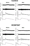
Figure 2.. Sample recordings of mechanically-activated (MA) inward currents before and after application of HEPES-buffered solution (Control) or TRPV4 antagonist solution (HC067047)
A, continuous sample recordings of MA current before and 5 min after the application of HEPES buffered solution (control solution). From top to bottom: raw current recordings in voltage clamp mode, representations of mechanical stimuli (see details in Methods) and step numbers of mechanical stimuli. B, C and D, samples of MA currents (rapidly adapting (RA); inter-mediately adapting (IA); and slowly adapting (SA)) at the same intensities that mechanical threshold was marked before the application of control solution, respectively. These currents were recorded from the different cells. E, continuous sample recording of MA current before and 5 min after the application of 1 µM HC067047 solution (TRPV4 antagonist solution). F, G, and H, samples of MA currents (RA, IA, SA) at the same intensities that mechanical threshold was marked before the application of HC067047 solution, respectively. These currents were recorded from the different cells.
Figure 3.
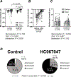
Figure 3.. Average changes in MA current amplitude (A and B), average changes in mechanical threshold (C) and population changes in mechanical threshold (D) in small DRG neurons from before to 5 min after application of HEPES-buffered solution (control) or TRPV4 antagonist solution (HC067047)
B, enlarged view of MA current amplitude of HC067047 trials in A. D, DRG neurons were classified by the difference in mechanical threshold (ΔMt) from the value before test solutions were applied compared to after the test applications were applied: decreased, ΔM ≤−2 steps; unchanged, ΔMt ≤ ±1 step; and increased, ΔMt ≥ 2 steps. Data are shown as the mean. ± SD. Two-way ANOVA was performed, followed by the Bonferroni’s multiple comparison test (A and C). Fischer’s exact test was performed (D). DRGs were collected from 10 rats and 42 small DRG neurons were assessed.
Figure 4.
Figure 4.. Response of group IV muscle afferents to mechanical stimulation before and 10 min after intramuscular injection of Krebs buffer solution (control) or TRPV4 antagonist solution (HC067047)
From top to bottom; the raw recording of individual group IV fibre activity and intensity of mechanical stimuli.
Figure 5.
Figure 5.. Average changes in mechanical threshold (A) and response of magnitude (B) in group IV muscle afferents from before to 10 min after intramuscular injection of Krebs buffer solution (control) or TRPV4 antagonist solution (HC067047)
Data are shown as the mean ± SD. Two-way ANOVA was performed, followed by the Bonferroni’s multiple comparison test. EDL muscle with the common peroneal nerve was isolated from 12 rats and 14 mechanosensitive group IV fibres were assessed.
Figure 6.
Figure 6.. Arterial blood pressure (ABP) and sympathetic responses to activation of the muscle mechanoreflex via passive stretch before and 5 min after intraarterial injection of saline (control) or TRPV4 antagonist solution (HC067047)
From top to bottom; the raw recording of ABP, raw and normalized renal sympathetic nerve activity (RSNA), and tension.
Figure 7.
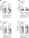
Figure 7.. Peak changes in mean arterial pressure (MAP) (A), heart rate (HR) (B), renal sympathetic nerve activity (RSNA) (C), and developed tension (D) during passive hindlimb muscle stretch before to 5 min after intraarterial injection of saline (control) or TRPV4 antagonist solution (HC067047)
Data are shown as the mean ± SD. Two-way ANOVA was performed, followed by the Bonferroni’s multiple comparison test. ABP in 16, HR in 16, RSNA in 11 and tension in 16 rats were assessed.

Similar articles

Cited by

References

    1. Ariano MA, Armstrong RB & Edgerton VR. (1973). Hindlimb muscle fiber populations of five mammals. J Histochem Cytochem 21, 51–55. - PubMed
    1. Barabas ME, Mattson EC, Aboualizadeh E, Hirschmugl CJ & Stucky CL. (2014). Chemical structure and morphology of dorsal root ganglion neurons from naive and inflamed mice. J Biol Chem 289, 34241–34249. - PMC - PubMed
    1. Bernardi MM & Neto JP. (1979). Effects of abrupt and gradual withdrawal from long-term haloperidol treatment on open field behavior of rats. Psychopharmacology (Berl) 65, 247–250. - PubMed
    1. Brohawn SG, Su Z & MacKinnon R. (2014). Mechanosensitivity is mediated directly by the lipid membrane in TRAAK and TREK1 K+ channels. Proc Natl Acad Sci U S A 111, 3614–3619. - PMC - PubMed
    1. Cao DS, Yu SQ & Premkumar LS. (2009). Modulation of transient receptor potential Vanilloid 4-mediated membrane currents and synaptic transmission by protein kinase C. Mol Pain 5, 5. - PMC - PubMed

Publication types

Substances